
Clinical Brain Imaging Stephen Salloway M D M
Clinical Brain Imaging Stephen Salloway, M. D. , M. S. Department of Clinical Neurosciences and Psychiatry Brown Medical School

Nobel Prize for Medicine 2003 Paul C. Lauterbur Sir Peter Mansfield
Objectives • Learn about the use of CT, MRI and SPECT scanning in clinical practice • Recognize key anatomical landmarks and begin to recognize and describe the appearance of common disorders Please look at the scans on your patients.

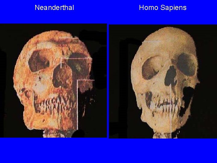
Neanderthal Homo Sapiens

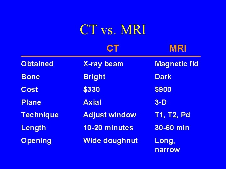
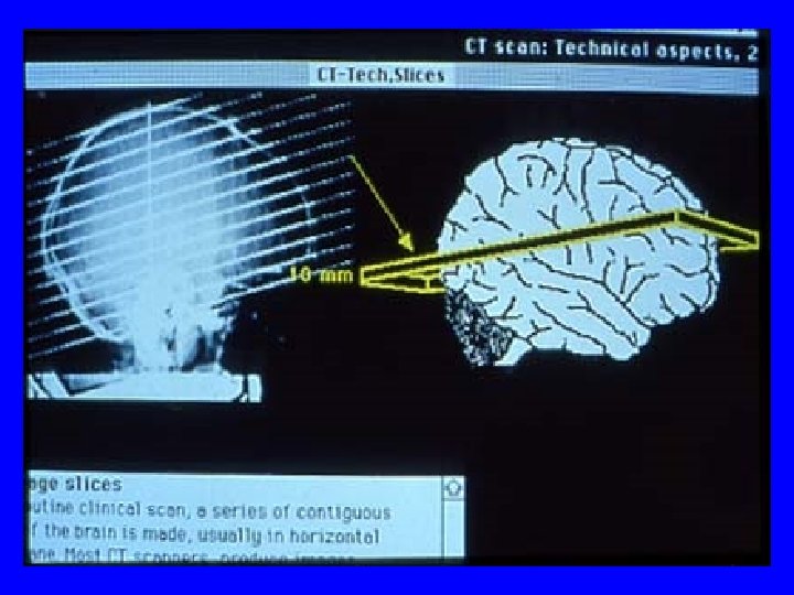
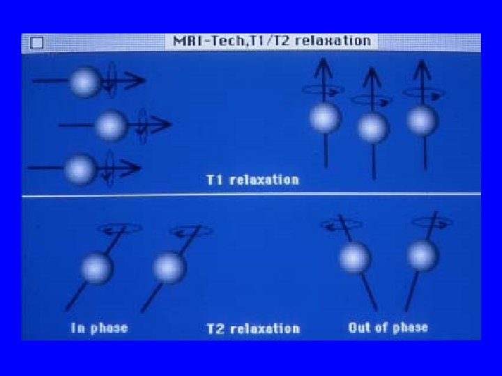
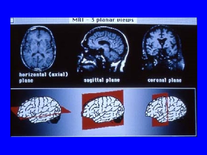
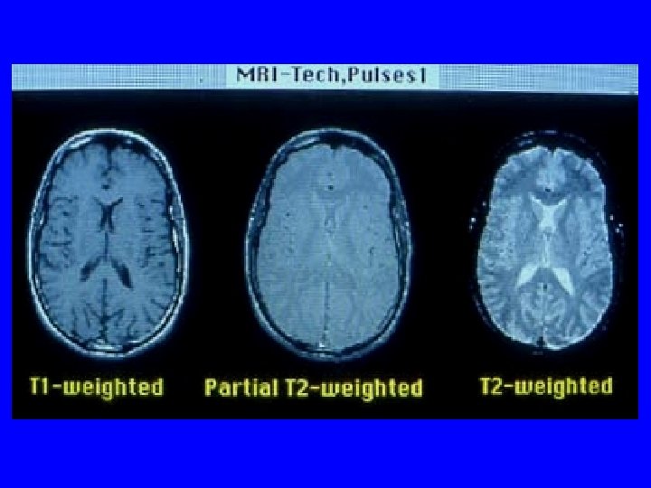
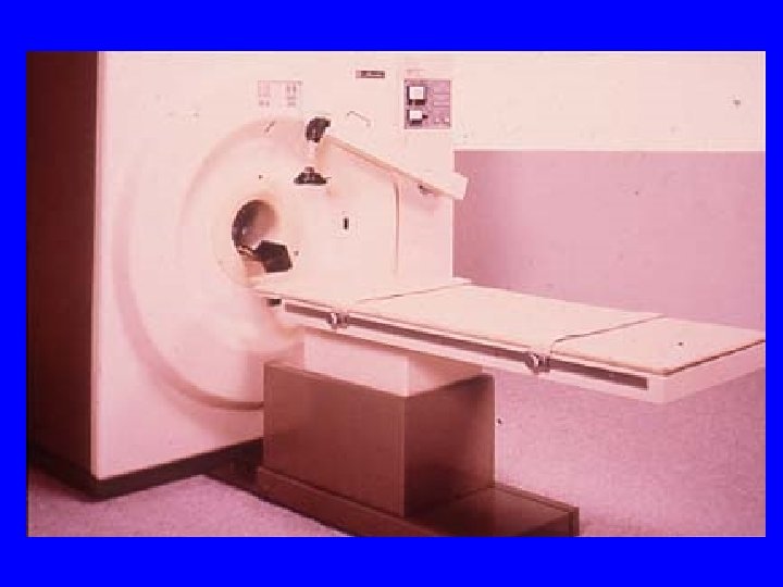
CT vs. MRI CT MRI Obtained X-ray beam Magnetic fld Bone Bright Dark Cost $330 $900 Plane Axial 3 -D Technique Adjust window T 1, T 2, Pd Length 10 -20 minutes 30 -60 min Opening Wide doughnut Long, narrow
Advantages to CT • • • Costs less than MRI Better access Shows up acute bleed A good quick screen Good visualization of bony structures and calcified lesions
Disadvantages to CT • Resolution • Beam-hardening artifact • Limited views of the posterior fossa and poor visualization of white-matter disease
Advantages to MRI • Good resolution—excellent view of brain structure • 3 dimensions • Good gray-white differentiation • Adjust settings based on characteristics of the lesion • Good view of the posterior fossa
Advantages to MRI • No radiation exposure • Gadolinium contrast is relatively nontoxic • Capacity for quantitative imaging, 3 -D reconstruction, angiography, spectroscopy
Disadvantages of MRI • Cost • Some patients ineligible because of pacemakers, other metal • Claustrophobia • Long exam • Access
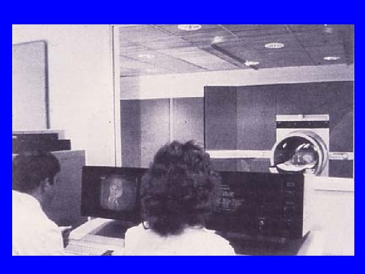
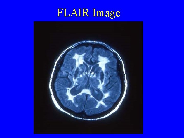
FLAIR Image
MRI Is the Test of Choice for Evaluating • Most lesions causing epilepsy—temporal lobe glioma, mesial temporal sclerosis • White-matter disease—subcortical dementia, HIV, MS • Lesions in the posterior fossa • TBI—axonal injury • Extent of anoxic injury, herpes encephalitis • Frontal atrophy, NPH • Other—brain anomalies, SLE, vasculitis, sagittal sinus thrombosis, pituitary lesions, AVM
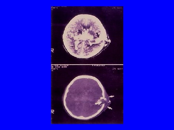
What Is Bright on CT? • • • Blood Contrast Bone Calcium Metal What Is Dark on CT? • Air • CSF/H 20

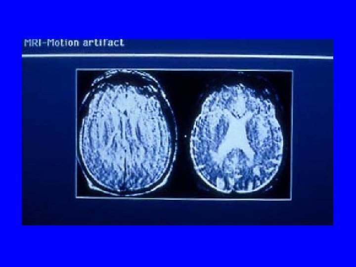
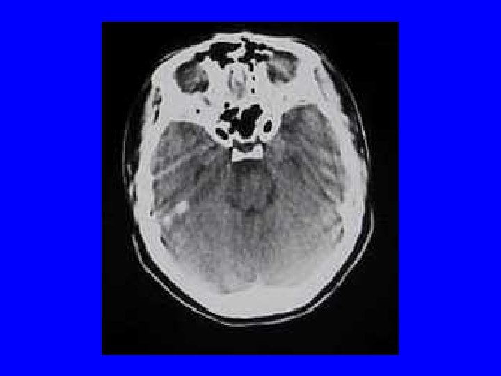
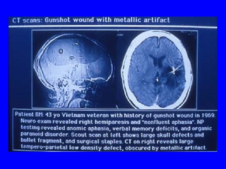
Artifacts • • Beam hardening Bone Foreign body Motion
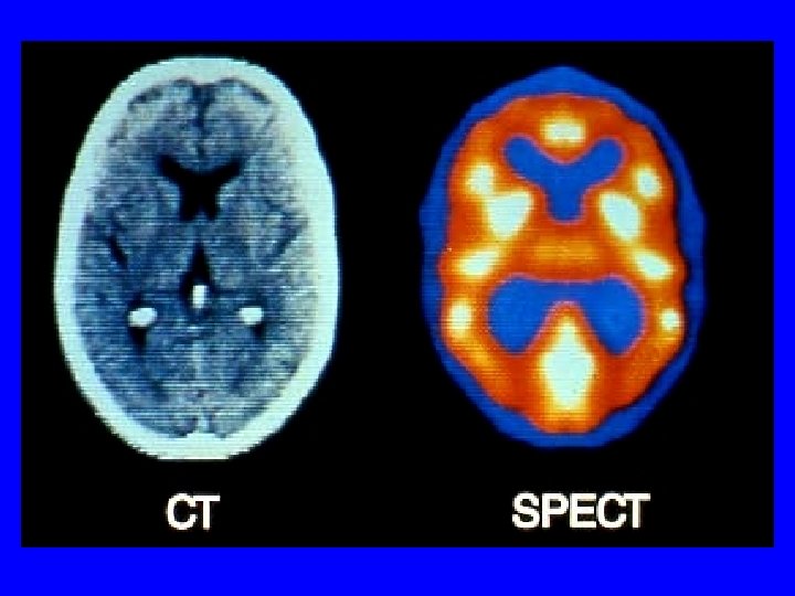
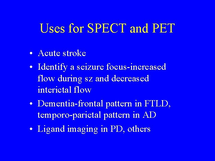
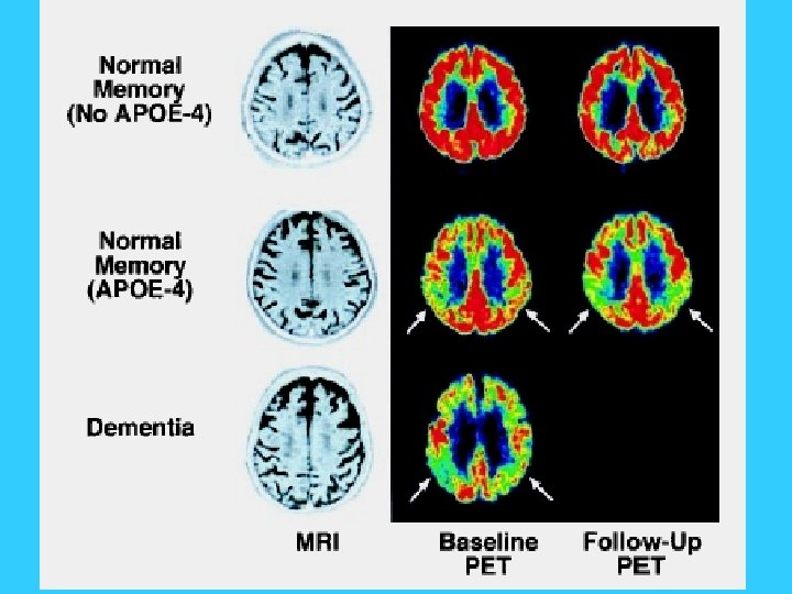
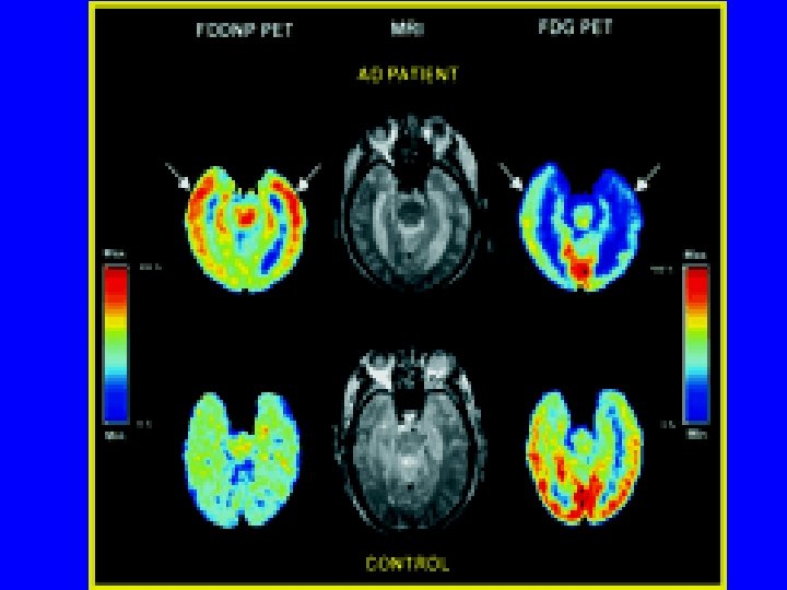
Uses for SPECT and PET • Acute stroke • Identify a seizure focus-increased flow during sz and decreased interictal flow • Dementia-frontal pattern in FTLD, temporo-parietal pattern in AD • Ligand imaging in PD, others
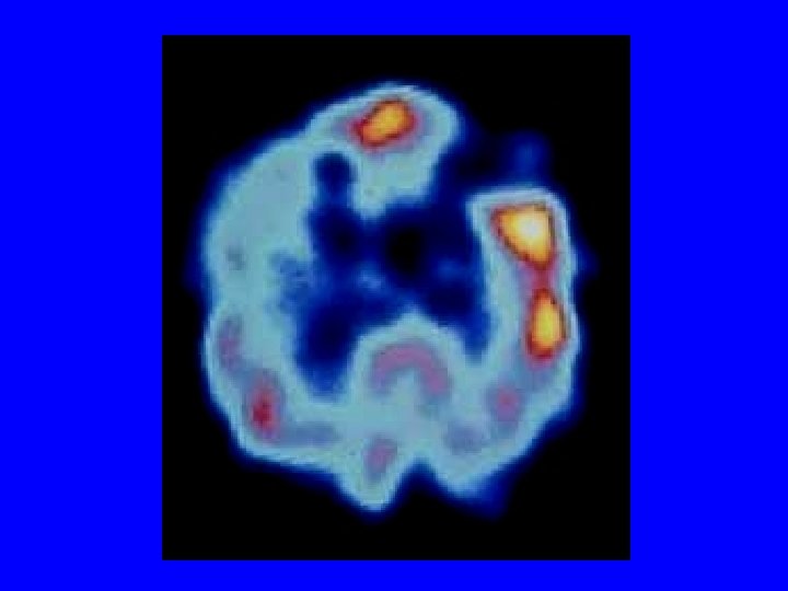
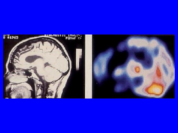
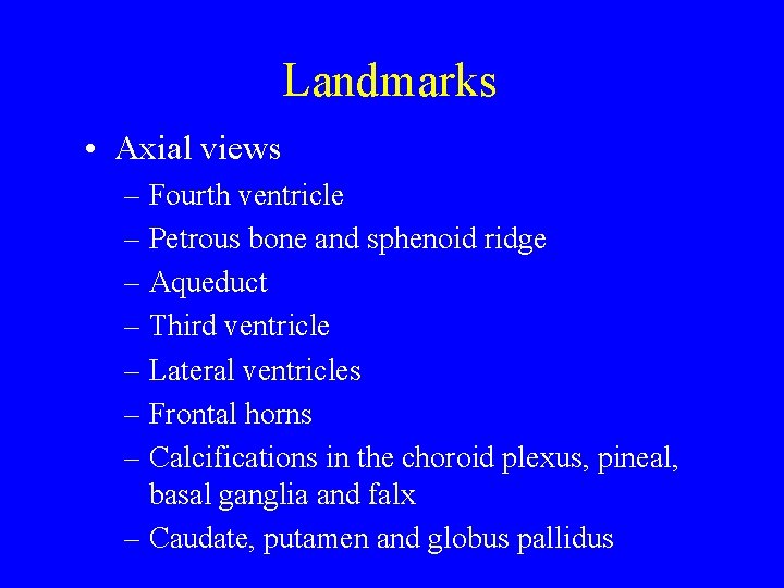
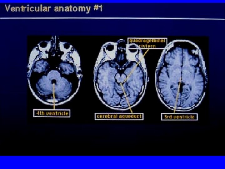
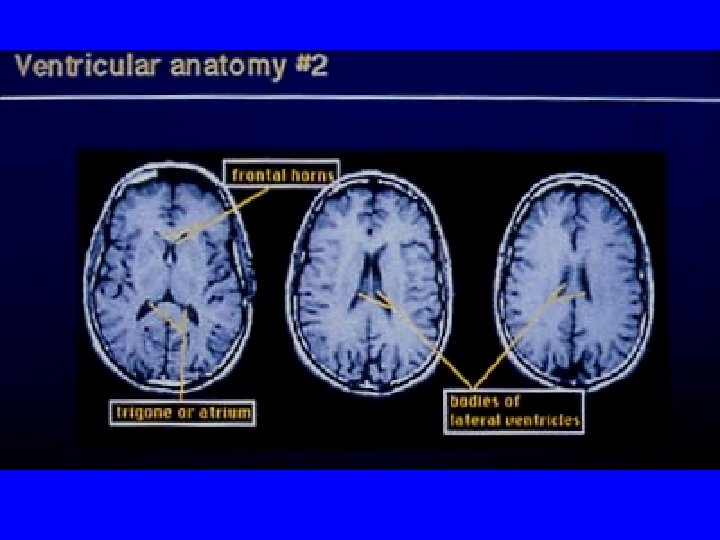
Landmarks • Axial views – Fourth ventricle – Petrous bone and sphenoid ridge – Aqueduct – Third ventricle – Lateral ventricles – Frontal horns – Calcifications in the choroid plexus, pineal, basal ganglia and falx – Caudate, putamen and globus pallidus
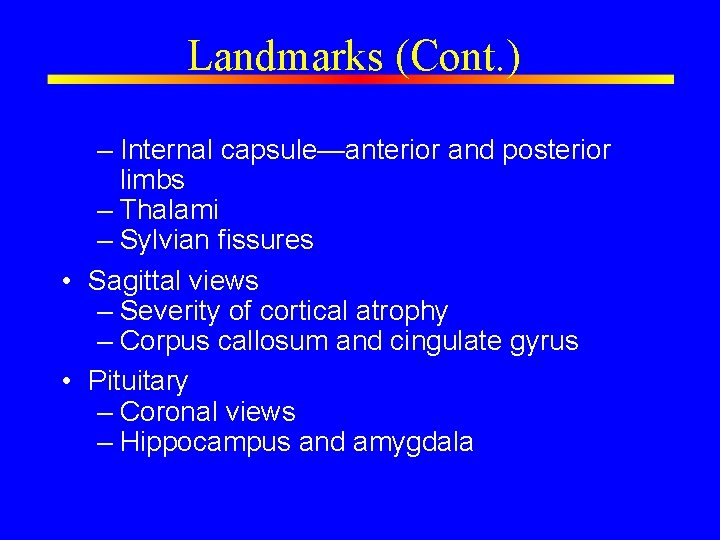
Landmarks (Cont. ) – Internal capsule—anterior and posterior limbs – Thalami – Sylvian fissures • Sagittal views – Severity of cortical atrophy – Corpus callosum and cingulate gyrus • Pituitary – Coronal views – Hippocampus and amygdala
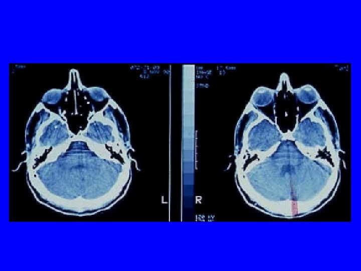
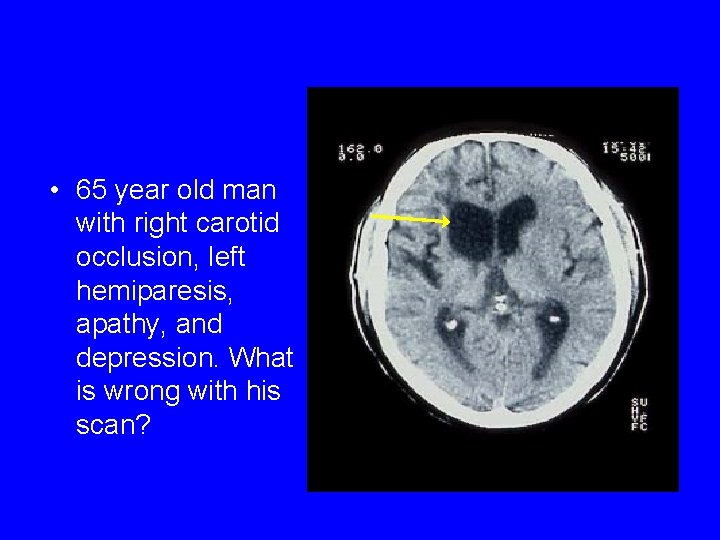
• 65 year old man with right carotid occlusion, left hemiparesis, apathy, and depression. What is wrong with his scan?
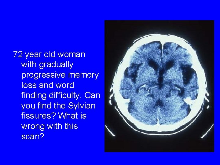
72 year old woman with gradually progressive memory loss and word finding difficulty. Can you find the Sylvian fissures? What is wrong with this scan?
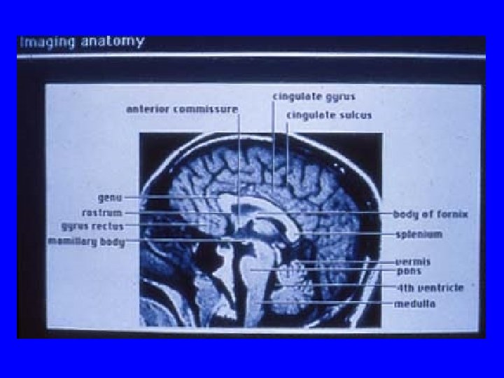

Normal Hippocampus
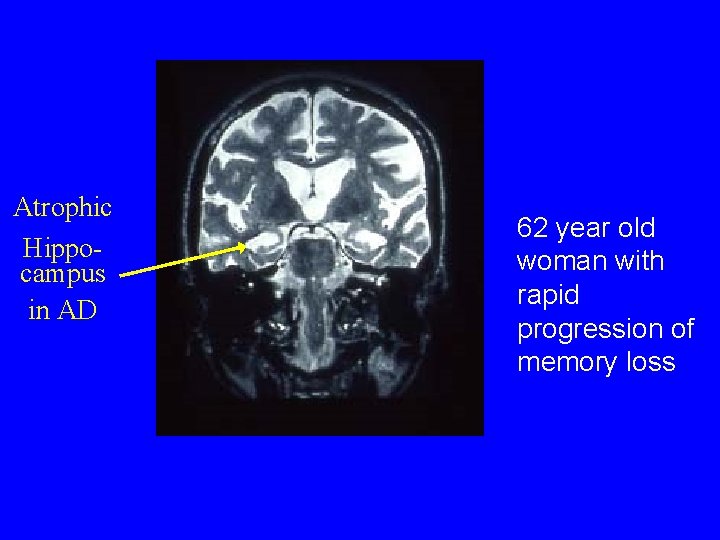
Atrophic Hippocampus in AD 62 year old woman with rapid progression of memory loss
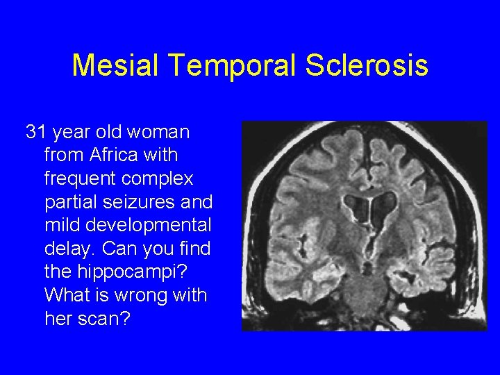
Mesial Temporal Sclerosis 31 year old woman from Africa with frequent complex partial seizures and mild developmental delay. Can you find the hippocampi? What is wrong with her scan?
Introduction to Scan Interpretation • Is the scan – Contrast or noncontrast? – Good quality? • Describe the abnormality – Size—small, punctuate, medium, large – Shape—round, well circumscribed, ovoid, irregular, patchy
Introduction to Scan Interpretation (Cont. ) • Signal intensity – High signal, hyperdense – Low signal, hypodense – Isointense, isodense – Mixed signal • Location

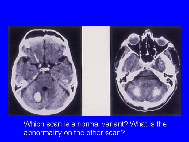
Which scan is a normal variant? What is the abnormality on the other scan?
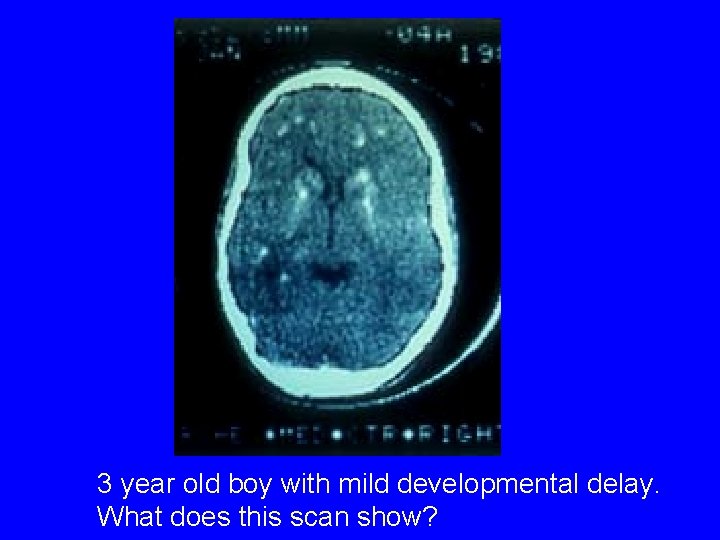
3 year old boy with mild developmental delay. What does this scan show?
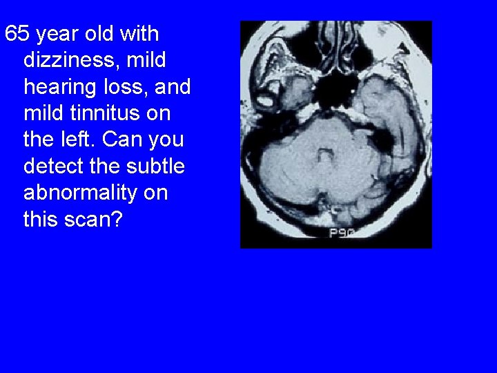
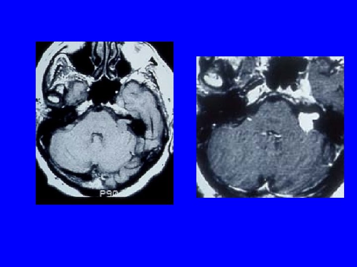
65 year old with dizziness, mild hearing loss, and mild tinnitus on the left. Can you detect the subtle abnormality on this scan?
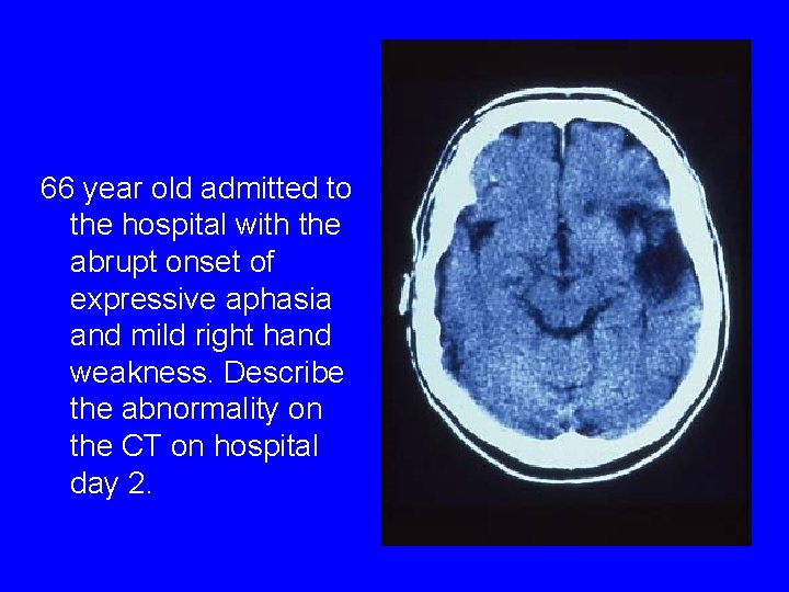
66 year old admitted to the hospital with the abrupt onset of expressive aphasia and mild right hand weakness. Describe the abnormality on the CT on hospital day 2.
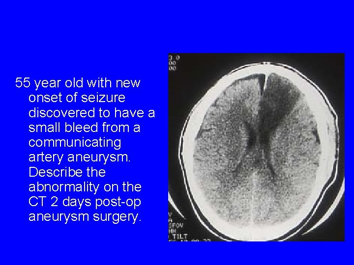
55 year old with new onset of seizure discovered to have a small bleed from a communicating artery aneurysm. Describe the abnormality on the CT 2 days post-op aneurysm surgery.
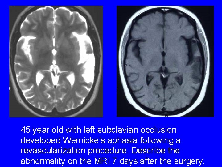
45 year old with left subclavian occlusion developed Wernicke’s aphasia following a revascularization procedure. Describe the abnormality on the MRI 7 days after the surgery.
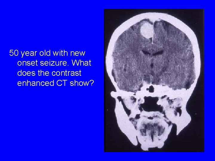
50 year old with new onset seizure. What does the contrast enhanced CT show?
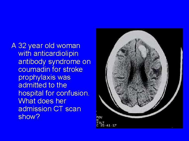
A 32 year old woman with anticardiolipin antibody syndrome on coumadin for stroke prophylaxis was admitted to the hospital for confusion. What does her admission CT scan show?
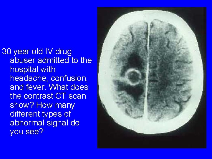
30 year old IV drug abuser admitted to the hospital with headache, confusion, and fever. What does the contrast CT scan show? How many different types of abnormal signal do you see?
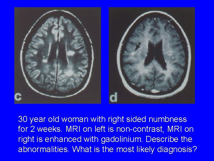
30 year old woman with right sided numbness for 2 weeks. MRI on left is non-contrast, MRI on right is enhanced with gadolinium. Describe the abnormalities. What is the most likely diagnosis?
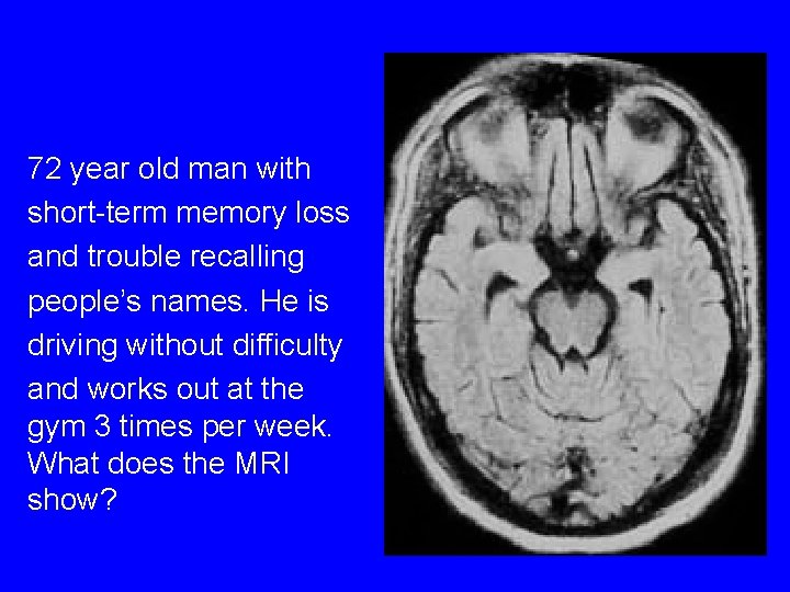
72 year old man with short-term memory loss and trouble recalling people’s names. He is driving without difficulty and works out at the gym 3 times per week. What does the MRI show?
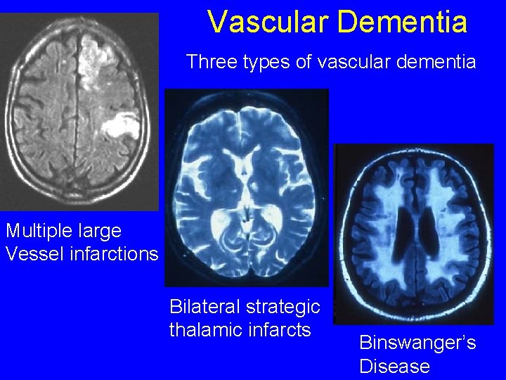
Vascular Dementia Three types of vascular dementia Multiple large Vessel infarctions Bilateral strategic thalamic infarcts Binswanger’s Disease
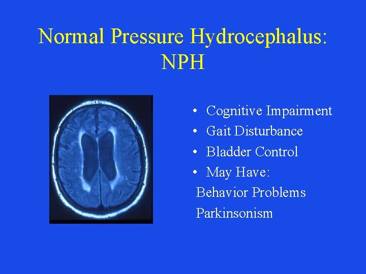
Normal Pressure Hydrocephalus: NPH • Cognitive Impairment • Gait Disturbance • Bladder Control • May Have: Behavior Problems Parkinsonism
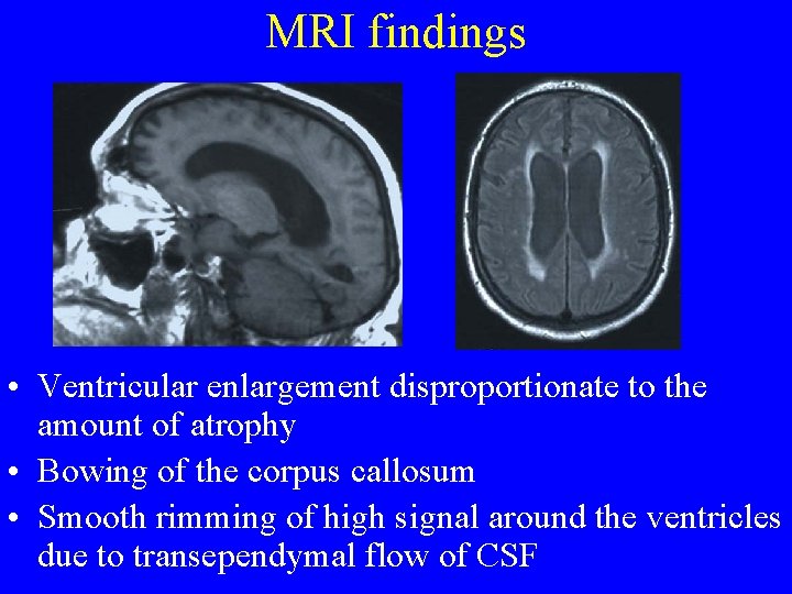
MRI findings • Ventricular enlargement disproportionate to the amount of atrophy • Bowing of the corpus callosum • Smooth rimming of high signal around the ventricles due to transependymal flow of CSF
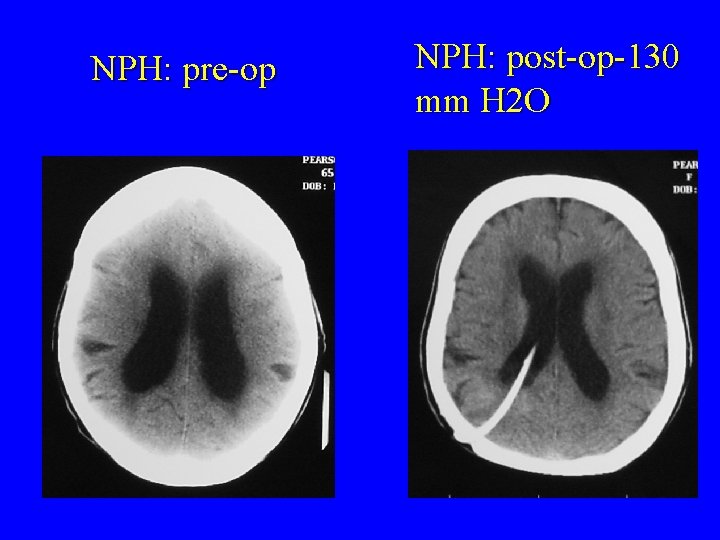
NPH: pre-op NPH: post-op-130 mm H 2 O

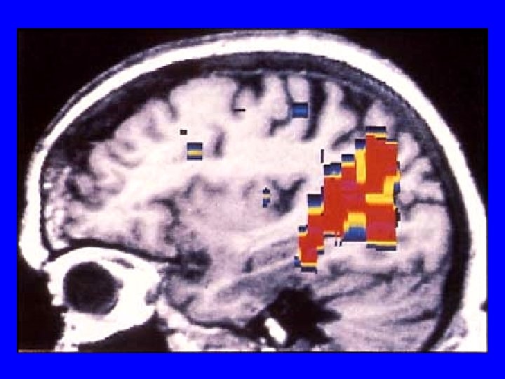
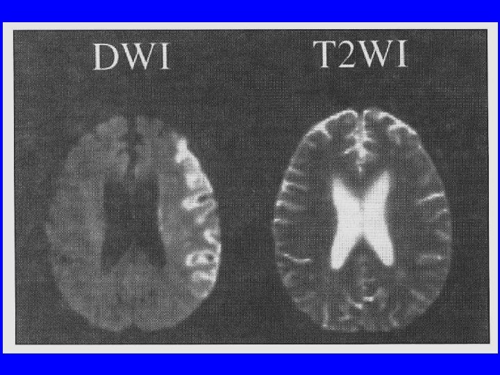
Types of f. MRI • BOLD-f. MRI which measures regional differences in oxygenated blood • Diffusion-weighted f. MRI which measures random movement of water molecules. Diffusion tensor imaging (DTI) measures diffusion of water in different directions and is a good test for studying white matter tracts. • MRI spectroscopy which can measure certain cerebral metabolites non-invasively
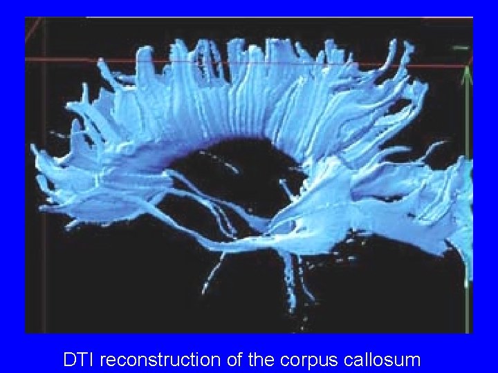
DTI reconstruction of the corpus callosum
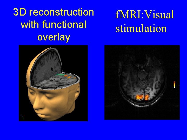
3 D reconstruction with functional overlay f. MRI: Visual stimulation
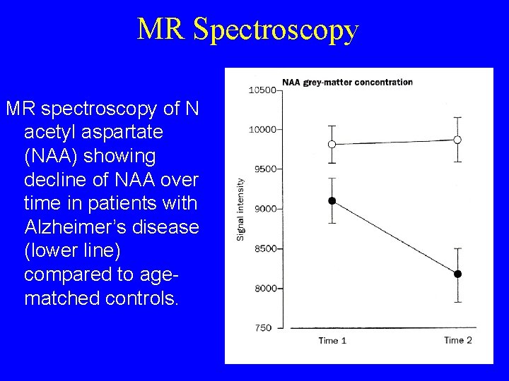
MR Spectroscopy MR spectroscopy of N acetyl aspartate (NAA) showing decline of NAA over time in patients with Alzheimer’s disease (lower line) compared to agematched controls.
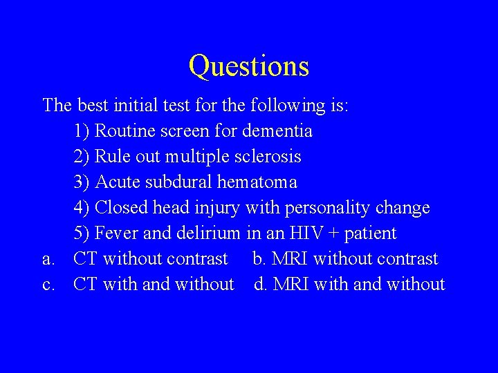
Questions The best initial test for the following is: 1) Routine screen for dementia 2) Rule out multiple sclerosis 3) Acute subdural hematoma 4) Closed head injury with personality change 5) Fever and delirium in an HIV + patient a. CT without contrast b. MRI without contrast c. CT with and without d. MRI with and without
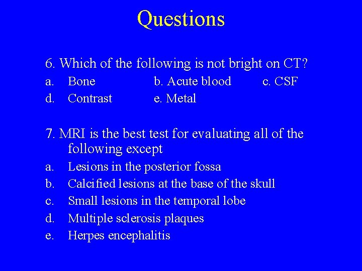
Questions 6. Which of the following is not bright on CT? a. Bone d. Contrast b. Acute blood e. Metal c. CSF 7. MRI is the best test for evaluating all of the following except a. b. c. d. e. Lesions in the posterior fossa Calcified lesions at the base of the skull Small lesions in the temporal lobe Multiple sclerosis plaques Herpes encephalitis

Answers 1. A 5. D 2. B 6. C 3. A 7. B 4. B
- Slides: 72