Clinical biochemistry The hypothalamus and pituitary gland Presanted

Clinical biochemistry The hypothalamus and pituitary gland Presanted by: Huda Jaber

GENERAL PRINCIPLES OF ENDOCRINE DIAGNOSIS A hormone can be defined as a substance secreted by an endocrine gland that is transported in the blood, therebyregulating the function of another tissue(s). Ex. GH, T 4, INSULIN and rout of secretion from glands Conversely, trophic hormones from the pituitary gland stimulate target endocrine glands to synthesize and secrete further hormones, which in turn partly control. trophic hormone release, usually by negative feedback inhibition • example, hypercalcaemia and parathyroid hormone (PTH), (inversely proportional)and plasma T 4 concentration inhibits the secretion of thyroid stimulating hormone(TSH) Endocrine glands may secrete excessive or deficient amounts of hormone • Abnormalities of target glands may be primary or secondary to dysfunction of the controlling mechanism, usually located in the hypothalamus or anterior pituitary gland.

dynamic’ tests Suppression tests are used mainly for the differential diagnosis of excessive hormone secretion The substance (or an analogue) that normally suppresses secretion by negative feedback is administered and the. response is measured Failure to suppress implies that secretion is not under normal feedback control.

Stimulation tests are used mainly for the differential diagnosis of deficient hormone secretion. The trophic hormone that normally stimulates secretion is administered and the response is measured. . A normal response excludes an abnormality of the target gland, whereas failure to respond confirms it.

HYPOTHALAMUS AND PITUITARY GLAND There are two lobes of pituitary gland anterior and posterior lobes. Both depend on hormones synthesized in the hypothalamus for normal function The hypothalamus also has extensive neural connections with the rest of the brain. Stress and some psychological disorders affect the secretion of pituitary hormones and of the hormones from other endocrine glands.
 ––")
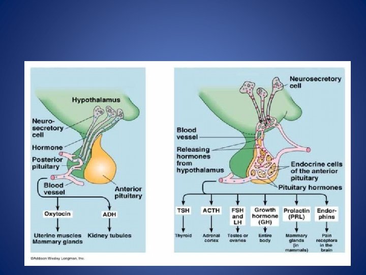
Control of posterior pituitary hormones Two structurally similar peptide hormones antidiuretic hormone (ADH) –– and oxytocin, are synthesized in the hypothalamus and transported to pituitary stalk attached to specific carrier proteins – neurophysins. . Then stored in the posterior pituitary gland are. released into the bloodstream under hypothalamic control, together with neurophysin Neurophysin has no apparent biological function and is rapidly cleared from plasma.
 is mainly synthesized in the hypothalamus and enhances water reabsorption")
Antidiuretic hormone (arginine vasopressin) is mainly synthesized in the hypothalamus and enhances water reabsorption from the collecting ducts in the kidneys. Oxytocin is synthesized in the hypothalamus. It controls the ejection of milk from the lactating breast and may have a role in initiating uterine contractions It may be used therapeutically to induce labour.

Anterior pituitary hormones There is no direct neural connection between the hypothalamus and the anterior pituitary gland. The hypothalamus synthesizes small molecules (regulating hormones or factors) that are carried to the cells of the anterior pituitary lobe by the hypothalamic portal system This network of capillary loops, which, after passing down the pituitary stalk, divide into a second capillary network in the anterior pituitary gland, from where hypothalamic hormones stimulate or inhibit pituitary hormone secretion into the systemic circulation.

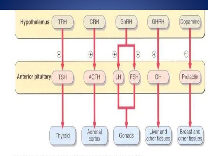
The cells of the anterior pituitary lobe can be classified simply by their staining reactions as acidophils, basophils or chromophobes. Acidophils are of two cell types: lactotrophs, which secrete prolactin somatotrophs, which secrete GH (somatotrophin). These hormones, which are simple polypeptides with similar amino acid sequences, mainly affect peripheral tissues directly. Stimulation and inhibition of secretion via the hypothalamus is influenced by neural stimuli.

Basophils secrete hormones that affect other endocrine glands. The hypothalamic control is mainly stimulatory. There are three cell types: Corticotrophs synthesize a large polypeptide (pro-opiomelanocortin), which is a precursor of both adrenocorticotrophic hormone (ACTH; corticotrophin) and βlipotrophin Secretion of these hormones occurs in parallel Adrenocorticotrophic hormone stimulates the synthesis and secretion of steroids, and maintains adrenal cortical growth. Gonadotrophs secrete the gonadotrophins, folliclestimulating hormone (FSH) and luteinizing hormone (LH), which act on the gonads Thyrotrophs secrete TSH (thyrotrophin), which acts on the thyroid gland. These hormones are structurally similar glycoproteins consisting of two subunits, α and β. The α -subunit is common to all three hormones; the β -subunit is important for receptor recognition. Chromophobes, once thought to be inactive, do contain secretory granules .
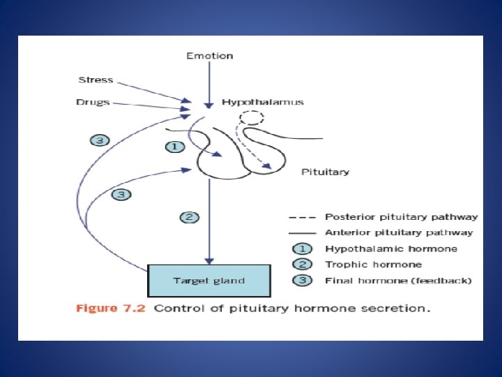

Extra hypothalamic neural stimuli modify, and at times over-ride, other control mechanisms Physical or emotional stress and mental illness may give similar findings to, and even precipitate, endocrine disease. The stress caused by insulin-induce hypoglycaemia is used to test anterior pituitary function. Stress may also stimulate the secretion of ADH from the posterior pituitary

Feedback control is mediated by the concentrations of circulating target-cell hormones; a rising concentration usually suppresses trophic hormone secretion This negative feedback may directly suppress hypothalamic hormone secretion or may modify its effect on pituitary cells (long feedback loop The secretion of hypothalamic hormones may also be suppressed by rising concentrations of pituitary hormone in a short feedback loop.

Evaluation of anterior pituitary function The diagnosis of suspected hypopituitarism is best excluded by the direct measurement of pituitary hormones after stimulation or by demonstrating target gland hyposecretion after the administration of the relevant trophic hormone. However, prolonged hypopituitarism may result in secondary failure of the target gland with diminished response to stimulation. Laboratory tests establish only the presence or absence of hypopituitarism, and the cause must be sought by other clinical means such as radiological imaging

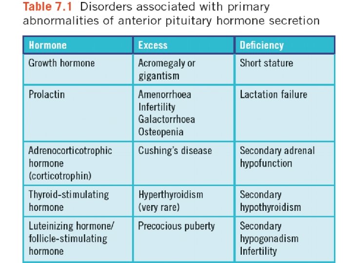
DISORDERS OF ANTERIOR PITUITARY HORMONE SECRETION The main clinical syndromes associated with excessive or deficient anterior pituitary hormone secretion are shown in Table bellow. Excessive secretion usually involves a single hormone, but deficiencies are often multiple. However, many pituitary tumours are nonsecretory and may present clinically with eye signs or headaches.

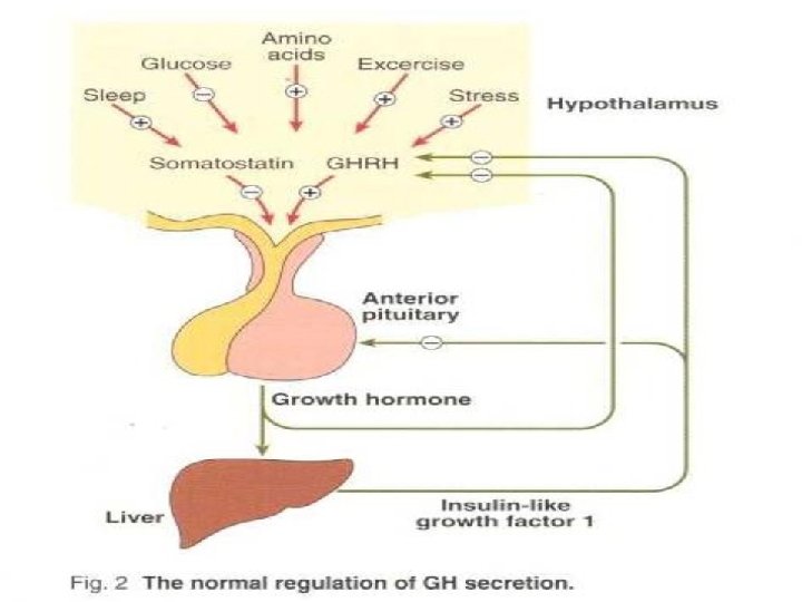
Growth hormone secretion from the anterior pituitary gland is mainly controlled by hypothalamic GH releasing hormone (GHR ) After synthesis by the hypothalamus, this is transported via the hypothalamic portal system to the somatotrophs of the anterior pituitary. Secretion of GHRH, and therefore of GH, is pulsatile, occurring about seven or eight times a day, usually associated with exercise, onset of deep sleep, in response to the falling plasma glucose concentration about an hour after meals. At other times, plasma concentrations are usually very low or undetectable, especially in children. Growth hormone release is inhibited in a negative feedback pathway by another hypothalamic hormone, somatostatin (GH-release inhibiting hormone). Insulin like growth factor 1 (IGF-1) acts by feedback to inhibit GHRH action.

stress, one cause of which is hypoglycaemia, glucagon, some amino acids, for example arginine, drugs such as levodopa and clonidine. All these stimuli have been used to assess GH secretory capacity, which may also be impaired in obese patients, in hypothyroidism and hypogonadism, in some cases of Cushing’s syndrome and in patients receiving large doses of steroids.

Actions of growth hormone The main function of GH is to promote growth. Its action is primarily mediated by IGFs, polypeptides that are synthesized in many tissues, where they act locally. Plasma concentrations of one of these, IGF-1 (also known as somatomedin C), correlate with GH secretion. Carbohydrate metabolism is affected by GH: GH antagonizes the insulinmediated cell uptake of glucose, and excess secretion may produce glucose intolerance. Fat metabolism is stimulated by GH: lipolysis is stimulated, with a consequent increase in the concentration of circulating free fatty acids. Free fatty acid antagonizes insulin release and action. Growth hormone enhances protein synthesis, in conjunction with insulin, to stimulate amino acid uptake by cells. The production of IGF-1 is also influenced by other factors, the most important

Growth hormone excess: gigantism and acromegaly Growth hormone excess causes gigantism during childhood and acromegaly in adults. Most patients with GH excess have acidophil adenomas of the anterior pituitary gland, which may be secondary to excessive hypothalamic stimulation. Rarely, malignant tumours may release GH or GHRH. The clinical manifestations of GH excess depend on whether the condition develops before or after fusion of the bony epiphyses. Gigantism is caused by excess GH secretion in childhood before fusion of the epiphyseal plates, which may be delayed by accompanying hypogonadism. Heights of up to about 2 metres may be reached. Acromegalic features may develop after bony fusion, but these patients may die in early adult life from infection or cardiac failure or as a consequence of progressive pituitary tumour growth

The features of acromegaly • An increase in the bulk of bone and soft tissues with enlargement of, for example, the hands, tongue, jaw and heart. Changes in facial appearance are often marked, due to the increasing size of the jaw and sinuses; the gradual coarsening of the features may pass unnoticed for many years. Thyroid gland enlargement may be clinically detectable, but the patient is usually euthyroid. • Excessive hair growth, hyperhidrosis and sebaceous gland secretion are common. • Menstrual disturbances are common in females. • Impaired glucose tolerance is present in about 25 per cent of patients, about half of whom develop symptomatic diabetes mellitus. In most cases the pancreas can secrete enough insulin to overcome the antagonistic effect of GH. • There is a predisposition to multiple pre-malignant colon polyposis and hypertension. • Hyperphosphataemia, hypercalcaemia and hypertriglyceridaemia may also be present.
 is more sensitive than computerized tomograph (CT) scanning.")
Diagnosis • Magnetic resonance imaging (MRI) is more sensitive than computerized tomograph (CT) scanning. • Plasma GH concentrations are usually higher than normal. • The diagnosis is confi rmed by demonstrating a raised plasma GH conc. that is not suppressed by a rise in plasma glucose concentration. In normal subjects, plasma GH concentrations fall to very low levels – to below 1 mg/L after a 75 g oral glucose load. • In acromegaly, the secretion of GH is autonomous and this fall may not occur or be only slight, or there may even be a paradoxical rise. Growth hormone secretion is inhibited by hyperglycaemia in the normal subject. Glucose suppression test for suspected acromegaly Procedure: After an overnight fast, insert an indwelling intravenous cannula. After at least 30 min, take basal samples for plasma glucose and GH estimation. The patient should drink 75 g of glucose dissolved in about 300 m. L of water, or an equivalent glucose load. Take samples for glucose and GH assays at 30, 60,

Treatment Surgery to remove the adenoma, Medical therapy, usually with either bromocriptine or cabergoline (dopamine receptor agonists) or somatostatin analogues (somatostati n itself has too short a half-life for effective therapeutic use). Octreotide or lanreotide, which bind to somatostatin receptors, can be used or pegvisomant (GH receptor antagonist) Radiation therapy. The aim of treatment is to ameliorate symptoms and to obtain an oral glucose suppressed GH concentration of less than 1 mg/L (this cut-off can be GH assay dependent – conversion factor for units: m. U/L = 2. 4 ¥ mg/L) and normalization of plasma IGF-1 concentrations.

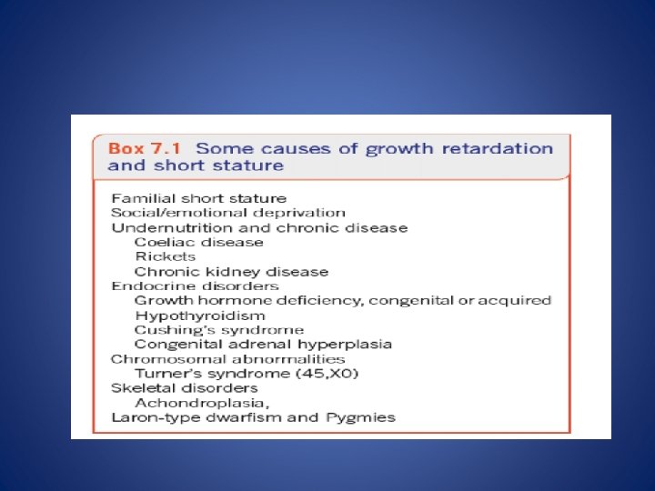
Growth hormone deficiency In adults, GH deficiency may cause clinical symptoms, such as tiredness, dyslipidaemia and increased cardiovascular disease. Growth hormone deficiency can cause short stature in children. It is important to investigate children with reduced growth rate to identify those who may benefit from recombinant human GH replacement treatment. Isolated GH deficiency is most commonly secondary to idiopathic deficiency of hypothalamic GHRH

Diagnosis • It is, important to exclude hypothyroidism, chronic diseases and malabsorption states, poor nutritional state. Clinical examination should assess for obvious syndromes, pubertal status, bone age, growth or growth velocity. • Karyotyping may be indicated if a chromosomal disorder such as Turner’s syndrome (45, X 0) is suspected. the end of prepuberty. Thus, in children with bone age more than 10 years, priming with sex hormones before investigation may be necessary. For example, ethinyloestradiol may be given to girls and testosterone to boys prior to testing

• A low plasma IGF-1 concentration may be a useful screening test. • Urinary GH excretion, either in 24 -h collections or timed overnight, may offer a relatively safe screening test. • If blood is taken at a time when physiologically high concentrations are expected, the need for stimulation tests may be avoided, for example 60– 90 min after the onset of sleep and about 20 min after vigorous exercise. • If GH deficiency is not excluded by the above measurements, it is necessary to perform one or more stimulation tests • The response of GH to insulin may be the most reliable to detect GH deficiency, but it is not without the risk of fatal hypoglycaemia. • Glucagon could also be used as an alternative • A GH absolute response of greater than 20 m. U/L (7 μg/L) makes GH deficiency unlikely. Other such stimuli include arginine, clonidine or the GHRH test. Once GH deficiency has been established, a cause should be sought by appropriate clinical and imaging means.

• Figure shows an algorithm for the investigation of short stature.

DISORDERS OF POSTERIOR PITUITARY • Disorders of the posterior pituitary are rare • compared with those of the anterior pituitary • Deficiency of ADH in diabetes insipidus may present as polyuria • In • the syndrome of inappropriate ADH, hyponatraemia due to water excess occurs. .

HYPOPITUITARISM • Hypopituitarism is a syndrome of deficiency of pituitary hormone production that may result from disorders of the hypothalamus, pituitary or surrounding structures. • Clinical features of deficiency are usually absent until about 70 per cent of the gland has been destroyed, unless there is associated hyperprolactinaemia, when amenorrhoea and infertility may be early symptoms. • Panhypopituitarism alludes to the involvement of all pituitary hormones; • alternatively, only one or more may be involved, as in partial hypopituitarism. • Suspicion of anterior pituitary hypofunction usually arises in patients presenting with various features such as clinical and radiological evidence of a pituitary or localized brain tumour, hypogonadism, adrenocortical insufficiency, short stature caused • by GH deficiency, and hypothyroidism • Gonadotrophins are often the first to decrease in hypopituitarism and it is unusual for the post-pituitary hormones such as ADH and oxytocin to be affected

Consequences of pituitary hormone deficiencies • Progressive pituitary damage usually presents with evidence of deficiencies of gonadotrophins and GH . Plasma ACTH and/or TSH concentrations may • remain normal, or become deficient months or even years later.

The clinical and biochemical consequences of the target-gland failure include the following: • Growth retardation in children This may be due to deficiency of GH; deficiency of TSH, and therefore of thyroid hormone, may contribute. • Secondary hypogonadism This is due to gonadotrophin deficiency, presenting as amenorrhoea, infertility and atrophy of secondary sexual characteristics with loss of axillary and pubic hair and impotence or loss of libido. Puberty is delayed in children. • Secondary adrenocortical hypofunction (ACTH deficiency) In contrast to the primary form (Addison’s disease), patients are not hyperpigmented because ACTH secretion is not raised. • The sodium and water deficiency and hyperkalaemia characteristic of Addison’s disease do not usually occur because aldosterone secretion (which is controlled by angiotensin and not by ACTH) is normal.

• However, cortisol is needed for normal free water excretion, and consequently there may be a dilutional hyponatraemia due to cortisol deficiency. • Cortisol is also necessary for the maintenance of normal blood pressure. Hypotension may be associated with ACTH deficiency. • Cortisol and/or GH deficiency may cause increased insulin sensitivity with fasting hypoglycaemia.
 This may sometimes be clinically indistinguishable from primary")
• Secondary hypothyroidism (TSH deficiency) This may sometimes be clinically indistinguishable from primary hypothyroidism. • Prolactin deficiency Associated with failure to lactate, this may occur after post-partum pituitary infarction (Sheehan’s syndrome). • However, in hypopituitarism due to a tumour, plasma prolactin concentrations are often raised and may cause galactorrhoea (secretion of breast fluid). • Patients with hypopituitarism, like those with Addison’s disease, may die because of an inability to secrete an adequate amount of cortisol in response to stress caused by, for example, infection or surgery. • Other life-threatening complications are hypoglycaemia and hypothermia.

Pituitary tumours The clinical presentation of pituitary tumours depends on the type of cells involved and on the size of the tumour (microadenomas less than 10 mm and macroadenomas more than 10 mm). • of secretory cells may produce the clinical effects of excess hormone secretion: Tumours • excess prolactin causes infertility, amenorrhoea and varying degrees of galactorrhoea • excess GH causes acromegaly or gigantism, • excess • ACTH causes Cushing’s syndrome. Large pituitary tumours may present with: • visual • disturbances caused by pressure on the optic chiasma or headache due to raised intracranial pressure, • deficiency of some or all of the pituitary hormones due to destruction of secretory cells in the gland. Non-secreting tumours are difficult to diagnose using biochemical tests, although the combined pituitary stimulation test may indicate subclinical impairment of function. Hyperprolactinaemia, which may be asymptomatic, is a valuable biochemical markerof the presence of a pituitary tumour. Prolactin may be secreted by the tumour cells or by unaffected lactotrophs if tumour growth interferes with the normal inhibition of prolactin secretion

Investigation of suspected hypopituitarism • Deficiency of pituitary hormones causes hypofunction of the target endocrine glands. • Investigation aims to confirm such deficiency, to exclude disease of the target gland then to test pituitary hormone secretion after maximal stimulation of the gland. • Measurement should be made of the plasma concentrations of: • LH, FSH and oestradiol (female) or testosterone (male), • total or free T 4 and TSH, • prolactin, to test for hypothalamic or pituitary stalk involvement, • cortisol at 09. 00 h, to assess the risk of adrenocortical insufficiency during later testing.

• If the plasma concentration of the target gland hormone is low and the concentration of trophic hormone is raised, the affected target gland should be investigated. • Conversely, if the plasma concentrations of both the target gland trophic hormones are low or low-normal, consider a pituitary stimulation test. • Investigation of the pituitary region using radiological techniques such as CT or MRI scanning may help elucidate a cause of the hypopituitarism. • the combined pituitary stimulation test (insulin or glucagon plus TRH and Gn. RH given as one test), this is rarely required, as useful information can be obtained from the basal pituitary hormones and, if indicated, an insulin stimulation/ hypoglycaemia test, although this is not without risk.

Insulin tolerance or insulin stimulation test • This test is potentially dangerous and must be done under direct medical supervision due to severe hypoglycaemia and the test should be carried out only in specialist units by experienced staff. • It is contraindicated in the following patient groups: the elderly, patients with ischaemic heart disease, epilepsy or severe panhypopituitarism, and patients in whom plasma cortisol at 09. 00 h is less than 100 nmol/L.

• A resting electrocardiogram should be normal. • Hypothyroidism should be treated before hand as this can impair the cortisol and GH responses. • However, note that treatment with thyroxine can precipitate an adrenal crisis in such patients and thus corticosteroid replacement is also necessary. • Indications of the insulin stimulation test may include: • assessment of GH in growth deficiency, assessment of ACTH/cortisol reserve • differentiation of Cushing’s syndrome from pseudo- Cushing’s syndrome, for example depression or alcohol excess. Both ACTH and GH are released in response to the stress of hypoglycaemia. • Fifty millilitres ofin 20 case per severe cent glucose for intravenous administration must be immediately available symptomatic hypoglycaemia develops. • Care should be taken not to induce severe hyperglycaemia during infusion, as it may cause hyperosmolality, which can be dangerous. • Plasma cortisol is usually measured as an index of ACTH secretion. • If glucose needs to be given, continue with the sampling.

Procedure • After an overnight fast, fter at least 30 min, take basal samples at time 0 min for cortisol, GH and glucose. • Inject soluble insulin in a dose sufficient to lower plasma glucose concentrations to less than 2. 5 mmol/L and evoke symptomatic hypoglycaemia. • The recommended dose of insulin must be adjusted for the patient’s body weight and for the suspected clinical condition under investigation. • The usual dose is 0. 15 U/kg body weight. • If either pituitary or adrenocortical hypofunction is suspected, or if a low fasting glucose concentration has been found, reduce the dose to 0. 1 or 0. 05 U/kg. • If there is likely toacromegaly be resistance to the action insulin Cushing’s syndrome, or obesity, 0. 2 orof 0. 3 U/kg because may be of needed. • Take blood samples at 30, 45, 60, 90 and 120 min after the injections for cortisol, GH and glucose assays.

Interpretation • Interpretation is not possible if hypoglycaemia is not attained, and the dose of insulin cautiously be repeated if this is not attained in the 45 -min blood sample. • If hypoglycaemia has been adequate, plasma cortisol concentrations should rise by more than 200 nmol/L and exceed 580 nmol/L, and an adequate GH response occurs with an absolute response of greater than 20 m. U/L (7 • In Cushing’s syndrome, neither plasma cortisol nor μg/L). GH concentrations rise signifi cantly, although they usually do in cases of pseudo-Cushing’s syndrome. • After the test, a supervised meal should be given and the patient should not drive for at least 2 hours.

Glucagon stimulation test of the hypothalamus– pituitary axis • This test is useful if the insulin hypoglycaemic test is contraindicated. • However, it is essential that the test is carried out in a specialist unit by experienced staff. • The basic principle is that glucagon stimulates GH and ACTH release probably via a hypothalamic route. • The test is contraindicated if there is severe adrenal failure, for example if cortisol at 09. 00 h is less than 100 nmol/L or in hypothyroidism. • It is also unreliable in the presence of diabetes mellitus.

• Procedure • Patients should fast overnight, although they can drink water. • For adults, 1 mg of glucagon is injected subcutaneously at 09. 00 h. • Blood samples are taken at 0, 90, 120, 150, 180, 210 and 240 min for cortisol and GH.

• Interpretation • Plasma cortisol should normally rise by at least 200 nmol/L to more than 580 nmol/L, and an adequate GH response occurs with an absolute response of greater than 20 m. U/L (7 μg/L).

• Treatment of hypopituitarism • This consists of specific therapy depending on its cause and may include surgical removal of a large adenoma. • If the ACTH axis is impaired, it is essential to prescribe a glucocorticoid, for example hydrocortisone in the acute situation or prednisolone for maintenance. • Secondary hypothyroidism will need thyroid replacement. Adrenal replacement should precede T 4 therapy to avoid an Addisonian crisis Gonadotrophin deficiency may require testosterone in males and oestrogen replacement in • In children women, with or without asbe appropriate. and sometimes in progesterone adults, GH may indicated.
- Slides: 48