Clinical approach management of fractures Open fractures followup


Clavicle: Clavicle fractures are classified into 3 groups")


- Slides: 25
Clinical approach & management of fractures: Open fractures & follow-up Presented By: Laith Sorour
General terms: • Fracture A Break in the structural continuity of bone 1. Open (Compound) Fracture : the skin is breached 2. Closed (Simple) Fracture : overlying skin remains intact • Reduction Maneuver to restore proper alignment to fracture or joint • Closed reduction (Conservative) Reduction done without surgery (e. g. , casts, splints) • Open reduction (Operative) Surgical reduction • Fixation Stabilization of a fracture after reduction by means of surgical placement of hardware that can be external or internal (e. g. , pins, plates, screws)
Open Fractures • Definition : Wound overlying fracture, through which fracture fragments are in continuity with outside environment; high risk of infection (And this is what we fear from). • All open fractures assumed to be Contaminated so we MUST Prevent INFECTION ! • Which fracture has the highest mortality? Pelvic fracture (up to 50% with open pelvic fractures) • The treatment goals for open fractures are ? : 1. To prevent infection 2. To allow the fracture to heal 3. To restore function in the injured limb
Initial management of Open fractures At the scene of the accident or ER • ALWAYS Do ATLS protocol (ABCDE), especially Many patients with open fractures have multiple injuries and severe shock; for them, appropriate treatment at the scene of the accident is essential. • So first we care about 1)Life then 2)The limb(Splint the limb), 3)the wound(cover with sterile material) and lastly 4)the fracture itself
Classifying Gustilos classification of open fractures : • Type 1 is a low-energy fracture with a small, clean wound and little soft-tissue damage. • Type II is a moderate-energy fracture with a clean wound more than 1 cm long, but no skin flap, not much soft-tissue damage and no more than moderate comminution of the fracture. • Type III is a high-energy fracture with extensive damage to skin, soft tissue and neurovascular structures, and contamination of the wound. A. Type III A: the fractured bone can be adequately covered by muscle or skin, despite the laceration. B. C. Type III B: there is extensive periosteal stripping and fracture cover is not possible without the use of local or distant flaps. Type III C: if there is an arterial injury which needs to be repaired, regardless of the amount of other soft-tissue damage. • *** The incidence of wound infection correlates directly with the extent of soft-tissue damage, rising from less than 2% in Type I to over 10% in Type III fractures. The risk of infection also rises with increasing delay in obtaining soft-tissue coverage of the fracture
Type 1 type 2 TYPE IIIC TYPE IIIA TYPE IIIB
Tx of Open Fractures in the hospital What are the five steps in the initial treatment of an open fracture After ABCDE? 1. Prophylactic antibiotics to include IV gram-positive ± anaerobic coverage 2. Surgical débridement 3. Inoculation against tetanus 4. Early definitive wound cover & Lavage wound <6 hours postincident with sterile irrigation 5. Open reduction of fracture and stabilization (e. g. , use of external fixation) ***Repeated examination of the limb because open fractures can also be associated with compartment syndrome.
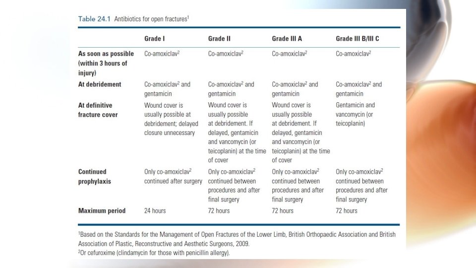
Sterility and A. B cover The wound must be kept covered until the patient reaches the operating theatre. Antibiotics are given as soon as possible, no matter how small the laceration, and are continued until the danger of infection has passed. • Usually we give: co-amoxiclav or cefuroxime • If the patient is allergic to penicillin and flucloxacillin give clindamycin
Debridement and wound excision • In the Operating theatre, never in ER. WHY? ! • should be performed by someone skilled in dealing with both skeletal and soft tissues; ideally this should be a joint effort by orthopaedic and plastic surgeons. • Under generalized anesthesia • Maintain traction on injured limb and hold it sill • Remove clothing • Replace dressing with sterile pad, clean and shave surrounding skin • Remove pad and irrigate wound with a lot of warm normal saline • DO NOT use a tourniquet • Extend wound and excise ragged margins but only enough to leave healthy skin edges • Remove foreign materials and tissue debris • Wash out wound again with warm NS (6 -12 L), A common mistake is to inject syringefuls of fluid through a small aperture – this only serves to push contaminants further in. Adding antibiotics or antiseptics to the solution has no added benefit • Remove devitalized tissue, devitalized tissue provides a nutrient medium for bacteria. All doubtfully viable tissue, whether soft or bony, should be removed • Best to leave cut nerves and tendons alone
Wound closure • A small, uncontaminated wound in a Grade I or II fracture may (after debridement) be sutured, provided this can be done without tension. • In the more severe grades of injury, fracture stabilization and wound cover using split-skin grafts, local or distant flaps is ideal, provided both orthopaedic and plastic surgeons are satisfied that the wound is clean and viable after debridement. (Delayed PrimaryTertiary healing) • If a combined plastic-and-orthopaedic approach is not available at the time of debridement, the fracture is stabilized by external fixation and then left open and dressed with an impervious dressing.
Tx of the Fracture • We begin with the fracture after finishing with wound. • Fracture Stability is important in – 1)Reducing the likelihood of infection 2) Assisting in recovery of the soft tissue • Method of fixation depends on: – Degree of contamination – Length of time from injury to operation – Amount of soft tissue damage • For all grades up to 3 A We follow the suggested rules for closed fractures, The only difference is that in open fractures there should be greater reluctance to resort to operative methods of fixation, especially if there seems to be serious risk of infection • Metallic internal fixation must be employed the metal should be placed well away from the wound • If fracture is unstable & unsuitable for tx by traction or by simple splintage alone, external fixation by pins inserted into the bone fragments and fixed to a rigid external bar is often the method of choice rather than internal fixation.
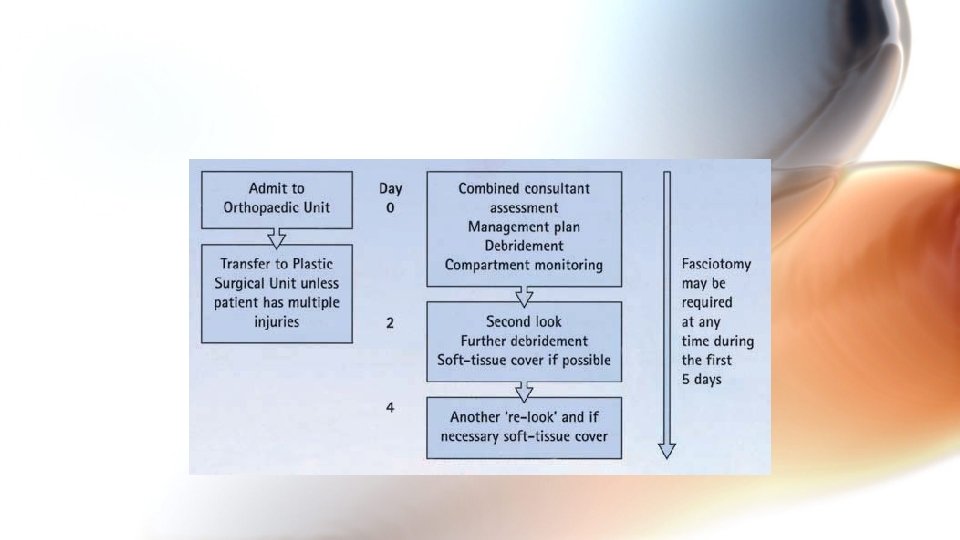
Aftercare and Team Work Post-op – Limb is elevated – Circulation carefully monitored – Antibiotic cover continued; swab samples will dictate whether a diff. antibiotic is needed – If wound has been left open, inspect in 2 -3 days. Delayed primary suture is then often safe or, if there has been much skin loss, plastic surgery for grafting may be necessary Teamwork – For optimal results, open fractures with skin and soft-T damage are best managed by a partnership of ortho and plastic surgeons, ideally from the outset rather than by later referral – If no plastic surgeon on site, use a digital camera for image transmission by internet to communicate and consult.
Pain • How to reduce pain? Fracture immobilization, ice, elevation of the affected limb and analgesia. *About Analgesia? -For minor fractures NSAIDs or Acetomenophane -For traumas with severe pain Short acting Opoids + NSAIDS/Acetomenphane (to lower the dose of opoid) **Acute fracture pain usually lasts from a few days to a week, if pain persists or increases after that there must be something wrong, like what? -Acute Compartment syndrome, pressure related injury to the skin or soft tissue, movement or displacement of fracture due to inadequate immobilization , complications due to cast or splint
Pharmacologic management • Vit D. is usually prescribed or recommend sun bathing since a lot of people are deficient. • Patient are encouraged to reduce smoking and alcohol because they impair healing. • It is still not clear if some drugs accelerate healing or not, some of them are being studied like GH, PTH, BMP, PDGF and bisphonates. • Vit. C is used to prevent Regional pain syndrome?
Follow up and patient education • We don’t treat the fracture, we treat the patient. So we need to educate him and follow up him even after the cast is removed. • Interval between visits depend on 1)type of fracture 2)type of cast 3)patient compliance • As general rule states, lower limb casting needs more immobilization time than upper. • We must tell the patient if he will get any of these he will need immediate reevaluation: -new or worsening pain –diminished circulation, signs? –diminished motor function? – bleeding or discharge from the cast –feeling pressure or numbness –significant damage to the splint or cast
Follow up for Stable fractures • Initial follow up after casting is usually after 3 -7 days and even earlier if the patient feel pain, swelling or any acute symptoms. • What we need to check? -signs of wear and proper fit of the cast. -teach the patient rehabilitation methods (exercise, compliance with activity restriction).
Follow up for unstable fractures • It depends on fracture type & age • Children need 2 -3 times a week for first 3 weeks of the injury onset, why? • In adults once every 7 -10 days • We must do an x-ray over the cast to check if the alignment is good.
Examples on management of common fractures 1)Clavicle: Clavicle fractures are classified into 3 groups based on site of fracture, where each group have it own treatment: • Group I fractures: Middle third injuries • Group II fractures: Distal third injuries • Group III fractures: Medial (proximal) third injuries *vast majority of clavicle fractures heal with nonoperative management.
Clavicle Surgical indications include the following: • Complete fracture displacement • Severe displacement causing tenting of the skin with the risk of puncture • Fractures with 2 cm of shortening • Comminuted fractures with a displaced transverse "zed" (or Z-shaped) fragment • Neurovascular compromise • Displaced medial clavicular fractures with mediastinal structures at risk • Polytrauma (with multiple fractures): To expedite rehabilitation • Open fractures • An inability to tolerate closed treatment • Fractures with interposed muscle • Established, symptomatic nonunion • Concomitant glenoid neck fracture (floating shoulder)
Cont. . In addition; to an orthopedic surgeon if the fracture requires surgical fixation, consultations include the following: • General or thoracic surgeon: If an associated pneumothorax is identified • Vascular surgeon: For a suspected subclavian vessel injury • Group 1: Nonoperative treatment can be divided into the following 2 categories: Simple support of the extremity - As in a sling or a sling and swath Reduction and immobilization - Typically with figure-of-eight brace, gives advantage of using two hands. ***Healing time may be as short as 2 weeks for infants, with most adults healing in 4 -6 weeks. Immobilization should continue until repeat radiographs show callus formation and healing across the fracture site.
Fractures of forearm in Adults • Unless the fragments are in close apposition, reduction is difficult and re-displacement in the cast almost inevitable. Consequently, most surgeons opt for open reduction and internal fixation from the outset. The fragments are held by plates and screws. The deep fascia is left open, Why? and only the skin and subcutaneous tissues are sutured. After the operation, the arm is kept elevated until the swelling subsides, and during this period active exercises of the hand are encouraged. If the fracture is not comminuted and the patient is reliable, early range-of-movement exercises are commenced, but lifting and sports are avoided. • It takes 8– 12 weeks for the bones to unite
Phalangeal fractures ***Phalangeal fractures usually result from direct trauma and therefore any part of the bone may be broken. Treatment: • Open wounds should always be treated first. Skin must be preserved and carefully sutured, and wound healing must not be jeopardized by the treatment of the fractures. ***Undisplaced fractures: • These need the minimum of splintage. The type of splint depends on the fracture and the direction of potential instability. This may necessitate strapping the finger to its uninjured neighbour (‘buddystrapping’) or a splint on the digit alone. Early movement, without compromising stability, is encouraged. ***Displaced fracture of the proximal or middle phalanx: • The bone should be straightened under local anaesthesia, carefully avoiding malrotation; the injured finger is then splinted, leaving the other fingers free. The splint can be discarded after 3 weeks. If reduction cannot be held in this way, fixation with K-wires, a small plate or screws is indicated depending on the type of fracture. Surgery is challenging and invites stiffness. Meticulous attention to surgical technique and postoperative rehabilitation is needed. ***Fractures of the distal phalanx *-*Distal phalangeal fractures are usually due to crushing injuries or a blow from a hammer. The soft-tissue damage must be treated; the fracture can be ignored.
Femoral shaft fracture • The risk of systemic complications in these highenergy injuries can be significantly reduced by early stabilization of the fracture. Traction can reduce and hold most fractures in reasonable alignment and certainly the patient should be transported from the scene of the accident with the limb splinted – many emergency splints incorporate a facility to apply traction as well. Definitive treatment will depend on the type of fracture and patient. 1)Intramedullary nailing • Intramedullary nailing is the method of choice for most femoral shaft fractures, The basic implant system consists of an intramedullary nail (in a range of sizes) which is perforated near each end so that locking screws can be inserted transversely at the proximal and distal ends; this controls rotation and ensures stability. 2)Traction and bracing • The main indications for traction are: (1) fractures in children; (2) contraindications to anaesthesia; and (3) lack of suitable skill or facilities for internal fixation. • poor choice for elderly patients, for pathological fractures and for those with multiple injuries • While the patient is in traction, joint mobility must be preserved by encouraging movement and exercises. The position of the fragments should be checked repeatedly by x-ray 3) Open reduction and plating: • High complication rate, including implant failure. The main indications today are (1) the combination of shaft and femoral neck fractures and (2) a shaft fracture with an associated vascular injury • 4) External fixation • The main indications for external fixation are: (1) the treatment of severe open injuries; (2) management of patients with multiple injuries (damage control orthopaedics); and (3) dealing with severe bone loss