Click to edit Master title style Edit Master

 They have heteroxenous life cycle 2) They need vector")
 They have different morphological forms TRYPOMASTIGOTE TRYPANOSOMAL FORM EPIMASTIGOTE FORM CRITHIDIAL FORM -Promastigote")


& Polymorphic. Trypanosoma (Trypanosoma brucei complex) are of the same")

 The caustive species : -Trypanosoma brucei complex: Trypanosoma")


 The causative species : -Trypanosoma cruzi 1 -")


 Organisms")
 in histeocytes and")


. C-Animal inoculation. D-Xenodiagnosis:")


, doesn’t multiply in peripheral blood but")

- Slides: 62
Click to edit Master title style • Edit Master text styles • Second level • Third level • Fourth level • Fifth level Parasitology Department
Genus: Trypanosoma Genus: Leishmania 1) They have heteroxenous life cycle 2) They need vector for transmission.
3) They have different morphological forms TRYPOMASTIGOTE TRYPANOSOMAL FORM EPIMASTIGOTE FORM CRITHIDIAL FORM -Promastigote leptomonad form Amastigote leishmania form
Learning objectives By the end of this lecture you should be able to: • List Trypanosoma species. • Determine the Definitive host, Reservoir host and habitat. • Mention the mode of infection and infective stage (s). • Describe the life cycle of Trypanosoma species. • Correlate clinical picture to the pathogenesis and pathology. • Discuss management as regards both the diagnosis, with emphasis on diagnostic specimens, diagnostic stage and methods of diagnosis, as well as the treatment options. • Outline prevention and control methods.
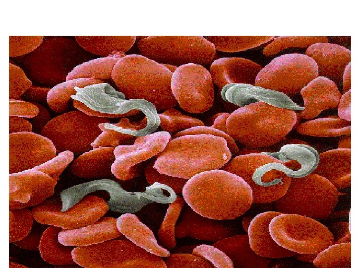
Trypanosomes are haemoflagellates that live in the blood &tissues. They take Trypomastigote form (Trypanosomal form ) in the blood of their vertebrate hosts. Morphological criteria of Trypomastigote (Trypanosomal form ) -It is spindle shaped 15 -35 u, has posterior kinetoplast. -They may be : - 1 -With variation in length , without free flagellum Polymorphic trypomastigote. 2 -with the same size , C shaped d with prominent kinetoplast Monomorphic trypomastigote.
Trypomastigotes are classified into : Polymorphic Trypanosoma • They take different shapes and size. • They include Trypanosoma brucei gambiense and Trypanosoma brucei rhodesiense. • They cause African. Trypanosomiasis (Sleeping sickness). Monomorphic Trypanosoma • They take the same size &shape. • They include Trypanosoma cruzi • They cause American Trypanosomiasis (Chagas disease)
Monomorphic Trypanosoma (Trypanosoma cruzi )& Polymorphic. Trypanosoma (Trypanosoma brucei complex) are of the same genus …… but the Geographical distribution, Insect vector, Life cycle , and Clinical picture , are quite different ……………
Polymorphic Trypanosomiasis African Trypanosomiasis Sleeping sickness
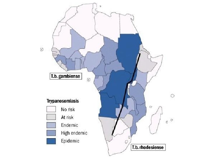
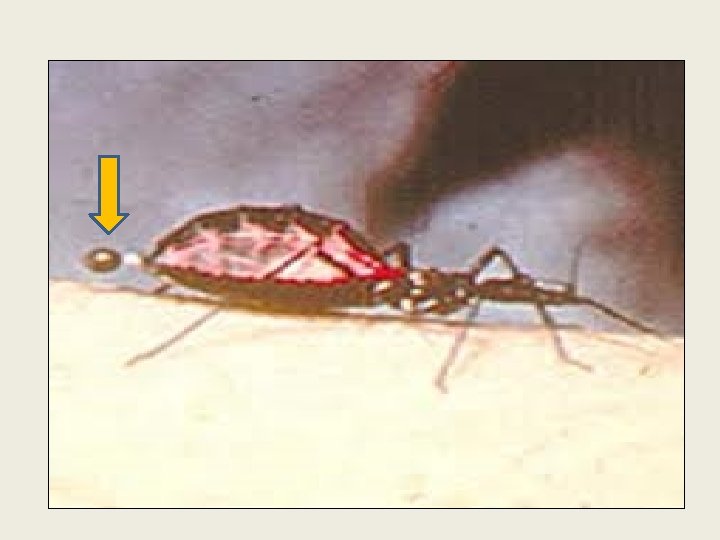
Polymorphic Trypanosomiasis African Trypanosomiasis (Sleeping Sickness) The caustive species : -Trypanosoma brucei complex: Trypanosoma brucei gambiense Trypanosoma brucei rhodesiene 1 - Both species are pathogenic to humans causing sleeping sickness, transmitted by Glossina fly (Tsetse fly ). They differ in: 1 - Geographical distribution within Africa. 2 - Species of Glossina fly. 3 - Growth rate & pathogenesis.
Vector born disease
West Africa East Africa
INFECTIVE STAGE CYCLOPROPAGATIVE DIAGNOSTIC STAGE
Morphological forms seen in the life cycle of African Trypanosomiasis : - 1 2 Two forms: 1 -Polymorphic trypomastigote form. 2 -Epimastigote form. TRYPOMASTIGOTE TRYPANOSOMAL FORM EPIMASTIGOTE FORM CRITHIDIAL FORM are seen either in the vector or in the vertebrate host -Promastigote leptomonad form leis
Morphological forms seen in The vector Tsetse fly 1 - Trypomastigote form In midgut 2 -Epimastigote (crithidial form) In hypopharynx 3 - Metacyclic trypomastigote In salivary gland It is short stumpy trypomastigote It is the infective stage The vertebrate host R. H: - Cattle , goats D. H: - Man 1 -Polymorphic trypomastigote It may be long slender , intermediate , or Short and stumpy.
CLINICOPATHOLOGICAL CORRELATION 1 - Trypanosoma chancre : Multiplication of the inoculated trypomastigote at the site of the bite local inflammatory response ending in chancre formation (local red painful indurated swelling ).
CLINICOPATHOLOGICAL CORRELATION 2 -Haemolymph stage : Trypomastigotes invade blood &lymphatic system to various organs multiply and induce inflammatory reaction and degenerative changes. Toxic mainfaestation: - irregular fever, headache, anorexia &loss of weight Other toxic manifestations: -myocarditis, glomerulonephritis , pneumonia &meningititis. Bone marrow affection : -Anemia , leucopenia & thrombocytopenia. Lymphocytic hyperplasia: -hepatosplenomegaly & generalized lymphadenopathy. Enlargment of lymph node of posterior ▲of neck (winterbottom sign).
CLINICOPATHOLOGICAL CORRELATION 3 -Meningoencephalitic stage Invasion of blood brain barrier: Panencephalitis, leptomeningetitis, infiltration by plasma Cells, plasma cells& morula cells. Brain damadge , personality changes , day time sleeping , Paralysis , convulsions, drowizness & coma.
African trypanosomiasis Sleeping sickness Gambian Trypanosomiasis Caustive Parasite Habitat Hosts Vector Rhodesian Trypanosomiasis Trypanosoma rhodesiense Trypanosoma gambiense They inhabit blood , C. T, L. N &C. S. F. (Tissue spaces , extra cellular ) Humans are chief reservoir Glossina palpalis Geographically West ¢ral Africa Both sexes Mainly wild and domestic animals ( Zoonotic) Glossina morsitans East & south Africa Cyclopropagative, Transmission Blood transfusion, congenital infection&organ transplantation
Clinical picture in Gambian & Rhodesian species Gambian Trypanosomiasis Rhodesian Trypanosomiasis -Course Mild , chronic & long I. P Severe , acute & Short I. P. -Chancre Less prominent -Haemolymph Stage -Meningoencephalitic Stage More severe Less prominent toxic -Toxic syptoms are more symptoms &Winterbottom frequent &less glandular sign IS MORE FREQUENT Enlargement. Death from myocarditis, before Meningoencephalitic stage. Slowly progressive Rarely manifested
Evasion of immune response by Polymorphic Trypomstigote: - 1 - Trypanosome evade the host defense by production of variant antigenic types (VATs) formed of (VSG) on the Trypomastigote surface. 2 -Host antibodies recognize surface glycoprotein and kill the organisms. However some protozoa escape destruction and change their glycoprotein coat so can evade the immune system. Again the immune system recognizes the new antigens and forms protective antibodies. The protozoa expresses another (VATs) and so on.
1 - Residence , history & Clinical picture ; 2 - Laboratory diagnosis: - Direct methods : A-Demonstration of the parasites in specimen from : Chancre , Lymph node , blood , bone marrow & C. S. F.
B- Culture on citrated blood medium epimastigote form. C-Animal inoculation specially used in Rhodesian type nucleated form. posterior D- C. S. F. Examination : - Is a must to decide for the treatmemt. 1 -Detection of the parasite. 2 -High Ig. M & ↑ Protein content. 3 -Increase cell count: lymphocytes, plasma cells & Mott’s Morula cells(pathognomonic).
-INDIRECT METHODS: -I. F. A. T, I. H. A. T & E. L. I. S. A -MOLECULAR DIAGNOSIS
Early stage Haemolymphatic stage Late stage Meningoencephalitic stage Diamidine compound as : - Arsenical compound as: - - Pentamidine -Tryparasamide - Melarsoprol Urea compound as: -Antrypol (suramine) - Eflornithine
1 - Treatment of patients. 2 - Antiglossina measures : A- For glossina palpalis , treatment of river banks with insecticides , clearing of vegetation along villages. B-For glossina morsitans , complete destruction of grass lands and spraying of savannah by insecticides. 3 - Protection against flies by using of repellent. 4 - Measures against wild and domestic reservoir animals.
Monomorphic Trypanosomiasis American Trypanosomiasis Chagas Disease
Monomorphic Trypanosomiasis American Trypanosomiasis (Chagas disease) The causative species : -Trypanosoma cruzi 1 - They are pathogenic to humans causing Chagas disease, transmitted by Triatoma megista (winged bug). Geographically the disease is present in Central &South America.
Infective stage CYCLOPROPAGATIVE Diagnostic stages
Trypanosoma cruzi American trypanosomiasis, Chagas ‘ disease • Epidemiology • Is a zoonotic disease in south and central America. • It passes its life cycle in two hosts , humans and reduviid bugs. • Reservoir hosts : rodents , armadillos , dogs and cats. • Vectors : Reduviid bugs(triatomid bugs) ; These are night biting bugs which typically defecate while feeding.
Posterior station Epimastigote form Metacyclic trypomastigote Monomorphic trypomastigotes Amastigote form
Trypanosoma cruzi American trypanosomiasis, Chagas , disease Infective stage : Metacyclic trypomastigotes Mode of Infection : Rubbing of feces into the bite wound or through mucosal surfaces , particularly the conjunctiva ( by persons fingers ). Parasites can be transmitted through blood transfusion , organ transplantation or congenitally.
Trypanosomes The parasite undergoes development and multiplication inside vector = Cyclopropagative transmission. • Organisms migrate to the Hindgut of the vector(Posterior station) • Organisms are passed in feces • Infection is transmitted by rubbing the feces into the wound of its bite • The trypanosomes follow Stercorarian development Kissing bug or Cone nosed bug or Triatomine bug
1 -Intracelllular 2 -Multiplying 1 -Extracellular 2 -Non multiplying 3 -Present in peripheral blood BIG KINETOPLAST C SHAPED Big nucleus
It is the infective stage passed in the faeces of the vector.
Trypanosoma cruzi American trypanosomiasis, Chagas ‘ disease Habitat : • Intracellular amastigote stage in cells of Reticuloendothelial system and Nonreticuloendothelial cells as cardiac , skeletal and smooth muscle cells and other tissues. • Extracellular trypomastigotes in the circulating blood.
CHAGAS DISEASE D. H : - Man R. H : - Dogs , cats , and rodents. Habitat: -Amastigotes are intracellular parasites found inside REC , mononuclear phagocytes and muscles. - Monomorphic trypomastigotes are extracellular in peripheral blood. Vector : winged bug (Triatoma megista ). Geographically : Central &South America. . Transmission : Cyclopropagative transmission, Blood transfusion , organ transplantation &Congenital infection.
Trypanosoma cruzi American trypanosomiasis, Chagas , disease Pathogenicity and clinical features Incubation period 1 - 2 weeks A ) Organisms proliferate in the cells at the site of infection Induce a local inflammatory reaction , with invasion of histeocytes , adipose cells of the subcutaneous tissue and the adjacent muscle cells swelling at the site of entry reachs centimeters and painful. chagoma ( on face or anywhere), it
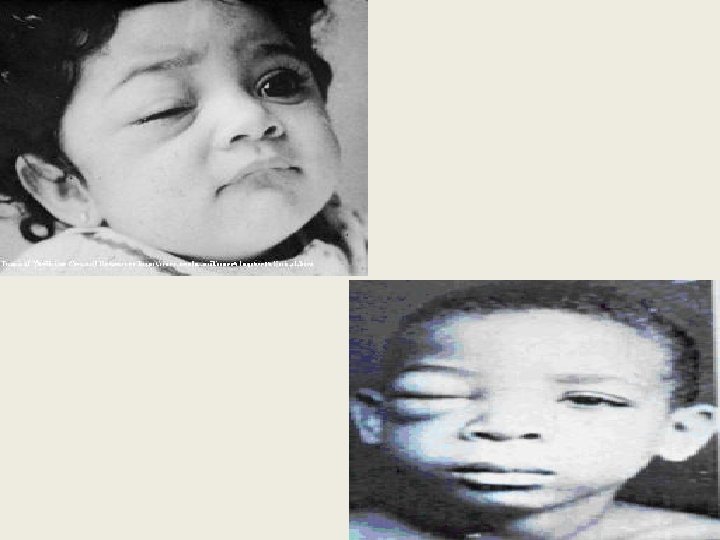
Trypanosoma cruzi American trypanosomiasis, Chagas , disease Pathogenicity and clinical features Incubation period 1 - 2 weeks A ) Organisms proliferate in the cells at the site of infection When they enter through the conjunctiva causing unilateral cojunctivitis with a unilateral edematous swelling of the eyelids results , conjunctivitis & Romana ‘ sign
Trypanosoma cruzi American trypanosomiasis, Chagas , disease Pathogenicity and clinical features Incubation period 1 - 2 weeks B)The parasite spread through the lymphatic system involving various tissues and cells through reticuloendothelial system. Inside these cells they get transformed into amastigote forms which divide by binary fission.
Trypanosoma cruzi American trypanosomiasis, Chagas , disease Pathogenicity and clinical features C ) Organisms appear in blood in 10 days Generalized malaise , chills , fever , muscle aches , generalized glandular enlargement , mild splenic and hepatic enlargement , red spots on chest and abdomen and epistaxis especially in young children.
CLINICOPATHOLOGICAL CORRELATION 1 - Local multiplication of the parasites (amastigotes ) in histeocytes and different cells chagoma ( local cutaneous induration ). 2 -If portal of entry eyes unilateral orbital oedema. (ROMANA’S SIGN ). Unilateral oedema &conjunctivitis
CLINICOPATHOLOGICAL CORRELATION 3 - Parasitisation of all tissue cells ( especially cardiac muscles , its conducting fibres ), smooth muscles(and the autonomic nerve ganglia) , glia cells of the brain & reticuloendothelial cells destruction of cells by multiplying amastigotes. Multiplying amastigotes in pseudocyst.
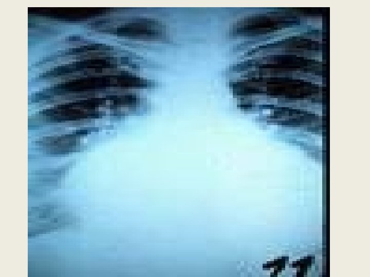
Trypanosoma cruzi American trypanosomiasis, Chagas , disease Pathogenicity and clinical features The disease manifests clinically in two forms : A ) Acute form Usually in children, present with fever and generalised nonpitting oedema of the body. The disease lasts for 3 to 4 weeks and sometimes ends fatally with myocarditis or meningoencephalitis. B )The chronic form Found in adults presents as neurotropic , cardiotropic or viscerortropic forms and may lasts for several years.
Clinical forms of Chagas disease: Acute form of Chagas disease 1 -Mainly affect infants & young children. 2 - Chagoma is the typical lesion , appears at the site of parasite entry. Unilateral palperal edema & cojunctivitis develops if the portal entry is eye (Romanas sign). 3 - Malaise , fever , oedema of the face with affection of R. E. C. moderate hepatomegaly, anemia & lymphadenopathy. The acute infection may Resolve spontaneously Chronic Myocarditis Meningoencephalitis Death.
Trypanosoma cruzi American trypanosomiasis, Chagas , disease Chronic form of Chagas disease. Chronic stages Occur many years after initial infection. Depends on the Intracellular multiplication of amastigote form damage of the cells and tissues. EX. Myocardium, skeletal muscles, neuroglial cells& reticuloendothelial system.
Trypanosoma cruzi American trypanosomiasis, Chagas , disease Pathogenicity and clinical features Chronic stages Damage of myocardium conduction defect (especially right bundle branch block , ventricular arrhythmias , cardiac enlargement and heart failure) Great reduction of autonomic cells in muscle layers of viscera and heart loss of autonomic function fibrosis and hypertrophy of the organ. Inflammation followed by
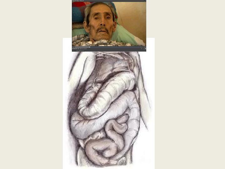
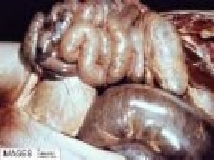
Trypanosoma cruzi American trypanosomiasis, Chagas , disease Pathogenicity and clinical features Chronic stages Damage of autonomic nerve cells mega-disease ( mega oesophagus , mega colon , mega ureter ) Damage to the autonomic nervous system of the heart parallels that to Auerbach’s plexus in the walls of the digestive tract. Hypertrophy of the muscle layers and diminution in number of the ganglion cells are seen in the affected portions of the digestive tract , most frequently in the esophagus and colon.
Chronic form of Chagas disease 1 -Affects older children or young adults with or without history of acute attack. 2 -The parasites present in different organs at low numbers : 1 -Heart affection: - Myocarditis , Heart failure, arrhythmia & cardiomegaly. 2 -C. N. S. : - Meningitis 3 -Neuromuscular system , destruction of Autonomic ganglia in smooth muscle of hollow organs: - Gastrointestinal manifestations : Mega organ disease. Megaoesophagus : - Regurgitation & dysphagia Megacolon : - Abdominal pain & chronic constipation
1 - CLINICAL PICTURE : RESIDENCE & HISTORY 2 - LABORATORY DIAGNOSIS - DIRECT METHODS : A- Demonstration of the parasites Amastigote in tissues Chagoma, L. N , Muscle biopsy : Monomorphic Trypomastigote in Blood & aspirate of LN&chagoma N. B : - Monomorphic trypomastigotes are much smaller or nearly absent in chronic chagas.
B- Culture on citrated blood medium : Epimastigote (Crithidial form ). C-Animal inoculation. D-Xenodiagnosis: Feeding of lab. Bred(parasite free) winged bug on suspected person , then dissect the gut for detection of Epimastigote & Metacyclic trypomastigote.
-INDIRECT METHODS: I. F. A. T, I. H. A. T & E. L. I. S. A CRUZIN TEST : INTRADERMAL TEST, killed epimastigotes are used as antigen injected intradermally Positive reaction in the form of red indurated area. . -MOLECULAR DIAGNOSIS - E. C. G. -IMAGE DIAGNOSIS
1 - Treatment of patient by Primaquine. 2 - Antitriatoma measures. 3 - Control measures against reservoir hosts.
• MONOMORPHIC 1 - Ex. (Trypanosoma cruzi), doesn’t multiply in peripheral blood but multiply intracellulary in amastigote form. • POLYMORPHIC 1 - EX (Trypanosoma brucei complex), multiply in the peripheral blood. 2 - Transmitted by faeces after insect bite (contaminative)=Stercorarian. 2 -Transmitted by Saliva (inoculative) 3 -Show posterior station development 3 - Show anterior station development = Salivarian. African trypanosomiasis American trypanosomiasis Chagas disease Sleeping sickness
Click to edit Master title style • Edit Master text styles Thank you • Second level • Third level • Fourth level • Fifth level