Cleft Lip and palate Dr Nitin Sharma MBBS
, MS, MCh(Gold Medalist), FMAS,")
Cleft Lip and palate Dr Nitin Sharma MBBS( Gold Medalist), MS, MCh(Gold Medalist), FMAS, FISPU AIIMS, New Delhi Assistant Professor(Pediatric Surgery)

PRESENTATI ONS Is it possible to be normal with treatment?

PRESENTATI ONS Why did happened, Who has fault? How should I feed my baby?

FUTURE ? ? ?
: Group I – Pre alveolar clefts: � Unilateral")
CLASSIFICATION SYSTEMS DAVIS AND RITCHIE CLASSIFICATION(1922): Group I – Pre alveolar clefts: � Unilateral cleft lip � Bilateral cleft lip � Median cleft lip Group II - Post alveolar clefts: � Cleft hard palate alone � Cleft soft palate and hard palate � Sub mucous cleft
: Group III-Alveolar clefts: � Unilateral alveolar cleft �")
CLASSIFICATION SYSTEMS DAVIS AND RITCHIE CLASSIFICATION(1922): Group III-Alveolar clefts: � Unilateral alveolar cleft � Bilateral alveolar cleft � Median alveolar cleft
 Group I (A) - Defects of the soft palate only Group")
VEAU CLASSIFICATION (1931) Group I (A) - Defects of the soft palate only Group II (B) - Defects involving the hard palate and soft palate extending not further than the incisive foramen, thus involving the secondary palate alone. Group III (C) – Complete unilateral cleft, extending from the soft palate to the alveolus, usually involving the lip Group IV (D) - Complete bilateral clefts, resembles Group III but is bilateral. When cleft is bilateral, pre-maxilla is suspended from the nasal septum.

ARTURO SANTIAGO CLASSIFICATION: Santiago A proposed a classification in 1969 He used four digits to indicate presence of cleft and its location. Each digit is followed by letter to indicate condition of cleft (complete, incomplete or sub mucous) Four digits represent the following four structures affected by cleft. � The first digit refers to the lip. � The second digit refers to the alveolus. � The third digit refers to the hard palate. � The fourth digit refers to the soft palate.

ARTURO SANTIAGO CLASSIFICATION: The numbers used as digits represents the condition of cleft 0= No cleft � 1= Midline cleft � 2= Cleft on right side � 3= Cleft on left side � 4= Bilateral cleft � The letters indicate more specifically the type of cleft A = An incomplete midline cleft � B = An incomplete cleft of right side � C = An incomplete cleft of left side � D = Bilateral incomplete cleft � E = Sub mucous cleft �
")
LAHSAL CLASSIFICATION OF CLEFT LIP AND PALATE: Kreins O (cited by Hodgkinson et al) proposed LAHSHAL system for classification of cleft lip and palate patients which was modified on the recommendation of Royal College of Surgeons Britain in 2005

LAHSAL CLASSIFICATION OF CLEFT LIP AND PALATE:

ELNASSRY CLASSIFICATION: Elnassry proposed following classification in 2007. He divided cleft lip and palate patients in to seven classes. � Class I: Unilateral cleft lip � Class II: Unilateral cleft lip and alveolus � Class III: Bilateral cleft lip and alveolus � Class IV: Unilateral complete cleft lip and palate � Class V: Bilateral complete cleft lip and palate � Class VI: Cleft hard palate � Class VII: Bifid uvula

Embryology Primary Palate Forms during 4 th to 7 th week of Gestation Defect anterior to incisive foramen prepalatal alveol, maksilla, lip, nose and palatine bone Unilateral or bilateral Cleft severity varies Complet ( all skin, muscle, mucosa, maksillary and nasal bones, nasal cartilages) Incomplet (intact nasal sill, minimally seperated, only small scar) Secondary Palate Forms in 6 th to 9 th weeks of gestation � � Palatal shelves change from vertical to horizontal position and fuse Tongue must migrate antero-inferiorly Face is formed at intrauterin at 10 th week


Palate Devolopment Primary palate median palatine processes premaxilla Secondary palateral palatine processes
")
Palate Devolepment(6 -12. Weeks)
")
Palate Devolopment (6 -12. Weeks)

NORMAL LIP MUSCULAR ANATOMY CLEFT LIP ANATOMY

Problems in Cleft Lip and Cleft Palate • • • Feeding Frequent upper respiratuary tract infection Frequent gas regurgitation Otitis media Nasal regurgitation of food Aspiration pneumenia Growing retardation Other anomalies Psycological problems (family)

Feeding Rules Swallowing is not impaired, oral feeding is possible Feeding with a spoon/palade. The child should be held in a head-up position at about 45 º during and after feeding Lateral position during sleeping
 Weight > 10 pound (4500 gr) Hb")
When to Operate Generally (Rules of 10’s) Weight > 10 pound (4500 gr) Hb > 10 gr Age > 10 weeks Cleft lips between 3 -6 months Cleft palate between 12 -18 months (preferred before speech devolops)

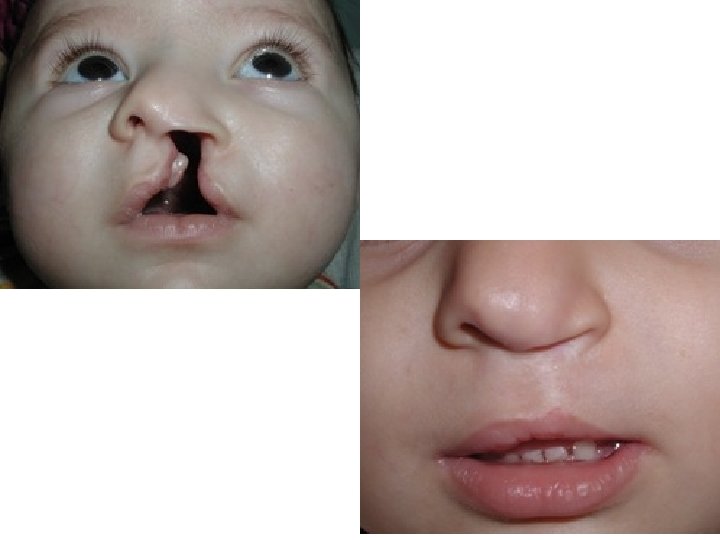
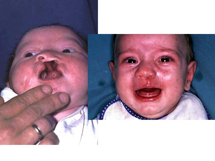
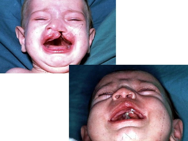
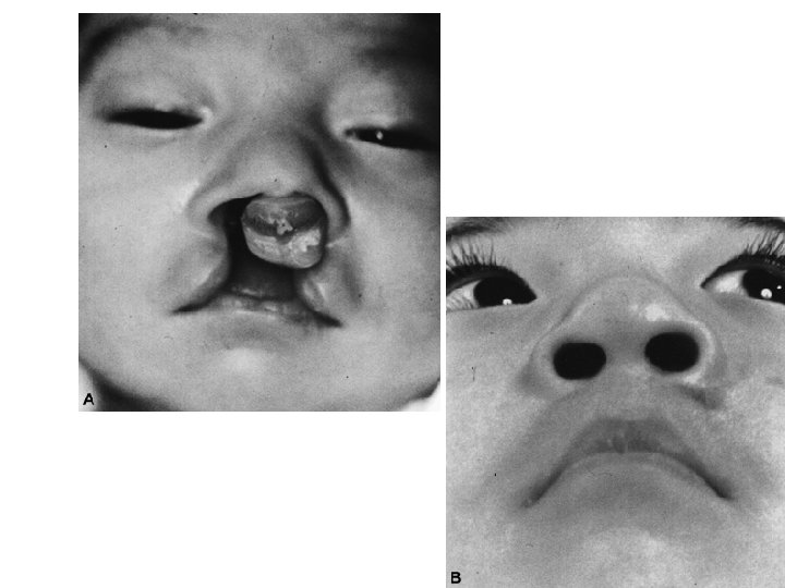
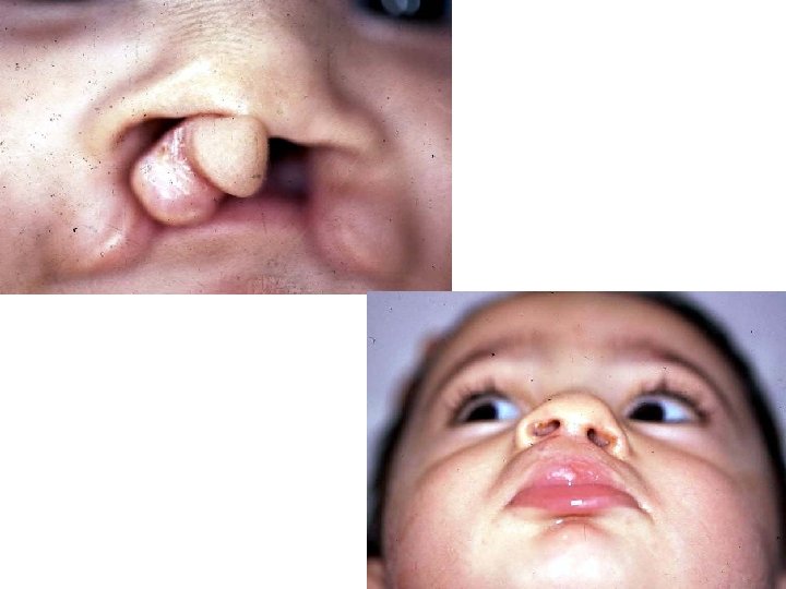
Cleft Lip Treatment Cleft lip Mikroform cleft lip Unilateral cleft lip Bilateral cleft lip Associated nasal deformity is classified as mild, moderate or severe Alveolar arc position evaluated. If necessary “presurgical maksiller orthodontics” applied
")
Operation technique in Microform cleft (Straight line closure)
")
Surgical technique for unilateral cleft lip (Millard Rotation-Advancement)
")
Surgical technique for unilateral cleft lip (Tennison Triangular Flap)

Surgical technique for unilateral cleft lip and palate Millard techniques provides primary lip and nasal repair. It is possible “gingivoperiostoplasy” after “Presurgical maksiller ortopedics”

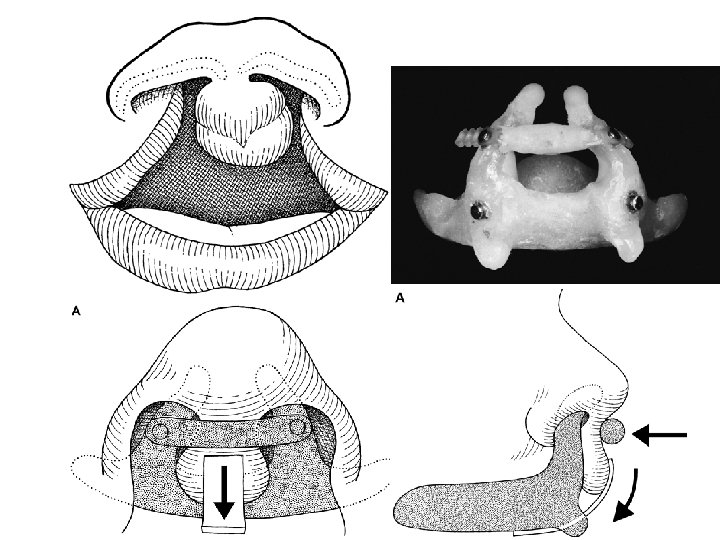
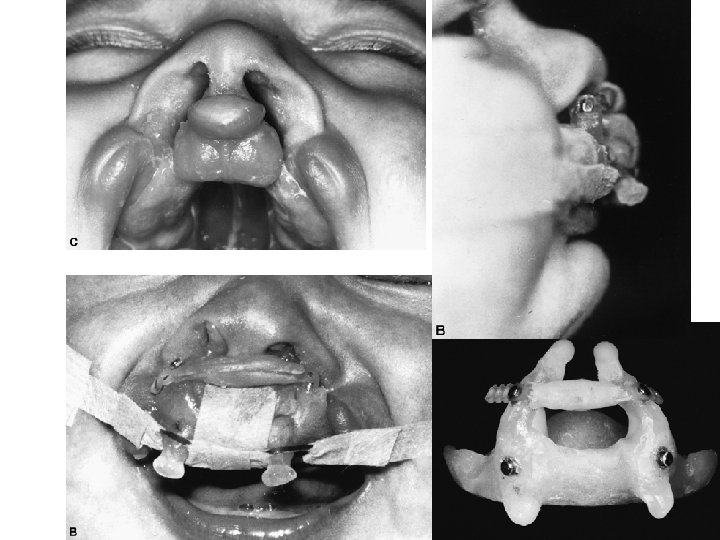
Pre -Orthodontic treatment After 3 months of Grayson molding plate application

A. M. Kul, right unilateral primary and secondary cleft palate Pre -Orthodontic therapy After 3 months of Grayson molding plate application

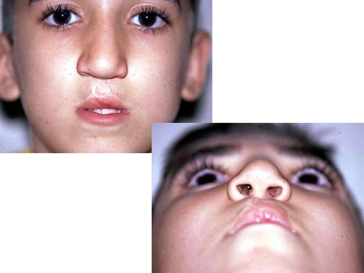
Postoperative 6 months

Postoperative 1, 5 years
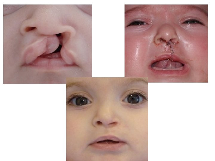
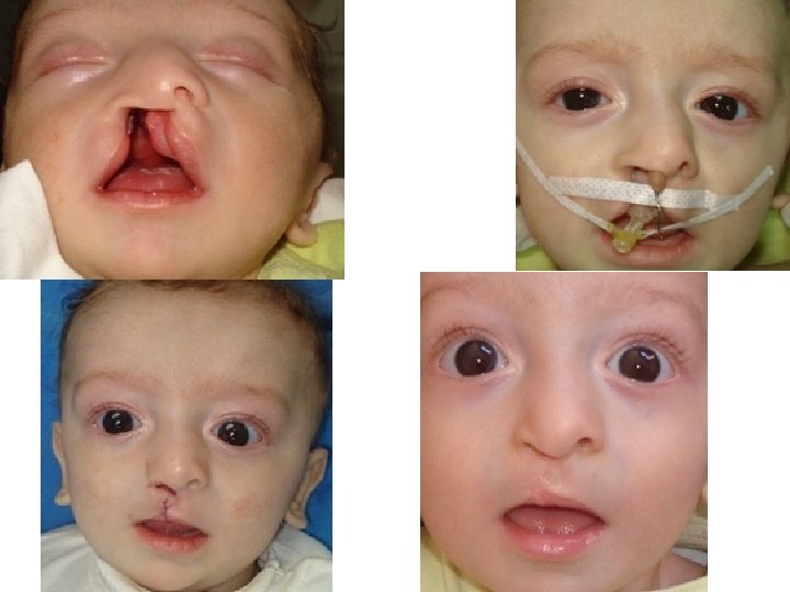
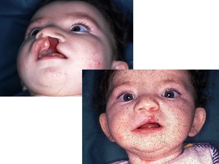
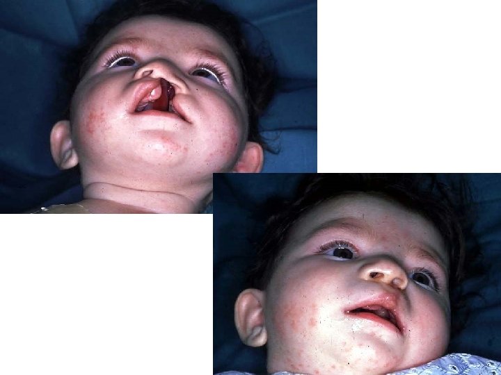

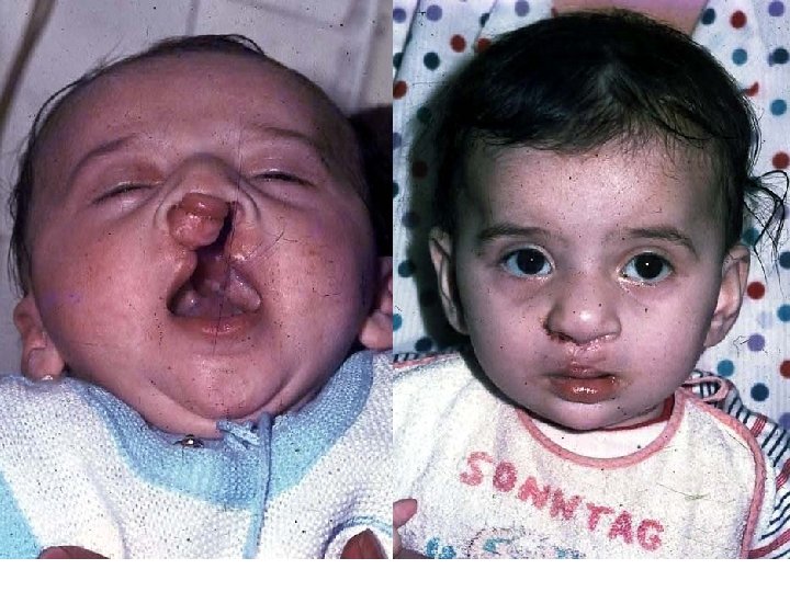
Bilateral Cleft Lip More complex and difficult to treat Projectile premaxilla Broad and flared nasal tip Prolabium Short columella or absent columella Incomplete or complete It is important to retropositon the premaxilla with presurgical orthopedic treatment Surgical techniques used for unilateral cleft lip repair are used for bilateral cleft lip repair in one or two stage operation (Millard, Tennison. . . )
")
Treatment of Premaxilla Lip repair or “Lip-adhesion” Elastic traction ( with a Head Bonnett) Premaxillary retantion (Latham) Nasoalveoler molding (Grayson) Surgical premaxilla excision or set-back (severe maxillary retrusion)
")
Bilateral Incomplete Cleft lip Operation Technique Millard (Two stage)
")
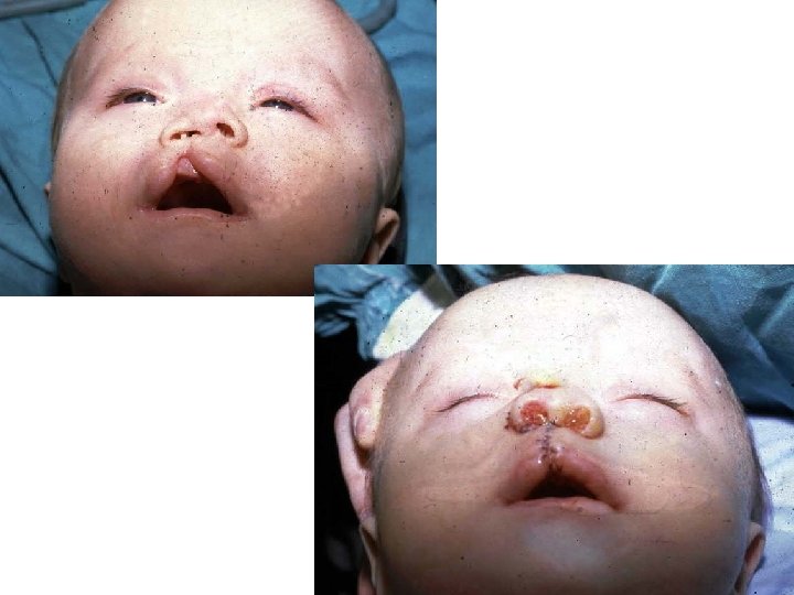
Bilateral Incomplet Cleft lip Operation Technique Straight Line Closure (One stage)

Cleft Palate

Cleft Palate and palatal muscles close the velopharengeal valve Velopharengeal closure can not be done in cleft palate patient. Patient can not create intraoral pressure Feeding and speech are effected

Anatomy Soft palate muscles insert on posterior margin of remaining hard palate rather than midline raphe

Cleft Palate Affects 1/2500 living births More often in girls Heredity is less affects Complete up to incisive foramen İncomplete Only soft palate cleft

Problems with cleft palate Feeding Speech Hearing and middle ear problems Additional anomalies (% 30) Psychological problems

Goal of Palatal Repair Understandable speech No maxillary retrusion No hearing problem Good occlusion

Submucous Cleft Palate Anatomic problem Mostly asymptomatic % 15 velopharengeal insufficiency Muscles are not fused middle of palate (muscular diastasis) notch at the back of the hard palate Bifid uvula Persistent ear disease swallowing difficulties Short soft palate Limited motion Easy to get tired while speaking When light goes through nose, light can be seen from oral side It is not necessary surgical treatment until child growth enough to cooperate

Treatment of Submucous Cleft Palate Submucous cleft palate only requires surgery if it is causing problems for the individual The most common reason for treating a person with a submucous cleft palate is because of abnormal, nasal-sounding speech

Von laganbeck palatoplasty

VWK palatoplasty

Two flap palatoplasty
 Breathing and")
Pierre Robin Sequence Micrognathy Glossoptosis Airway obstruction Cleft palate( % 50 ) Breathing and feeding problem

Complications Acute Period bleeding, Airway obstruction Infection Wound seperatiom Late maksillary hipoplasia, dental oklusion problems) Hearing problems velopharyngeal insufficency Fistula formation

Cleft lip and palate treatment time table

Velopharyngeal Insufficency The inability of the velopharyngeal sphincter to sufficiently separate the nasal cavity from the oral cavity during speech Speech problem (hypernasality, nasal emission, consanant production difficulty, decrese in voice strength and short phrases) swallowing problems

Treatment of Velopharyngeal Insufficency Patient should evaluate by speech terapist before any treatment Nasendoscopic evaluation and Multiview videofluoroscopy is importany diagnostic tests Goal is to provide normal velopharyngeal anatomy

Pharyngeal wall motion. B: Markings for a proposed tailor-made pharyngeal flap. The 2. 5 cm width is one half the width of the posterior pharynx 5 and would be appropriate for a patient whose pharyngeal wall motion ranges from 3– 3. 5. Pharyngeal wall motion. A: Frontal view of the oropharynx showing gradations of medial motion of the lateral pharyngeal walls. 0 = no motion, 5 = maximal motion to the midline.
 Pharyngoplasty (Hynes, Orticochea) Soft")
Surgical Treatment of Velopharyngeal Insufficency Pharyngeal Flaps (Superior, inferior pedicled) Pharyngoplasty (Hynes, Orticochea) Soft palate lengtening and levator muscle repair Posterior wall augmentation (teflon, proplast)
 Secondary Onarımlar (preschool age) Alveolar")
Other Operations Fistula Repair Velopharyngeal Insufficency correction (5 yeras) Secondary Onarımlar (preschool age) Alveolar bone grafting (before canine theth eruption) Orthodontic Surgery (12 -14 years) (Le-Fort I Maksillary osteotomy, Mandibular split ramus osteotomy) Rhinoplasty (16 -18 years)
- Slides: 67