Classification of Epithelial Tissue a Layer Simple One
 Layer - Simple - One Layer Stratified -")
 • always has a free")
,")
 Stratified Squamous Location - surface of skin, lining")


 Ground")
 Cells – * Resident – stable numbers, always")
 • Fibroblasts – most common type of resident")
 • Specific Cells: chondrocytes – cartilage cells adipocytes")
 Matrix: Ground Substance: Fluid, Semifluid or Gel-like (which")
 Fibers (cont. ): * Elastic – Structure: thin")

- Slides: 18
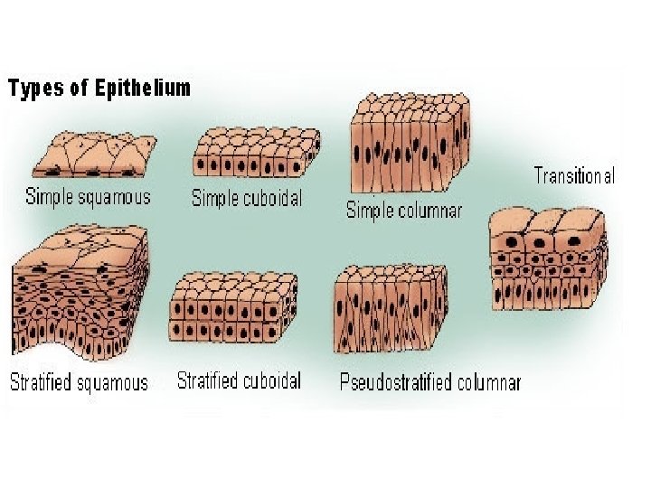
Classification of Epithelial Tissue • (a) Layer - Simple - One Layer Stratified - Many Layers Pseudostratified - One layer but it looks like more than one layer because the cells are different shapes and the nuclei are located at different levels • (b) Shape - Squamous - flat Cuboidal - cubed/square Columnar - column/rectangular Transitional - changes shape due to amount of pressure exerted on it contains the ability to stretch and expand as a response to pressure
Characteristics of Epithelial Tissue • cells packed together (cellularity) • always has a free surface exposed to outside or empty internal space (apical surface) & attached to underlying tissue (basal surface) (polarity) • anchored to connective tissue by a thin membrane called the basement membrane (attachment) • lacks blood vessels - blood diffuses from connective tissue (avascularity) • cells reproduce quickly - as a result, healing occurs rapidly (more quickly than any other tissue) (regeneration) • acts as a barrier
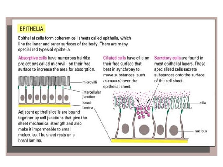
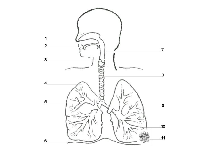
Specific Types of Covering & Lining Epithelial Tissue Simple Squamous Location: blood vessels (capillaries), alveoli (air sacs) of lungs, kidney tubules Function: (very thin tissue) diffusion & filtration Simple/Stratified Cuboidal Location: glands and ducts, kidneys Function: secretion &/or absorption Simple Columnar Location: lining of stomach, intestine, & gallbladder Function: protection, secretion, & absorption Pseudostratified Ciliated Columnar Location: respiratory system (nasal cavity & bronchi) Function: protection & secretion
Covering & Lining Epithelial (cont. ) Stratified Squamous Location - surface of skin, lining of mouth, throat, esophagus, rectum, anus, and vagina Function - provides physical protection against abrasion, pathogens, and chemical attack (protection against wear and tear / friction) Transitional Location - urinary bladder Function – permits expansion and recoil after stretching
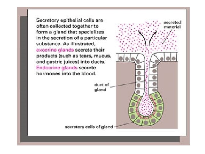
Glandular Epithelium Composed of cells that specialize in production and secretion of specific substances • Endocrine - secrete their products within/into tissue and blood (hormones) (ductless glands) • Exocrine - secrete their products into ducts opening onto internal or external surfaces (sweat, saliva)
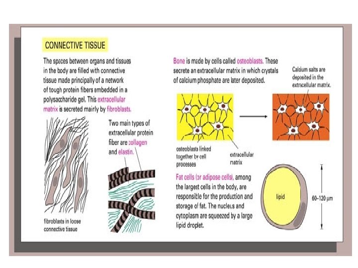
Characteristics of Connective Tissue • • • binds/connects structures provides support & protection insulates fills spaces within body cavities stores fat transports produces blood cells protects against infection helps repair tissue damage cells farther apart & intercellular material (matrix) between cells • usually reproduce well & good blood supply so well nourished (healing generally occurs quickly)
Structure of Connective Tissue LIVING PORTION NONLIVING PORTION Cells Matrix Resident Wandering (1) Ground Substance (2) Fibers
Structure of Connective Tissue (cont. ) Cells – * Resident – stable numbers, always there, don’t move. . . Fibroblasts, Mast Cells, Specific Cells * Wandering – move temporarily into tissue from bloodstream, usually as a response to infection/injury. . . White Blood Cells/Macrophages (perform phagocytosis to clear foreign particles)
Structure of Connective Tissue (cont. ) • Fibroblasts – most common type of resident cell, large and usually starshaped, produce fibers by secreting proteins • Mast Cells – large & widely distributed, near blood vessels secrete two types of chemicals: heparin – prevents blood clotting & histamine – promotes reactions related to allergies & inflammation
Structure of Connective Tissue (cont. ) • Specific Cells: chondrocytes – cartilage cells adipocytes – adipose cells osteocytes – bone cells
Structure of Connective Tissue (cont. ) Matrix: Ground Substance: Fluid, Semifluid or Gel-like (which is semisolid), Rigid Fibers: * Collagenous Structure: thick threads of collagen protein grouped in long, parallel bundles (WHITE) Function: provide great strength Location: important components of body structures that hold things together (for example: tendons & ligaments)
Structure of Connective Tissue (cont. ) Fibers (cont. ): * Elastic – Structure: thin fiber of elastin protein that branches (YELLOW) Function: weaker, but stretch easily & snap back into place Location: vocal cords & elastic cartilage * Reticular – Structure: very thin fiber that is highly branched (BROWN) Function: form delicate supporting networks Location: walls of blood vessels, basement membrane
Types of Connective Tissue Rigid/Supporting CT: Bone Cartilage – Fibrocartilage, Hyaline, Elastic CT Proper: Loose Fibrous CT – Areolar, Adipose, Reticular Dense Fibrous CT – Tendons & Ligaments Fluid CT: Blood Lymph