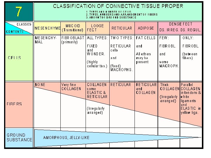
CLASSIFICATION OF CONNECTIVE TISSUE Classification depends on the


")


: • Closely-packed")


- Slides: 16
CLASSIFICATION OF CONNECTIVE TISSUE Classification depends on the I- Embryonic connective proportion of cells to fibers, and on tissue it includes: the arrangement, and the types of q. Mesenchymal CT. fibers. q. Mucoid CT. Three categories can be defined: II- Connective tissue proper: it includes: III- Specialized connective tissue; it q Loose areolar connective tissue. includes: q Dense irregular connective tissue ﺛﻼﺛﺔ ﺃﻤﻮﺭ ﺗﻤﻴﺰ ﺍﻝ q. Cartilage. q Dense regular connective tissue. : C. T q. Bone. q Elastic connective tissue. Cell , fibers q. Blood. , ground q Reticular connective tissue. substance q Adipose connection tissue.
Classification of C. T. Depend on ground substance q Embryonic C. T q Mucoid C. T. Abundant G. S Little fiber or reticular F ﺍﻟﻤﻘﺼﻮﺩ ﺑﻼﺯﻣﺎ ﺍﻟﺪﻡ Jelly like C. T. Proper Ø Loose C. T. Ø Dense C. T. or Special type of c. t Modified types v Blood =fluid v Cartilage= firm v Bone = hard or calcify : ﻻﺯﻡ ﻧﻌﺮﻑ C. T ﻷﻲ ﻧﻮﻉ FIBERS ﻧﻮﻉ ﺍﻝ Most common type of cell ﺍﻟﻤﻜﺎﻥ -
Embryonic connective tissue Mesenchymal CT Mucoid CT Site: it is found in the umbilical cord and pulp of growing teeth. Site: it is found in embryo. Histological structure: it consists Histological structure: It consists of: q Abundant ground substance Most cell (Wharton's jelly) composed Ø Undifferentiated mainly of hyaluronic acid. It mesenchymal cells (UMCs) appears homogeneous and with their processes come in basophilic. contact with each other forming a network. q Spindle-shaped UMCs that are widely separated and fibroblasts. Ø A gel-like, amorphous ground substance. q Unapparent fine collagen fibers(Type 2) that have the same Ø Scattered reticular fibers. Most fiber refractive index as the matrix.
Mucoid C. T. = Embryonic C. T • Mucoid connective tissue (or mucous tissue) is a type of connective tissue found during fetal development. • It is composed mainly of ground substance with few cells & fibers • It is most easily found as a component of Wharton's jelly. • Cells : UDMC, Fibroblasts • Fibers : present but not apparent collagen type II • Ground substance : Abundant Sites: • Mucous connective tissue forms the umbilical cord. • The vitreous of the eyeball is a similar tissue. ﻣﻮﺟﻮﺩ ﻭﺭﺍﺀ ﺍﻟﻌﺪﺳﺔ ﻭﻭﻇﻴﻔﺘﻪ support of eye
Connective tissue proper according to main component ﺍﻟﺘﺴﻤﻴﺔ Loose: connective tissue is relatively cell rich, soft. It is also rich in vessels and nerves. Loose connective tissue may occur in some special variants: • LACT: connective tissue Loose areolar CT • Reticular: connective tissue • Adipose: tissue. Collagen type 1 Dense C. T. : connective tissues are completely dominated by fibres. They are subdivided according to the arrangement of the fibres in the tissue. Dense irregular: connective tissue the fibres do not show a clear orientation within the tissue but instead form a densely woven three-dimensional network (dermis). Dense Regular 1. White fibrous C. T. 2. Elastic C. T.
Connective tissue proper Loose areolar CT: Site: it is the most widely distributed connective tissue in the body. It binds body parts together while allowing them to move freely over one another. It contains many small blood vessels coursing through this tissue. • Loose CT is found in the following sites: It is present beneath the epithelium in all mucous membranes forming the lamina propria. Because it avascular It forms the papillary layer of dermis which attaches the skin epidermis to underlying structures. It surrounds glands, small blood vessels, and nerves.
Histological structure • All types of fibers; collagen, elastic and a small proportion of the reticular fibers. All types of connective tissue cells with predominance of fibroblasts and macrophages. Good amount of ground substance. Function: a. Supports and binds other tissues (by its fibers). b. Holds body fluids and provide nutrition (by its ground substance). c. Defends against infection (by its white blood cells, plasma cells, mast cells and macrophages).
Loose Areolar C. T. CT A L SKIN
Reticular CT: • Histological structure: Reticular fibers, forming a network. Reticular cells, these are the fibroblasts of reticular connective tissue, that synthesize the reticular fibers. • Site & function: reticular tissue is limited to certain sites. It forms the supporting stroma for: Hemopoietic tissue in the bone marrow. Lymphoid tissue in lymph nodes and spleen. Hepatocytes in liver. Liver (silver stain)
Adipose C. T. Unilocular adipose C. T. Yellow fat • • Unilocular fat cells C. T. fibers: collagenous F. rich in blood supply Carotenoids ﻫﻲ ﺍﻟﺘﻲ ﺃﻜﺴﺒﺘﻪ ﺍﻟﻠﻮﻥ ﺍﻻﺻﻔﺮ Sites: • Subcutaneous tissue • Around vital organs LARGE AND FLATTEN ﻋﺒﺎﺭﺓ ﻋﻦ ﺧﻼﻳﺎ ﺩﻫﻨﻴﺔ Multilocular adipose C. T. Brown fat • • Multilocular fat cells C. T. fibers – collagenous F. rich in blood supply Many blood vessels , numerous mitochondria , cytochrome pigment ﺃﻜﺴﺒﺘﻪ ﺍﻟﻠﻮﻥ ﺍﻟﺒﻨﻲ Sites: • Back & neck of newborne + SHOULDER
Adipose C. T. Function: Storage of energy in the form of triglycerides. Subcutaneous adipose tissue shapes the body. Pads of fatty tissue in palms and soles act as shock absorber. Thermal insulation of the body; due to the poor heat conduction of adipose tissue. Fixation of the vital organs as heart and kidney, thus keeping them in position.
Dense regular C. T. White Fibrous C. T. Histological structure (Figure 84): • Closely-packed wavy bundles of collagen fibers running in the same direction and parallel to the direction of pull. • Rows of fibroblasts (tendon cells) with flattened nuclei aligned between the collagen bundles. • Little amount of ground substance. Sites & function: • Unlike areolar CT, this tissue is poorly vascularized. This type of tissue forms white flexible structures with great resistance to pulling forces wherever it is exerted in a single direction. It is found in: • Tendons, which attach muscles to bones. • Ligaments, which bind bones together at joints. § sclera of the eye COLLAGEN TYPE 1 ﻓﻴﻬﺎ
Dense Irregular CT: • Histological structure: Thick bundles of collagen fibers arranged irregularly (running in more than one plane). Little amount of ground substance with few fibroblasts. • Sites & function: this type of tissue forms sheets in body areas where tension is exerted from many different directions. It is found in the reticular layer of dermis of the skin. It forms the capsules of fibrous joints. It forms the capsules of body organs e. g. kidney, spleen, lymph nodes and liver.
Elastic C. T. • Histological structure: the elastic fibers predominate; they run in all directions, also they may form fenestrated membranes. • Site & function: this tissue is present where flexibility and elastic recoil are needed; it is found in: Elastic laminae of arteries. True vocal cords. Few ligaments in the body are very elastic such as ligamenta flava and ligamenta nuchae connecting adjacent vertebrae. • ﻭﻳﻮﺟﺪ ﺃﻴﻀﺎ ﻓﻲ ﺍﻟﺸﺮﻳﺎﻥ ﺍﻻﻭﺭﻃﻲ
CONNECTIVE TISSUE • Connective tissues are the most abundant of the primary tissues. • The cells of the connective tissues are far apart, separated by an abundant amount of extracellular material, also called extracellular matrix Function : 1. Binding, support and packaging: 2. Protection, defense and repair: 3. Insulation: Fat cells or adipose tissue, is a connective tissue which not only cushions body organs but also insulates them and provides reserve energy fuel. 4. Transportation: Blood is a connective tissue and it carries and delivers oxygen and nutrient to tissues.