Classification Etiopathogenesis pathophysiology of STEMI Dr Shahinlal N


 Acute myocardial injury with atleast one")


 increased vascular permeability, leukocyte")


 MI with angiographically normal or coronary")


 Infarct expansion acute dilatation & thinning of area of infarction not")

Ventricular dilatation • Dilatation of viable portion of ventricle • compensatory mechanism to maintain")


- Slides: 107
Classification Etiopathogenesis & pathophysiology of STEMI Dr. Shahinlal. N SR , Department of Cardiology Govt medical college, Kozhikode
Fourth. UNIVERSAL DEFINITION OF MYOCARDIAL INJURY & INFARCTION MYOCARDIAL INJURY Elevated Cardiac Troponin values > 99 th percentile of URL. Myocardial injury ---- acute --rise and /or fall of c. Tn values.
CRITERIA FOR ACUTE MYOCARDIAL INFARRCTION(TYPES 1, 2, 3) Acute myocardial injury with atleast one of the following *symptoms of myocardial ischaemia *new ischaemic ECG changes *development of pathological Q waves *imaging evidence of new loss of viable myocardium /new RWMA consistent with ischemia *identification of a coronory thrombus by angiogram (not for type 2 or 3)
TYPES OF MI Type 1 Type 2 Type 3 Type 4 a ype 4 b Type 5
TYPE 1 Atherosclerotic plaque rupture /erosion Intraluminal thrombus in one /more coronary arteries Decreased myocardial blood flow Myocardial injury
TYPE 2 Condition other than plaque rupture contributing an imbalance B/W myocardial O 2 supply and demand Coronary artery spasm Coronary endothelial dysfunction Tachyarrhythmias, Bradyarrhythmias Anemia Respiratory failure Drugs & toxins Hypotension/ severe Hypertension
TYPE 3 Sudden cardiac death with symptoms of ischemia, new ST elevation/LBBB/VF Die before blood samples for biomarkers can be obtained/ increase in biomarkers can be identified.
TYPE 4 a/ TYPE 4 b/TYPE 5 Type 4 a> MI associated with PCI Type 4 b> MI associated with stent thrombosis Type 5 > MI associated wit CABG.
TYPE 4 A PCI relted MI CTn values > Five times the 99 th percentile URL----Pt with norml base line values. Pts with elevated pre procedure CTn , Post procedure value must raise by >20%
In addition one of the following *symptoms of myocardial ischaemia *new ischaemic ECG changes *development of pathological Q waves *imaging evidence of new loss of viable myocardium /new RWMA consistent with ischemia *identification of a angiographic complications (coronary dissection/occlusion of a major epicardial artery/ thrombus)
TYPE 4 B Stent/scaffold thrombosis. Acute ------- 0 -24 hr Sub acute ------- > 24 hr-30 days Late ------- > 30 days- 1 year Very late ------- > 1 year
Criteria for TYPE 4 B CTn value more than 99 th percentile URL with atleast one of the following *symptoms of myocardial ischaemia *new ischaemic ECG changes *development of pathological Q waves *imaging evidence of new loss of viable myocardium /new RWMA consistent with ischemia *identification of a coronory thrombus by angiogram
TYPE 5 CABG related MI Elevated c. Tn values > 10 times the 99 th percentile URL. (Normal base line c. Tn) Elevated pre procedure value----post procedure c. Tn must rise by >20%.
TYPE 5. In addition one of the following. 1. Development of new pathological Q waves 2. Angiographic evidence of new graft occlusion/ new native coronary artery occlusion. 3. Imaging evidence of new loss of viable myocardium/New RWMA.
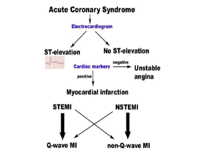
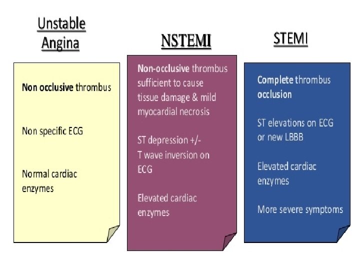
ACUTE CORONARY SYNDROME Spectrum of conditions occurs due to acute myocardial ischemia/ infarction Abrupt reduction in coronary blood flow
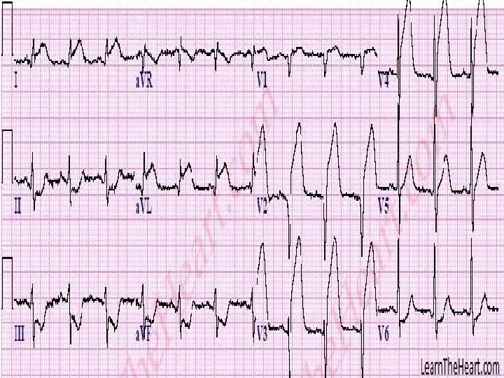
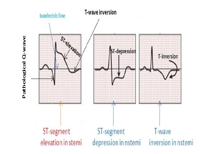
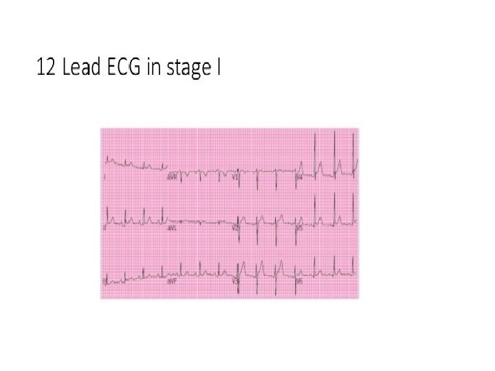
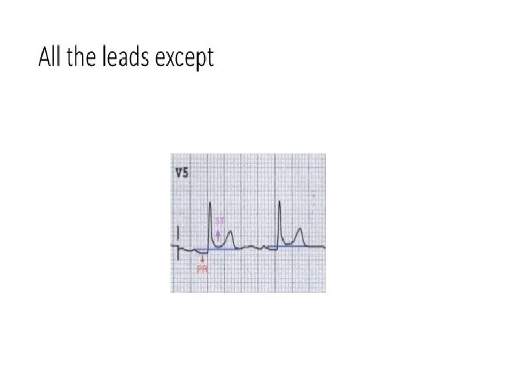
ECG & MI ST Elevation New ST elevation at j point in two contiguous leads >0. 1 mv in all leads except V 2/V 3 in V 2/V 3 >0. 2 mv in men>40 yrs >0. 25 mv in men<40 yrs >0. 15 mv in woman
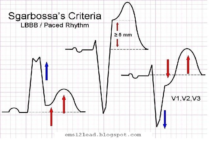
In setting of LBBB ST elevation > 1 mm & concordant QRS complex-------5 points ST segment depression > 1 mm in V 123 -----3 points Discordant ST sement elevation> 5 mm -----2 points Score >3 -----98% specific for acute MI
ST depression & T wave changes New horizontal/downslopping ST depression >0. 05 mv in two contiguous leads T inversion >0. 1 mv in two contiguous leads
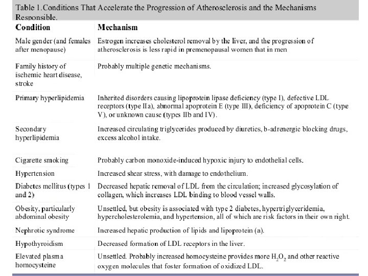
ATHEROSCEROTIC DISEASE > 90 % CAD Culprit cause is found to be ATHEROSCLEROTIC DISEASE
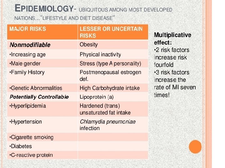
INTERHEART TRIAL 52 countries Africa/Asia/North and south America. Identified 9 easily measured risk factors for acute MI. Smoking/ Hyperlipidemia/HTN/DM/Obesity/Diet/Physical activity/Alcohol consuption/Psychosocial factors.
ATHEROMATOUS PLAQUEPATHOGENESIS • 1. Chronic endothelial injury (HTN/Hyperlipidemia/smoking/Toxins/virus/ immune reactions) increased vascular permeability, leukocyte adhesion, thrombosis. 2. Accumulation of lipoprotein oxidised LDL 3. Monocyte adhesion to endothelium macrophages & T cells absorb oxidised LDL and HSP------- foam cells
• 4. Platelet adhesion • 5. Factor release platelet , macrophages, endothelium……. . >PDGF , FGF , TGFα……. . >induce SMC recriutment from media 6. SMC proliferation and ECM 7. Lipid accumulation
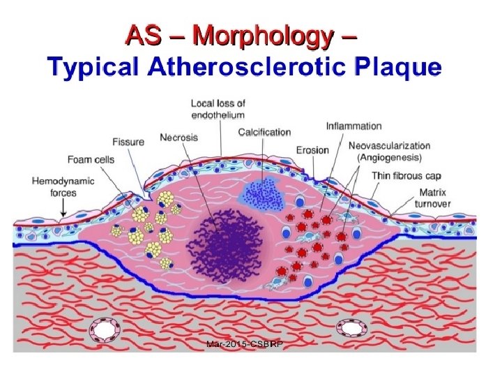
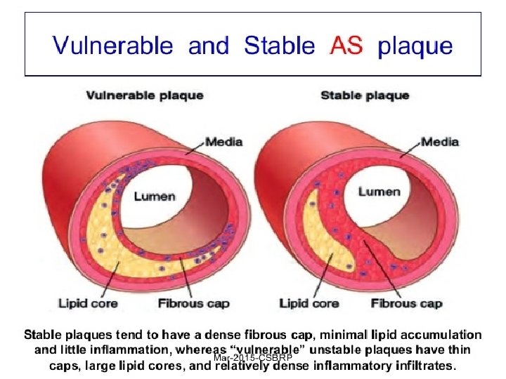
ATHEROSCLEROTIC PLAQUECONTENTS • 1. CELLS Smooth muscle cells, macrophages, T cells • 2. ECM collagen, proteoglycans , elastic fibres 3. LIPIDS
Atherosclerotic plaque changes • • Rupture, Ulceration, erosion Hemorrhage in to a plaque Athero embolism Aneurysm formation
NON ATHEROSCLEROTIC CAUSES OF MI • 1. Embolisation into coronary artery infective endocarditis/NBTE prosthetic valve neoplasm air induced at cardiac surgery hypercoagulable states 2. Spontaneous coronary artery dissection
Continues- • 3. syphilitic aortitis occlusion of coronary ostium 4. vasculitis Takayasu’s arteritis, PAN, Kawasaki, SLE, Giant cell arteritis 5. Meadiastinal irradiation 6. Hurler’s syndrom
---- • 7. Pseudoxanthoma elasticum • 8. Homocystinuria • 9. cocaine
MI NOCA (MYOCARDIAL INFARCTION WITH NONOBSTRUCTIVE CORONARY ARTERIES) MI with angiographically normal or coronary artery stenosis less than 50% Young females CAUSES coronary vasospasm dissection coronary emboli
continues Hematological disorders Polycythemia rubra vera Sickle cell anemia DIC TTP Augmented oxygen demand Thyrotoxicosis Amphetamine
continues Hypotension sepsis , blood loss, drugs Anatomical variations anomalous origin of coronary artery, coronary AV fistula, myocardial bridge
Continue— Prognosis…. >Good compared to Atherosclerotic MI Most common mimickers of MI……Acute myocarditis, Acute stress cardiomyopathy(Tako tsubo)
PATHOPHYSIOLOGY OF MI LV Dysfunction • Systolic dysfunction 4 abnormal contraction pattern Dysynchrony Hypokinesia Akinesia Dyskinesia Severe depression of myocardial contractility Increased end systolic volume Decreased CO Decreased BP • Diastolic Dysfunction
Ventricular Remodelling • Change in Size, shape, thickness involving both infarcted & non infarcted segments of ventricle A) Infarct expansion B)Ventricular dilatation
………. . A) Infarct expansion acute dilatation & thinning of area of infarction not explained by additional myocardial necrosis. slippage b/w muscle bundles disruption of normal myocardial cells destruction of extra cellular matrix.
Continue • Degree of infarct expansion ……pre infarction wall thickness existing hypertrophy possibly protect against infarct thinning. Apex---Thinnest vulnerable for infarct expansion. • High mortality…. Heart failure/ V. aneurysm/ Arrhythmias
--B)Ventricular dilatation • Dilatation of viable portion of ventricle • compensatory mechanism to maintain stroke volume • Start immediately after STEMI can progress for months---years
EFFECT OF Rx • Acute reperfusion------Restrict extent of myocardial necrosis • Steroids & NSAIDS----early MI----Scar thinning & infarct expansion. • RAAS inhibitors/ ARB/Aldosterone antagonist-------reduce remodelling.
Pathophysiology of other organ systems • LUNG Increased pulmonary capillary hydrostatic pressure-------interstitial edema----arteriolar & bronchiolar compression---hypoxemia Hyperventilation------Respiratory alkalosis TLC , FRC, RV, VC
Endocrine Pancreatic blood flow Insulin Sympathetic activity catecholamines Glycogenolysis hyperglyce mia Safest post MI glucose level <180 mg/dl
• Adrenal medulla------ increased Catecholamine release serious arrhythmias, extensive myocardial damage, cardiogenic shock. increased thromboxane. A 2……. >platelet aggregation • RAAS activation
• NATRIURETIC PEPTIDE ANP, BNP-------from atrias BNP & NT- pro BNP ----from ventricles rise in BNP and NT-pro. BNP after STEMI correlates with infarcts size and RWMA
RENAL FUNCTION CO AKI
HAEMATOLOGICAL • • • * propensity for platelet aggregation * leucocytes * viscosity
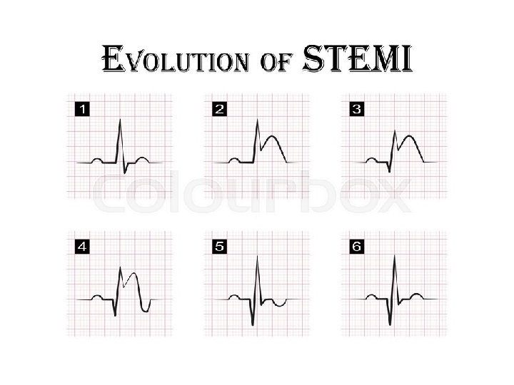
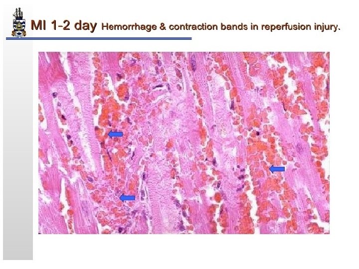
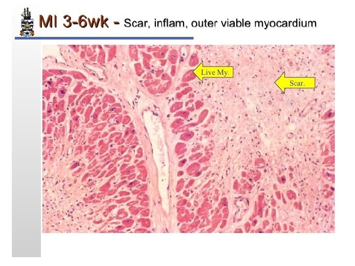
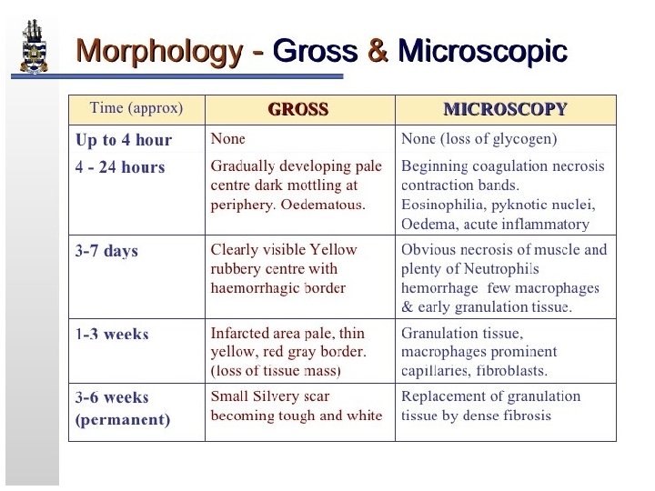
Pathological changes after MI • GROSS <4 hrs-----NONE 4 -24 hrs------Dark mottling 1 -3 days------ Infarct centre …yellow 3 -7 days-------Hyperemia at borders 7 - 14 days----- Red gray& depressed borders 2– 8 weeks--- Gray white granulation tissue. > 2 month----completed scarring
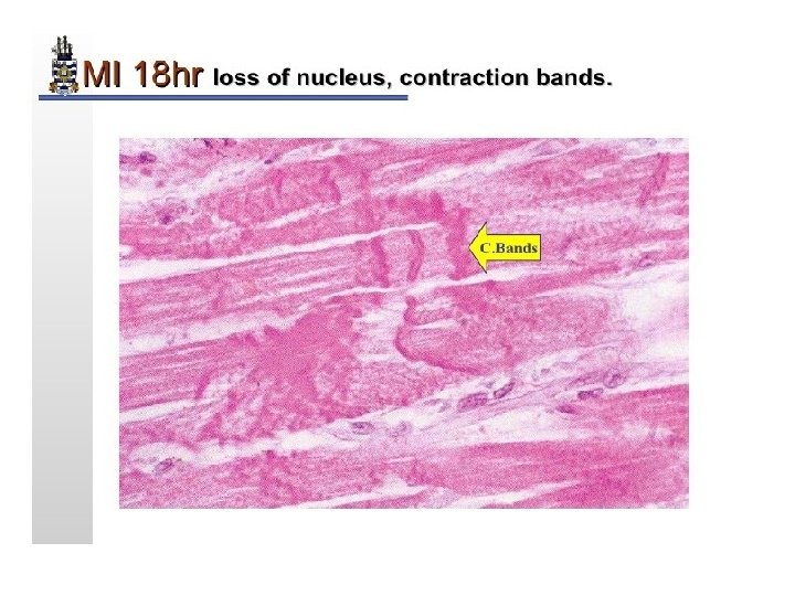
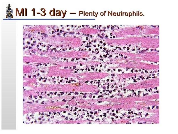
Microscopic • 30 min-4 hrs----- Glycogen depletion, intracellular edema • 4 -12 hrs---Disruption of mitochondria, coagulation necrosis • 12 -24 hrs--Contraction band necrosis Neutrophilic and eosinophilic infiltration
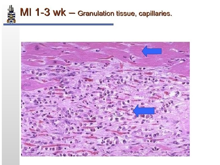
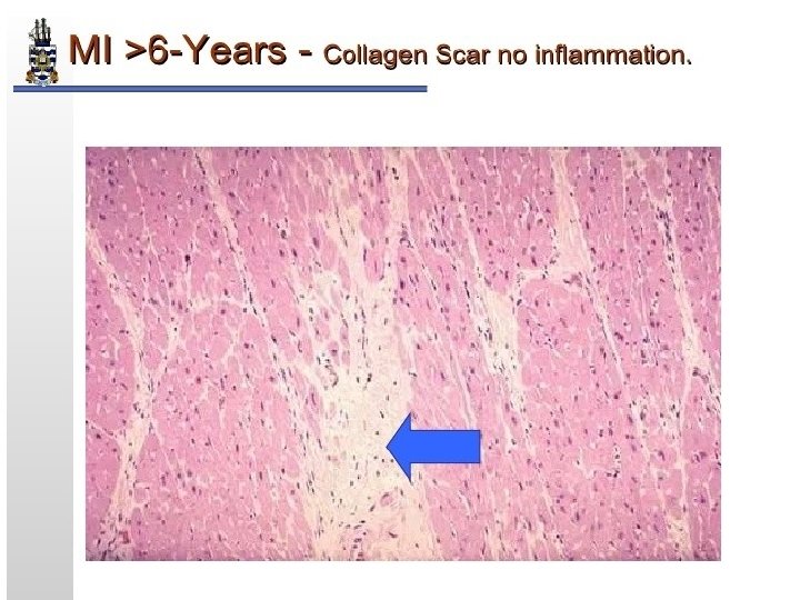
Microscopic 1 -3 days-----more neutrophils 3 -7 days-----apoptosis of neutrophils 7 -10 days ----granulation tissue formation 10 -14 days----mature granulation tissue with type 1 collagen • >2 month-----dense collagen scar • •
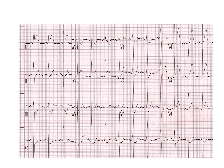
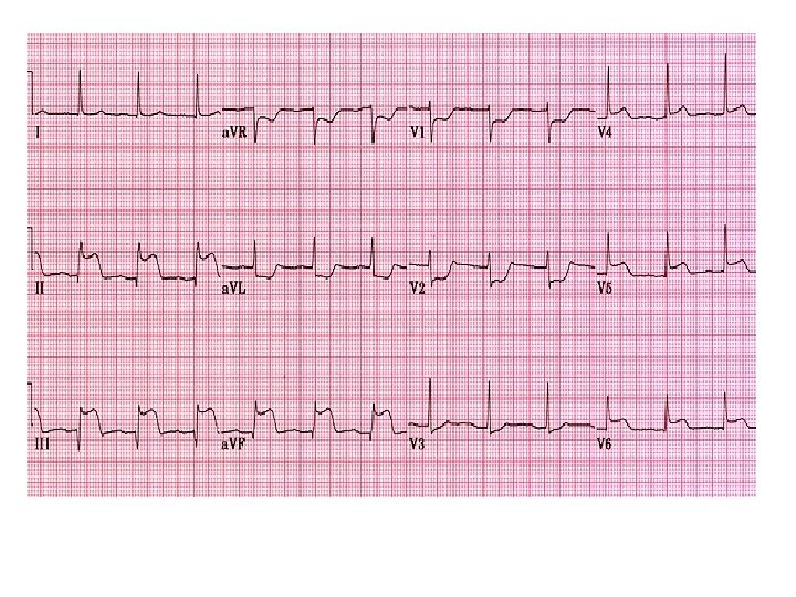
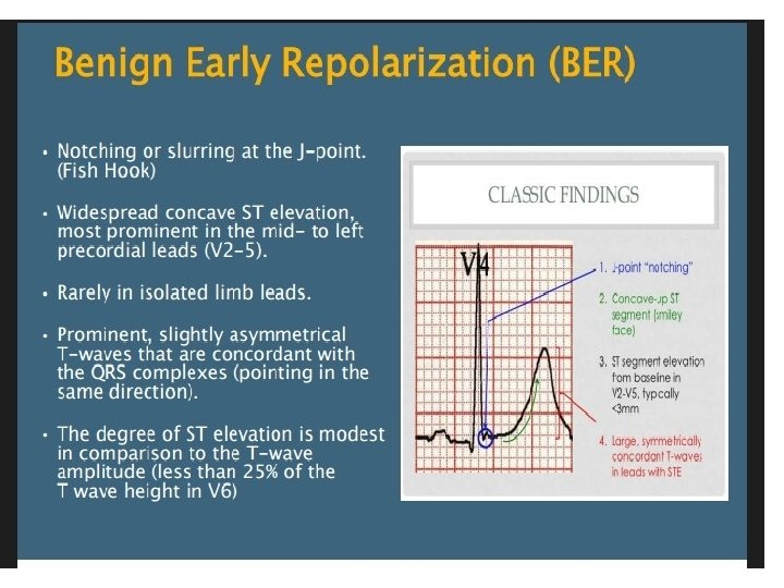
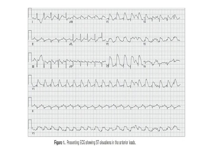
Conditions mimicking STEMI • PERICARDITIS • BER • VENTRICULAR ANEURYSM • BRUGADA SYNDROME • AORTIC DISSECTION • PULMONARY EMBOLISM Neth Heart j. 2008 oc
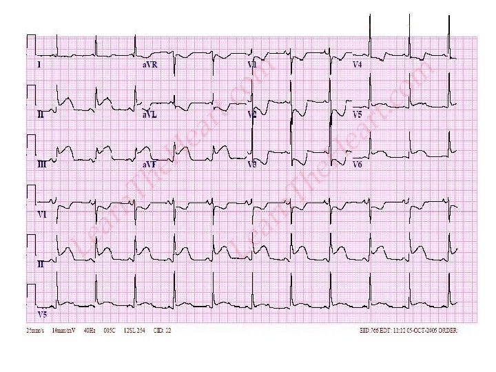
Continues- • • • LBBB LVH PRINZMETALS ANGINA HYPOTHERMIA HYPERKALEMIA
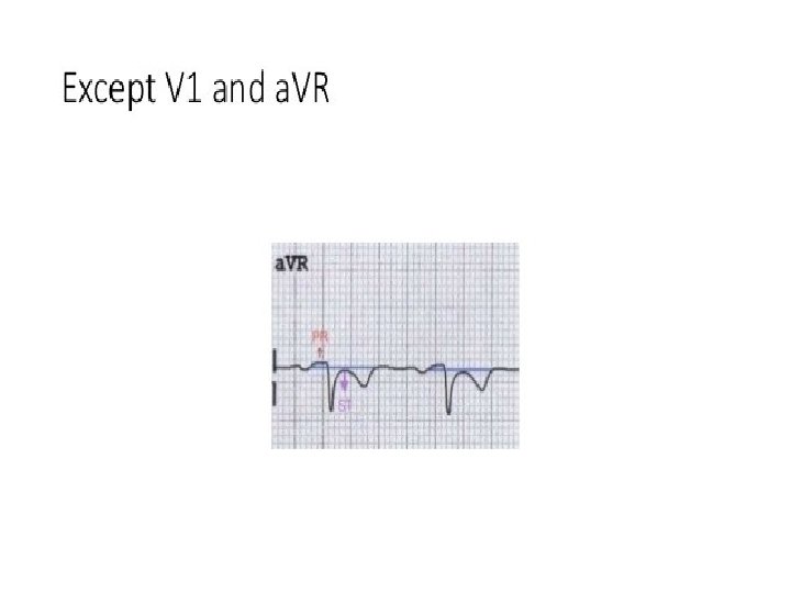
Pericarditis
Persistent ST elevation in LV aneurysm
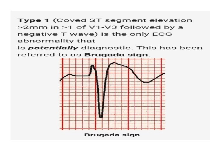
Brugada syndrome • • • Sodium channelopathy > 60 mutations 50% Spontaneous mutation Familial Brugada sign------ECG
ECG abnormality must be associated with one of the following • Polymorphic VT/VF • Family H/O sudden cardiac death • Inducibility of VT with programmed electrical stimulation • Syncope • Nocturnal agonal respiration
LVH
Prinzmetals angina
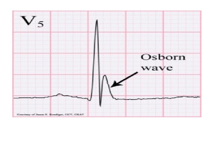
Hypothermia---Osborn wave
Question 1. Match the time after onset of MI with the histological features/clinical event 1. coagulation necrosis, wavy fibers, infiltration of interstitium by polymorphonuclear infiltrates 2. scar with dense collagen fibers 3. thrombotic coronary artery with no histopathological changes. 4. Rupture of ventricular wall (A)2 hr ( B)12 hr (C) 4 -7 days (D) 6 weeks
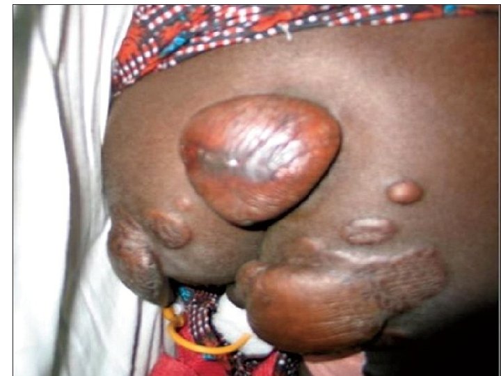
Questions 2 • a 16 year old boy presented with DOE and palpitation for the past 1 month. He had no other respiratory symptoms or fever. He was a non smoker , non hypertensive and non diabetic. His younger brother died suddenly a year earlier
2 D Echo/CAG • 2 D Echocardiogram dilated LV with severe LV systolic dysfunction and akinetic interventricular septum, moderate MR. CAG----- 90% discrete, ostialstenosis of the left main coronary artery and severe proximal LAD disease.
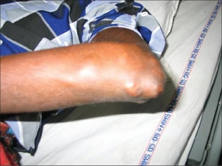
Answer • child had FAMILIAL HYPERCHOLESTROLEMIA • Clinically suspected because of tendinous and tuberous xanthomas with strong family history • Total cholestrol---440 mg/dl • LDL -------398 mg/dl • HDL --------30 mg/dl • TG --------60 mg/dl
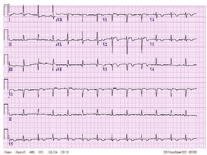
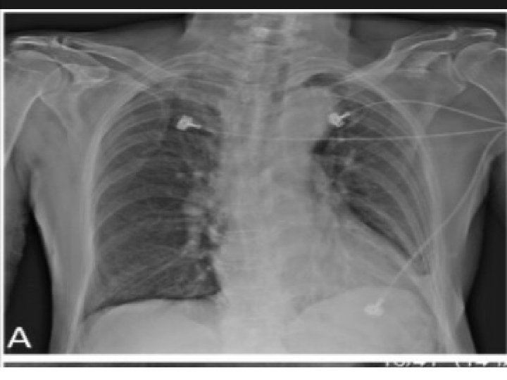
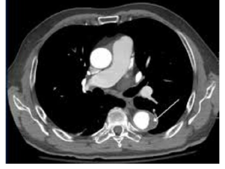
QUESTION 3 • A 69 yr old male with h/o HTN and t 2 DM presented with sudden onset of substernal chest pain and SOB. patient was anxious and diaphoretic in the emergency. vital signs showed a BP reading of 130/90 mm. Hg and HR of 68 bpm. Physical examination was otherwise unremarkable.
ANSWER • Aortic Dissection
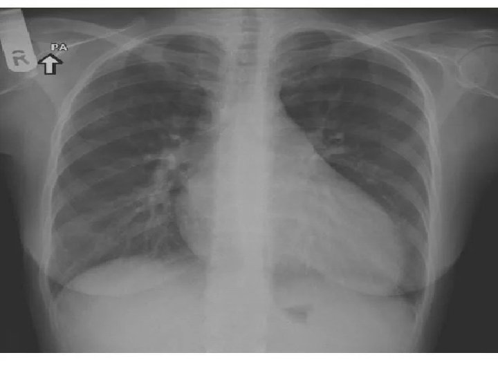
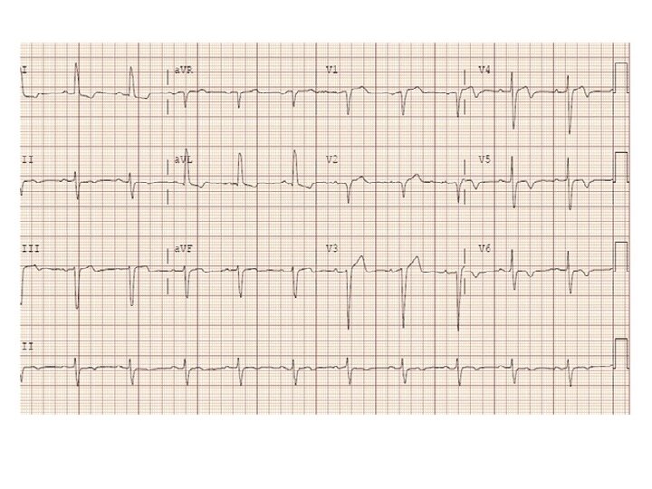
QUESTION 4 • A 63 yr old man was brought to emergency dept in cardiac arrest. he had a h/o HTN , DLP, CAD. pt c/o dizziness and dyspnoea prior to his syncope. his initial BP was 70/40 mm. Hg , HR 68 bpm , SPO 2 82% in RA.
------ • CAG Non obstructive epicardial coronary arteries and patent LAD 2 D ECHO Dilated Rt ventricle Rt sided heart catheterisation revealed RV pressure of 80/30 mm. Hg
Pulmonary angiogram disclosed filling defect s/o B/L pulmonary embolism