CLASS CESTODA TAPEWORMS General characters Tapeworms have flat
")


 • Host: Humans are the only definitive host. Cattle")

. • Ovary bilobed")


 - evaginated scolex (right) The scolex is similar")
 detach from the strobila")
.")


")


")
 in human")


 where infection with cysticerci")
. Cardiac involvement. Ophthalmic involvement.")


 Serology to detect ag or antibodies Stool for")


- Slides: 44
CLASS CESTODA (TAPEWORMS)
General characters: • Tapeworms have flat, segmented bodies, consisting of a head, known as the scolex, neck and a series of segments known as proglottids or strobila. • The scolex: Is the organ of attachment and provided with 4 suckers. Rostellum and hooks may be present in some species. • The strobila: consists of immature, mature and gravid segments.
General characters: • Cestodes are hermaphroditic worms, each mature segment possesses both male and female sex organs. • Tapeworms lack body cavity or digestive system. • Adult worms inhabit the small intestine of the definitive host. • They need intermediate host (except Hymenolepis nana).
Life cycle of cestodes: • Eggs contain hexacanth embryo i. e an embryo with 6 hooklets called (onchosphere). • When swallowed by intermediate host, the onchosphere changes into larval stage in the host tissues. • Larval stages of cestodes ( taenia) : • Cysticercus C. bovis : larval stage of Taenia saginata. C. cellulosae: Larval stage of T. solium. • Cysticercoid : Larval stage of Hymenolepis nana. • Hydatid cyst : Larval stage of Echinococcus granulosus
Human Taeniasis intestinal Taeniasis
1 -Taenia Saginata (Beef Tapeworm) • Host: Humans are the only definitive host. Cattle are intermediate host. • Habitat: Adult worms are found in small intestine of human. • Distribution: Cosmopolitan in countries in which beef is eaten raw or insufficiently cooked.
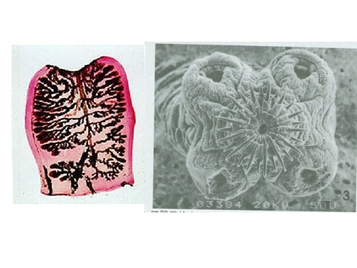
Morphology • Size: About 4 – 10 meters long. • Scolex: Globular in shape with 4 cup shaped suckers. No rostellum, no hooks.
Morphology • Mature segment: • Slightly broader than long (nearly squarish). • Ovary bilobed in the posterior part of the segment.
Morphology • Mature segment: • Uterus, simple tube in the median plane. • Vitelline glands compact and posterior to the ovary. Vagina opens in the genital atrium. • Testes numerous, spherical and scattered throughout the segment. .
Morphology • Gravid segment: 2 X 1 cm in size. Uterus consists of median longitudinal stem with 15 – 30 main uterine branches (on each side).
Morphology • Egg: Spherical 30 – 40 mic. In diameter. With an outer brownish, radially striated embryophore which surrounds a hexacanth embryo or onchosphere.
Cysticercius bovis - invaginated scolex (left) - evaginated scolex (right) The scolex is similar to that of adult worm in morphology Cysticercius bovis
Life cycle: • Single gravid segments (containing thousands of eggs) detach from the strobila and pass to outside with or without faeces.
• The eggs are ingested by intermediate host (cattle).
• • Onchosphere liberated, penetrates the intestinal wall and carried by the lymphatics or blood circulation to muscular tissues. It develops into Cysticercus bovis (infective stage).
• Humans become infected by ingesting cysticerci in raw or undercooked beef.
• In the intestine the scolex evaginates and the worm develops to maturity in 8 – 10 weeks.
Pathogenesis and symptoms: • Most infected people are asymptomatic. • Abdominal pain, diarrhoea, weight loss. • Moderate eosinophilia.
• Diagnosis: • Recovery of eggs or gravid segment (15 – 30 uterine branches) in the faeces. • Single gravid segments may crawl outside the anus (without stools).
• Treatment: • Niclosamide is usually effective in a single dose of 2 grams to be chewed on an empty stomach. • Praziquantel, also effective, the dose of 2. 5 mgkg has been recommended. • • • Prevention and control: Prevent soil contamination with human manure. Proper cooking of beef. Deep freezing of meat for one week. Proper meat inspection in slaughter houses.
Taenia Solium (Pork Tapeworm)
• Host: • Man is the only definitive host, pigs are the intermediate host. • Distribution: • Cosmopolitan in areas where pork is eaten raw or under cooked. • Morphology: • Scolex globular with 4 suckers and a rostellum armed with 2 rows of hooks.
T. Saginata T. solium Strobila 1000 – 2000 proglottides. 800 - 1000 proglottides Length 4 -10 m 3 -4 m Scolex 4 suckers without hooks 4 suckers with 30 hooklets uterine branches 15 -30 7 -12 Eggs 100. 000 / day 30. 000 -50. 000 / day Infective stage Cysticerci cysticerci – eggs Cysticercus C. bovis C. cellulosae Gravid proglottid 2 X 0. 7 cm 1. 5 – 0. 8
• Life cycle and pathogenesis: • It is similar to that of T. saginata except in: • Human becomes infected by ingesting raw or under cooked pork containing Cysticercus cellulosae. • Man sometimes acts as an accidental intermediate host for T. solium when eggs of this parasite are swallowed causing (Cysticercosis).
• Diagnosis: • Detection of eggs or gravid segments (with 7 – 13 uterine branches) in stool. Gravid segments usually come out in chains of 2 – 3 segments; only with faeces. • Treatment and prevention: • As T. saginata but antiemetic drug should be administered one hour before treatment to prevent the possible vomiting which might lead to cysticercosis.
Cystcercus cellulosae in muscles of pigs (measly pork)
Cysticercosis The presence of cysticercus cellulosae (the larval stage of T. solium) in human tissues.
Cysticercus cellulosae It is soybean-like in shape, has an small scolex invaginated into the translucent cyst. (left) The scolex evaginated from the cyst (right) Cysticercius cellulosae
• Mode of infection: • 1 - Accidental ingestion of eggs of T. solium. • 2 - Autoinfection: • external autoinfection; when infected person with adult T. solium contaminates his fingers with eggs in stools, and ingests these eggs with food (hand to mouth infection). • Internal autoinfection: When gravid segments of T. solium are regurgitated into the stomach due to antiperistaltic movement of the intestine. The segments disintegrate releasing eggs which hatch allowing hexacanth embryos to penetrate the gut. The embryos migrate to form larval stage in any organ or tissue as, eye, heart, liver, brain, lung, muscles.
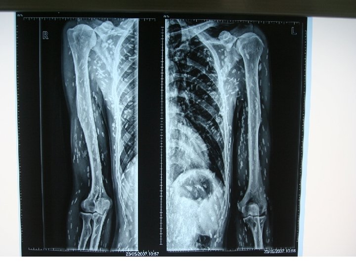
• Pathogenesis: • Depend on the tissue invaded by Cysticercus cellulosae. • Serious symptoms occur when these larvae invade vital organs as brain, eye. • Diagnosis: • X-ray in case of calcified larvae. • Radiographic, computed tomography (CT) or magnetic resonance imaging (MRI). • Eosinophilia. • Serological tests as ELISA and indirect haemagglutination.
CLINICAL MANIFESTATIONS symptoms may be different depending on the: (Site) where infection with cysticerci occurs (Number) how many cysts are there. (viability) whether the cyst is viable , dying or dead and calcified. Most cases are asymptomatic. Incubation: months~5 years •
Clinical features Subcutaneous and Muscular involvement. Neurocysticercosis (NCC). Cardiac involvement. Ophthalmic involvement.
1 -Subcutaneous and Muscular involvement: - 2/3 of patients have nodules. - Number of nodules: 1 -1000 sues: nodules on arms and chest. small, movable, painless - Muscular involvement: rarely painful; seen as calcifications following muscle bundles in thighs or arms. - Massive parasite burden : limb muscular enlargement (Pseudohypertrophy).
CLINICAL MANIFESTATIONS 2 - Cardiac involvement: 1 -5% of patients typically asymptomatic abnormal rhythms or heart failure (rare) 3 - Ophthalmic involvement: (1 -3% ). Intraocular cysts : a- solitary lesion: freely float in the vitreous humor b- large parasitic burden: visual disturbance and visual loss.
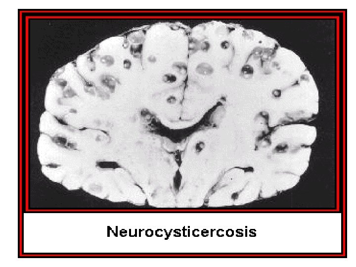
NEUROCYSTICERCOSIS NCC : frequently asymptomatic. • Symptoms : similar to those found with other intracranial mass lesions consistent with elevation of ICP. has gained increased recognition in the last 2 decades
• Diagnosis: • X-ray in case of calcified larvae. • Radiographic: computed tomography (CT) or magnetic resonance imaging (MRI). • Laboratory studies • Eosinophilia. • Serological tests as ELISA and indirect haemagglutination.
Laboratory studies Complete blood count (CBC) Serology to detect ag or antibodies Stool for ova and parasite Lab. studies : inferior to imaging in diagnosis but may play an adjunctive role.
• Treatment: • Surgical removal of cysticeri. • Therapeutic treatment as praziquantel 50 mg/kg, in divided doses for two weeks. • Steroides to prevent serious reactions of the body due to dying cysts. • Prevention: • Treatment of infected persons with T. solium. • Avoid using nauseating drugs in infected persons. • Avoid using human manure as fertilizer. • Periodic examination of food handlers.
How can we prevent cysticercosis ? Control the sources of infection Control transmission ways Educate the populations Avoid eating raw or undercooked pork Wash hands after using the toilet and before handling food Wash and peel all raw vegetables and fruits before eating
Thank you for attention!