CKD CHORONIC KIDNEY DISEASE What is CKD CKD


 <60 m. L/min/1. 73 m 2 �GFR is the best")


Using (Serum Creatinine Concentration , Age,")
 Cockcroft-Gaultequation")


patients with a silent primary glomerulopathy 2)patients in whom")
Excretion of metabolic waste products and foreign chemicals 2)Regulation of")


 b. Hypo-natremia(I) c. Hyper-kalemia(I) d. Hyper-phosphatemia(I)")
 2. Adynamic bone(D) 3. Vitamin.")
b 2. Sleep disorders(P) 3. Headache(P) 4. Impaired mentation (I)b")
 2. Congestive heart")
b 2. Hyperpigmentation (I, P, or D) 3. Pruritus(P)")
 2. Nausea and vomiting(I) 3. Gastroenteritis (I) 4. Peptic")
b 2. Lymphocytopenia (P) 3. Bleeding diathesis")


 Secretion of H+")


 Ischemic vasculardisease �The CKD-related risk factors comprise 1. anemia, 2.")
 Heart-failure �“bat wing” distribution -form of “low-pressure” pulmonary edema")
 Hypertension and left ventricular hypertrophy Ø anemia and the placement of an arterio-venous")


- Slides: 95
CKD CHORONIC KIDNEY DISEASE
What is CKD? CKD is defined bythe �–presence of kidney damageor decreased kidneyfunction �–forthree or moremonths, �–irrespective of thecause.
What is CKD? � The persistence of the damage or decreased Function for at least three months is necessary to distinguish CKD from acute kidney disease. � Kidney damage refers to pathologic abnormalities, whether established via: 1. renal biopsy or 2. imaging studies, or 3. inferred from markers such as a)urinary sediment abnormalities or b)increased rates of urinary albumin excretion
What is CKD? �Chronic kidney disease is defined based on the presence of either kidney damage or decreased kidney function for three or more months, irrespective of cause. Criteria: �Duration ≥ 3 months, based on documentation or inference �Glomerular filtration rate (GFR) <60 m. L/min/1. 73 m 2 �Kidney damage, as defined by structural abnormalities or functional abnormalities other
Duration≥ 3 months, based on documentation or inference �Duration is necessary to distinguish chronic from acute kidney diseases. 1. Clinical evaluation can often suggest duration 2. Documentation of duration is usually not available in epidemiologic studies
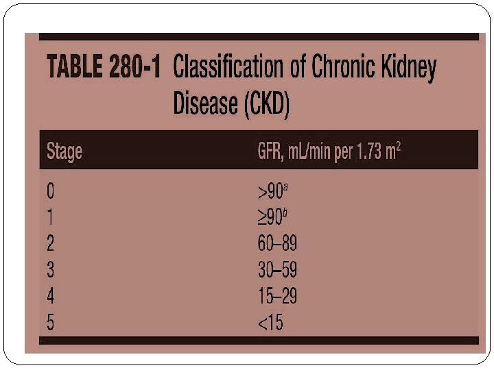
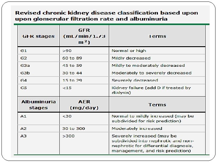
Glomerular filtration rate (GFR) <60 m. L/min/1. 73 m 2 �GFR is the best overall index of kidney function in health and disease. 1. The normal GFR in young adults is approximately 125 m. L/min/1. 73 m 2; GFR <15 m. L/min/1. 73 m 2 is defined as kidney failure 2. Decreased GFR can be detected by current estimating equations for GFR based on serum creatinine (estimated GFR) but not by serum creatinine alone 3. Decreased estimated GFR can be confirmed by measured GFR
Kidney damage, as defined by structural abnormalities or functional abnormalities other than decreased GFR � A) Pathologic abnormalities (examples). Cause is based on underlying illness and pathology. Markers of kidney damage may reflect pathology. 1. Glomerular diseases (diabetes, autoimmune diseases, systemic infections, drugs, neoplasia) 2. Vascular diseases (atherosclerosis, hypertension, ischemia, vasculitis, thrombotic micro angiopathy) 3. Tubulo interstitial diseases (urinary tract infections, stones, obstruction, drug toxicity) 4. Cystic disease (polycystic kidney disease)
Kidney damage, as defined by structural abnormalities or functional abnormalities other than decreased GFR � B) History of kidney transplantation. In addition to pathologic abnormalities observed in native kidneys, common pathologic abnormalities include the following: 1. Chronic allograft nephropathy (non-specific findings of tubular atrophy, interstitial fibrosis, vascular and glomerular sclerosis) 2. Rejection 3. Drug toxicity (calcineurin inhibitors) 4. BK virus nephropathy 5. Recurrent disease (glomerular disease, oxalosis, Fabry disease)
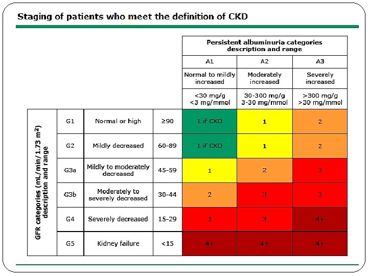
Kidney damage, as defined by structural abnormalities or functional abnormalities other than decreased GFR � C) Albuminuria as a marker of kidney damage (increased glomerular permeability, urine albumin-tocreatinine ratio[ACR] >30 mg/g). * 1. The normal urine ACR in young adults is <10 mg/g. Ø Urine ACR categories 10 -29, 30 -300 and >300 mg are termed "high normal, high, and very high" respectively. Ø Urine ACR >2200 mg/g is accompanied by signs and symptoms of nephrotic syndrome 2. Threshold value corresponds approximately to urine dipstick values of trace or 1+ 3. High urine ACR can be confirmed by urine albumin excretion in a timed urine collection
Kidney damage, as defined by structural abnormalities or functional abnormalities other than decreased GFR �D) Urinary sediment abnormalities as markers of kidney damage 1. RBC casts in proliferative glomerulo nephritis 2. WBC casts in pyelonephritis or interstitial nephritis 3. Oval fat bodies or fatty casts in diseases with proteinuria 4. Granular casts and renal tubular epithelial cells in many parenchymal diseases(nonspecific)
Kidney damage, as defined by structural abnormalities or functional abnormalities other than decreased GFR � E) Imaging abnormalities as markers of kidney damage (ultrasound, computed tomography and magnetic resonance imaging with or without contrast, isotope scans, angiography). 1. Polycystic kidneys 2. Hydronephrosis due to obstruction 3. Cortical scarring due to infarcts, pyelonephritis or vesico ureteral reflux 4. Renal masses or enlarged kidneys due to infiltrative diseases 5. Renal artery stenosis 6. Small and echogenic kidneys (common in later stages of CKD due to many parenchymal diseases)
PATHOPHYSIOLOGY OF CHRONIC KIDNEYDISEASE �Two broad sets of mechanisms of damage: 1. initiating mechanisms specific to the underlying etiology 2. a set of progressive mechanisms -hyperfiltration and hypertrophy of the remaining viablenephrons
PATHOPHYSIOLOGY OF CHRONIC KIDNEYDISEASE Increased intra renal activity of the reninangiotensin axis appears to contribute both to: �initial adaptive hyper filtration the subsequent mal -adaptive hypertrophy and sclerosis(TGF-β)
Left: Schema of the normal glomerular architecture. Right: Secondary glomerular changes
IDENTIFICATION OF RISK FACTORSAND STAGING OF CKD � Risk factors: � hypertension, � Diabetes mellitus, � Autoimmune disease, � Older age, � African ancestry, � a family history of renal disease, � a previous episode of acute kidney injury, � and the presence of a. proteinuria, b. abnormal urinary sediment , or c. structural abnormalities of the urinary tract
Recommended Equations for Estimation of Glomerular Filtration Rate (GFR)Using (Serum Creatinine Concentration , Age, Sex, Race, and Body Weight) 1) Equation from the Modification of Diet in Renal Disease study∗(MDRD)
2) Cockcroft-Gaultequation
IDENTIFICATION OF RISK FACTORSAND STAGING OFCKD �Chronic renal damage Ø Persistence in the urine of >17 mg of albumin per gram of creatinine in adult males and 25 mg albumin per gram of creatinine in adult females
ETIOLOGY AND EPIDEMIOLOGY Leading Categories of Etiologies of CKD∗ �Diabetic glomerular disease �Glomerulonephritis �Hypertensive nephropathy �Primary glomerulopathy with hypertension �Vascular and ischemic renal disease �Autosomal dominant polycystic kidney disease �Other cystic and tubulo interstitial nephropathy
ETIOLOGY AND EPIDEMIOLOGY �Newly diagnosed CKD: Ø present with hypertension �CKD is often attributed to hypertension: Ø When no overt evidence for a primary glomerular or tubulo interstitial kidney disease process is present
ETIOLOGY AND EPIDEMIOLOGY Two Categories: 1)patients with a silent primary glomerulopathy 2)patients in whom progressive nephrosclerosis and hypertension is the renal correlate of a systemic vasculardisease
Multiple Functions of Kidneys 1)Excretion of metabolic waste products and foreign chemicals 2)Regulation of water and electrolyte balances 3)Regulation of body fluid osmolality and electrolyte concentrations 4)Regulation of arterial pressure 5)Regulation of acid-base balance 6)Secretion, metabolism, and excretion of hormones 7)Gluco-neogenesis
PATHOPHYSIOLOGY AND BIOCHEMISTRY OF UREMIA �Elevated waste products: Hundreds of toxins, water-soluble, hydrophobic, protein-bound, charged, and uncharged compounds, guanidino compounds, urates and hippurates, products of nucleic acid metabolism, polyamines, myoinositol, phenols, benzoates and indoles ‘middlemolecules’
PATHOPHYSIOLOGY AND BIOCHEMISTRY OF UREMIA �A host of metabolic and endocrine functions normally performed by the kidneys is also impaired or suppressed: Ø anemia, Ø malnutrition, Ø and abnormal metabolism of carbohydrates, fats, and proteins
PATHOPHYSIOLOGY AND BIOCHEMISTRY OF UREMIA �Urinary retention, decreased degradation, or abnormal regulation of hormones Ø PTH, FGF-23, Ø insulin, glucagon, Ø steroid hormones including vitamin D and sex hormones, and prolactin
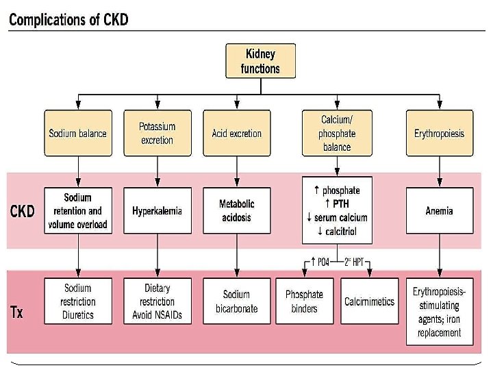
CLINICAL ANDLABORATORY MANIFESTATIONS OF CHRONIC KIDNEY DISEASE ANDUREMIA CLINICAL ABNORMALITIES INUREMIA 1. Fluid and electrolyte disturbances 2. Endocrine-metabolic disturbances 3. Neuro muscular disturbances 4. Cardiovascular and pulmonary disturbances 5. Dermatologic disturbances 6. Gastro intestinal disturbances 7. Hematologic and immunologic disturbances
1. Fluid and electrolyte disturbances a. Volume expansion(I) b. Hypo-natremia(I) c. Hyper-kalemia(I) d. Hyper-phosphatemia(I) improves with an optimal program of dialysis and related therapy; (P) persist or even progress, despite an optimal program; (D) develops only after initiation of dialysis therapy.
2. Endocrine-metabolic disturbances 1. Secondary hyperparathyroidism (I or P) 2. Adynamic bone(D) 3. Vitamin. D–deficient osteo-malacia (I) 4. Carbohydrate resistance(I) 5. Hyperuricemia (I or P) 6. Hypertriglyceridemia (I or P) 7. Increased Lp(a) level(P) 8. Decreasedhigh-density lipoprotein level(P) 9. Proteinenergymalnutrition (I or P) 10. Impaired growth and development(P) 11. Infertility and sexual dysfunction(P) 12. Amenorrhea (I/P) 13. β 2 -Microglobulin associated amyloidosis (P or D)
3. Neuro-muscular disturbances 1. Fatigue(I)b 2. Sleep disorders(P) 3. Headache(P) 4. Impaired mentation (I)b 5. Lethargy(I)b 6. Asterixis(I) 7. Muscularirritability 8. Peripheral neuropathy (I or P) 9. Restless legs syndrome(I or P) 10. Myoclonus (I) 11. Seizures (I or P) 12. Coma(I) 13. Muscle cramps (P or D) 14. Dialysis disequilibrium syndrome(D) 15. Myopathy (P or D)
4. Cardiovascular and pulmonary disturbances 1. Arterial hypertension (I or P) 2. Congestive heart failure or pulmonary edema(I) 3. Pericarditis(I) 4. Hypertrophic or dilated cardio myopathy (I, P, or D) 5. Uremic lung(I) 6. Accelerated atherosclerosis (P or D) 7. Hypotensionand arrhythmias (D) 8. Vascular calcification (P or. D)
5. Dermatologic disturbances 1. Pallor (I)b 2. Hyperpigmentation (I, P, or D) 3. Pruritus(P) 4. Ecchymoses(I) 5. Nephrogenic fibrosing dermopathy (D) 6. Uremic frost(I)
6. Gastrointestinal disturbances 1. Anorexia(I) 2. Nausea and vomiting(I) 3. Gastroenteritis (I) 4. Peptic ulcer (I or. P) 5. Gastrointestinal bleeding (I, P, or. D) 6. Idiopathic ascites(D) 7. Peritonitis(D)
7. Hematologic and immunologic disturbances 1. Anemia (I)b 2. Lymphocytopenia (P) 3. Bleeding diathesis (I or D)b 4. Increased susceptibility to infection 5. (I or P) 6. Leukopenia (D) 7. Thrombocytopenia(D)
FLUID, ELECTROLYTE, AND ACID-BASE DISORDERS
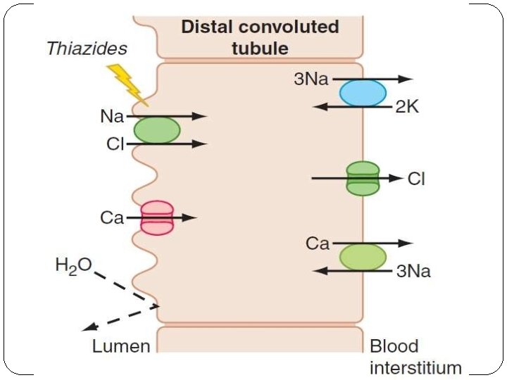
FLUID, ELECTROLYTE, AND ACIDBASE DISORDERS �Hypo-natremia –water restriction S�ECFV – salt restriction O�Thiazide : Little activity in CKD stage 3 and higher and Loop Diuretics are needed in higher doses D �Metolazone : is combined with loop diuretics , I which inhibits the sodium chloride co transporter U in the distil convoluted tubule can help in the renal M salt removal
FLUID, ELECTROLYTE, AND ACIDBASE DISORDERS • HYPERKALEMIA • Precipitated by • increased dietary potassium intake, • protein catabolism, • hemolysis, • hemorrhage, • transfusion of stored blood cells, • and metabolic acidosis • Medications
FLUID, ELECTROLYTE, AND ACIDBASE DISORDERS �Hypo-kalemia: • Not common in CKD • reduced dietary potassium intake • GI losses • Diuretic therapy • Fanconi’s syndrome • RTA • Hereditary or acquired Tubulointerstitial disease
FLUID, ELECTROLYTE, AND ACIDBASE DISORDERS • • • Metabolic acidosis common disturbance in advanced CKD combination of hyper-kalemia and hyper-chloremic metabolic acidosis is often present, even at earlier stages of CKD (stages 1– 3) Treat hyper-kalemia the p. H is rarely <7. 35 usually be corrected with oral sodium bicarbonate supplementation
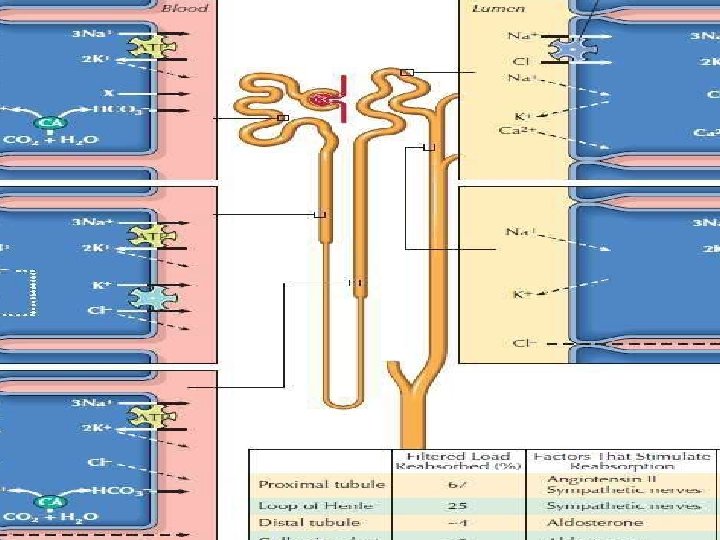
FLUID, ELECTROLYTE, AND ACIDBASE DISORDERS �Renal Control of Acid-Base Balance 1) Secretion of H+ and Reabsorption of HCO 3 by the Renal Tubules a. b. c. H+ is Secreted by Secondary Active Transport in the Early Tubular Segments Filtered HCO 3 is Reabsorbed by Interaction with H+ in the Tubules Primary Active Secretion of H+ in the Intercalated Cells of Late � Distal and Collecting Tubules 2) Combination of Excess H+ with Phosphate and Ammonia Buffers in the Tubule Generates “New” HCO 3 Phosphate Buffer System Carries Excess H+ into the Urine and � Generates New HCO 3 b. Excretion of Excess H+ and Generation of New HCO 3 by the Ammonia Buffer System a.
FLUID, ELECTROLYTE, AND ACIDBASE DISORDERS
To maintain euvolemia: Ø Adjustments in the dietary intake of salt Ø and use of loop diuretics, occasionally in combination with metolazone Hyponatremia: water restriction Hyperkalemia Ø Ø responds to dietary restriction of potassium, avoidance of potassium supplements use of kaliuretic diuretics potassium-binding resins, such as calcium resonium or sodium polystyrene The renal tubular acidosis and subsequent anion- gap metabolic acidosis Ø alkali supplementation, typically with sodium bicarbonate
DISORDERS OF CALCIUM AND PHOSPHATE METABOLISM The principal complications of abnormalities of calcium and phosphate metabolism in CKD Ø Ø occur in the skeleton and the vascular bed, with occasional severe involvement of extraosseous soft tissues Bone manifestations of CKD, classified as: Ø associated with high bone turnover with increased PTH levels Ø low bone turnover with low or normal PTH levels Ø
DISORDERS OF CALCIUM AND PHOSPHATE METABOLISM The patho-physiology of secondary hyper-parathyroidism: 1. Declining GFR leads to reduced excretion of phosphate 2. increased synthesis of PTH and growth of parathyroid gland mass 3. decreased levels of ionized calcium, resulting from diminished calcitriol production by the failing kidney
DISORDERS OF CALCIUM AND PHOSPHATE METABOLISM Calcium, phosphorus, and the cardiovascular system: Ø Hyper-phosphatemia and hyper-calcemia are associated with increased vascular calcification of the media in coronary arteries and even heart valves Ø ingested calcium cannot be deposited in bones with low turnover osteoporosis and vascular calcification Ø Hyper-phosphatemia can induce a change in gene expression in vascular cells
DISORDERS OF CALCIUM AND PHOSPHATE METABOLISM other complications of abnormal mineral metabolism: Ø Calciphylaxis (calcific uremic arteriolopathy) Ø Other etiologies Ø use of oral calcium as a phosphate binder Ø Warfarin
�Sevelamer and lanthanum –non calcium containing polymers �Calcitriol exerts a direct suppressive effect on PTH secretion and also indirectly suppresses PTH secretion by raising the concentration of ionized calcium �recommended target PTH level between 150 and 300 pg/m. L
CARDIO VASCULAR ABNORMALITIES 1) Ischemic vasculardisease �The CKD-related risk factors comprise 1. anemia, 2. hyperphosphatemia, 3. hyperparathyroidism, 4. sleep apnea. 5. Generalized inflammation �Cardiac troponin levels are frequently elevated in CKD without evidence of acute ischemia.
2) Heart-failure �“bat wing” distribution -form of “low-pressure” pulmonary edema
3) Hypertension and left ventricular hypertrophy Ø anemia and the placement of an arterio-venous fistula Ø low blood pressure actually carries a worse prognosis than does high blood pressure Ø Erythro-poiesis-stimulating agents
MANAGEMENT OF HYPERTENSION �Blood pressure should be reduced to 125/75 �Salt restriction should be the first line of therapy �MANAGEMENT OF CARDIO VASCULAR DISEASE �Lifestyle changes, including regular exercise �Manage dyslipidemia