Circulatory and Lymphatic System Contributions of the Circulatory

Circulatory and Lymphatic System
 • *Excretory:")
Contributions of the Circulatory System • *Nutritive: provides cells with food (nutrients) • *Excretory: provides for elimination of wastes • *Protection: provides for defense and resistance to disease • *Regulatory: provides for internal balance of heat and fluids • *Respiratory: provides cells with O 2 and the elimination of CO 2 **It’s the transportation system of the body!**

Characteristics of Blood • Blood is a fluid connective tissue • Composition of blood: • 45% blood cells (formed elements) • 55% plasma (liquid portion) • Blood contains 3 types of cells: • Red Blood Cells (RBC/erythrocytes) • White Blood Cells (WBC/leukocytes) • Platelets (thrombocytes) • Plasma • *Amount in an adult • 4 -6 quarts

Plasma • Liquid part of blood • 91. 5% water • 7% plasma proteins • Other 1. 5 %: salts, nutrients, electrolytes, hormones, respiratory gases, nitrogenous wastes, antibodies…. • Plasma also carries body heat
 • Normal range of RBC 4 -6 million cells • RBC’s contain")
Erythrocytes (RBC’s) • Normal range of RBC 4 -6 million cells • RBC’s contain protein hemoglobin: the part of the RBC that carries oxygen • Function of RBCs: carry oxygen to all body tissues • Normal range for hemoglobin 12 -18 grams per 100 cc’s of blood • Iron = mineral needed formation of hemoglobin
 • Normal WBC count = 5, 000 to 10, 000")
Leukocytes (white blood cells/WBC) • Normal WBC count = 5, 000 to 10, 000 cells per mm 3 • Function of WBCs: defend against infection and help provide immunity • Formed in red blood marrow and lymphatic tissue • Elevation of different WBCs help to diagnose specific problems
 • Platelets are not whole cells; they are fragments or pieces of")
Thrombocytes (Platelets) • Platelets are not whole cells; they are fragments or pieces of cells • Function of platelets: aid in blood clotting • Normal platelet count: 250, 000 to 400, 000 cells per mm 3 • Platelets are necessary for homeostasis (prevention of blood loss)

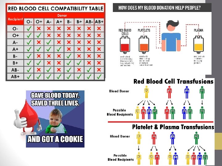
Blood Types • Our blood type is genetic • A type and cross match is done before a blood transfusion to prevent transfusing incompatible blood • Type A blood • Has A antigen on RBCs • Has anti-B antibodies in plasma • Type B blood • Has B antigen on RBC’s • Has anti-A antibodies in plasma

Blood Types • Type AB blood • Has both A&B antigen on RBCs • Has no antibodies in plasma (neither anti-A or anti-B antibodies) • ***Universal recipient (no antibodies)*** • Type O blood • Has no antigen on RBCs (no A or B antigen) • Has anti-A and anti-B antibodies in plasma • ***Universal donor (no antigens)***

Rh Factor • Rh factor is another antigen that may be present on RBCs • If the Rh factor antigen is present, the person is Rh+ • If the Rh factor antigen is not present, the person is Rh • Problem: Rh incompatibility between mother (-) and fetus (+): hemolytic anemia

Hemostasis • Involves 3 events: • Vascular spasm: a sudden, brief tightening of a blood vessel. Vascular spasms can temporarily reduce blood flow to tissues supplied by that vessel. • Platelet plugs: platelets clump together at the site of injury, swell, and stick to the injured area, acting as a plug to reduce the bleeding. • Chemical clotting: Platelets, which come from white blood cell fragments, immediately begin to adhere to the cut edges of the vessel and release chemicals to attract even more platelets. A platelet plug is formed, and the external bleeding stops.

Antigens and Antibodies • Antigen: Substance that stimulates the body to make antibodies, usually foreign, except proteins present on RBCs • Antibody: Proteins produced by the body to neutralize antigens

The Heart • Cone shaped, hollow muscular organ • Beats 100, 000 times/day • Located in the mediastinum • The thoracic cavity • Primary function: pump blood through arteries, capillaries, and veins • Animation
")
Pericardial Membranes • *The heart is enclosed in the pericardium (or pericardial membranes)

Walls of the Heart • 3 Distinct Layers • 1. Epicardium • 2. Myocardium- thick muscular wall of the heart • 3. Endocardium

Heart Chambers • Upper chambers of the heart – atria-Right and left atrium • Thin walls • Separated by interatrial septum • Common wall of myocardium • Atria are receiving chambers for blood • Lower chambers of heart – ventricles-Right and left ventricles Septum

Right Atrium • 2 large veins return blood from body to right atrium of the heart • Superior vena cava • Carries blood from upper body to heart • Inferior vena cava • Carries blood from lower body to heart • *Largest vein in the body

Left Atrium • *4 pulmonary veins return blood from the lungs to the left atrium of the heart

Right Ventricle • Receives blood from the right atrium • Pulmonary artery • *Pulmonary artery takes blood from the right ventricle to the lungs. It branches into left and right pulmonary arteries, one for each lung. *Tricuspid Value* Right atrium to right ventricle

Left Ventricle • Receives blood from left atrium • *Thickest walls • Pumps blood to body via aorta = largest artery of the body • Mitral valve • Blood is carried to the lungs by the pulmonary artery
 valve • Blood flows from right atrium through")
Heart Valves • Tricuspid/right atrioventricular (AV) valve • Blood flows from right atrium through this valve into right ventricle • Mitral / bicuspid / left AV valve • *Blood flow from left atrium (auricle) through this valve into left ventricle

Heart Valves • Pulmonary semilunar valve • Valve at the junction of the right ventricle and the pulmonary artery • Aortic semilunar valve • Valve at junction of aorta and left ventricle

Cardiac Conduction System • Cardiac cycle regulated by electrical activity of myocardium • Heart generates its own beat and the electrical impulses follow a specific route • SA Node in right atrium is the *pacemaker* • Initiates each heartbeat • *Conduction Pathway: SA Node-> 1. AV Node-> atrial myocardium (atria contract)-> 2. Bundle of HIS-> right and left bundle branches-> 3. Purkinje fibers-> ventricular myocardium (ventricles contract) • http: //www. mhhe. com/biosci/ap/dy namichuman 2/content/cardio/VPL 29. MOV

Nervous System Control of the Heart Cardiac muscle is involuntary in action Control center is in the brain (medulla) ANS = Sympathetic and Parasympathetic divisions *Sympathetic division of the ANS speeds the heart rate (adrenaline) • *Parasympathetic division of the ANS slows and strengthens the heart rate • •

Heart’s Pumping Cycle • Atria contract forcing blood into ventricles • Ventricles contract forcing blood out of the heart • Right Ventricle: blood pumped out of the pulmonary artery to lungs to be oxygenated • Left Ventricle: blood pumped out of the aorta to the body • Blood fills both atria • Right atrium: from body • Left atrium: from lungs

Heart Sounds • Each heartbeat produces 2 sounds: lub-dub • Lub-dub sound = closing of heart valves • *First sound: lub = closing of valves between atria and ventricles (AV valves) caused by ventricular sound • *Second sound: dub = closing of aortic and pulmonary semilunar valves • Heart murmur: an abnormal or extra heart sound caused by a malfunctioning heart valve

Cardiac Cycle • Sequence of events in 1 heartbeat • Simultaneous contraction of the two atria followed (the “lub” sound) by simultaneous contraction of the two ventricles (the “dub” sound) • *Systole: contraction (1 st sound heard)Lub • *Diastole: relaxation (2 nd sound heard) Dub

The Vascular System • Consists of arteries, capillaries, and veins • Arteries and veins transport blood between the capillaries and the heart • Capillaries exchange materials between blood and tissues

Arteries • *Carry blood away from heart • Blood is high in O 2 (oxygenated) • Highest blood pressure • Function as– collectors • Arterioles = small arteries • *Coronary arteries provide blood supply to the myocardial cells

Main Arteries Temporal Popliteal Dorsalis Pedis
,")
Veins • *Carry blood back to heart • Blood is low in oxygen (deoxygenated), high in CO 2 • Function as– distributors • Venules = small veins • Inner layer of veins is smooth but at intervals there are valves to prevent the backflow of blood

Capillaries • Carry blood from arterioles to venules • Site of exchange of materials between blood and tissue fluids surrounding cells • Gases (O 2 and CO 2) move by diffusion • Nutrients move by filtration • 2 way traffic – nourishment and exchange of wastes • *Walls are 1 cell thick, extensions of lining of arteries and veins

Distribution and Collection Routes • Distribution: • Heart to arteries to arterioles to capillaries • Distributes oxygen and nutrients • Exchange occurs in capillaries • Capillaries carry blood from arterioles to venules • Collection: • Capillaries to venules to veins to heart • Transports deoxygenated blood to heart • Then, blood is sent to the lungs for oxygen

Pathways of Circulation • 2 major pathways of circulation: • Pulmonary circulation • Systemic circulation

Pulmonary Circulation • Right-sided heart pump • Superior and Inferior Vena Cava return deoxygenated blood back to the heart into the right atrium • Blood passes from right atrium into the right ventricle going through the tricuspid valve • Right ventricle pumps deoxygenated blood into the pulmonary artery which branches into the right and left pulmonary arteries, one going to each lung • Pulmonary arteries are low in oxygen, high in carbon dioxide • Carbon dioxide will be exchanged for oxygen in the lungs and returned back to the heart via the pulmonary veins

Systemic Circulation • Left-sided heart pump • Pulmonary veins return oxygenated blood back to the heart and into the left atrium • Blood passes through the mitral valve to the left ventricle • Left ventricle pumps oxygenated blood into the aorta (passes through the aortic semilunar valve) and out the body

Hepatic Portal Circulation • Subdivision of Systemic Circulation • Blood from abdominal digestive organs and spleen circulate through the liver before returning to the heart • Veins from the spleen, stomach, pancreas, intestines do not drain into the inferior vena cava; they send their blood to the liver via the portal vein. Liver is a manufacturing plant. • Blood leaves the liver via the hepatic vein and then empties into the inferior vena cava; which goes to the right atrium of the heart

Cerebral Circulation • Part of the Systemic Circulation • Blood supplied to brain via 2 internal carotid arteries and 2 vertebral arteries. Jugular veins drain back to the superior vena cava. • Brain requires a constant flow of blood to supply oxygen and remove wastes. Blood flow to the brain doesn’t change with activity. • Circle of Willis (cerebral arterial circle) provides for a continuous blood supply

Renal Circulation • Part of Systemic Circulation • Renal circulation • Renal arteries bring blood to kidneys • Renal veins drain blood from kidneys

The Lymphatic System • Functions: • Returns lymph fluid to blood • Filters injurious agents and prevents them from entering the bloodstream • *Forms some WBCs (lymphocytes and monocytes) • Defends against infection
 that enters lymph capillaries • Filtration in")
Lymph • Name for tissue fluid (watery-like) that enters lymph capillaries • Filtration in capillaries creates tissue fluid, most is returned to the blood

Lymphatic Ducts • 2 ducts empty lymph into circulatory system • Thoracic duct • *Largest duct • *Drains ¾ of body and empties lymph into left subclavian vein • Right Lymphatic duct • Drains about ¼ of body and empties lymph into right subclavian vein • Flaps in both subclavian veins permit entry of lymph but prevent blood from flowing into lymph vessels

Lymph Nodes and Nodules • Masses of lymphatic tissue • Found in groups along pathway of lymph vessels • All located at junctions of head and extremities with trunk of body • Functions: • Filtration • Phagocytosis • WBC formation

Lymph Nodes • *Cervical lymph nodes • *Tonsils = lymphatic nodules of pharynx • *Axilla = armpit • Iliac=inner hip • Inguinal=groin

Spleen • *Located LUQ of abdominal cavity, just below diaphragm, behind the stomach • Functions: • *WBC production • *Defense = contains plasma cells that produce antibodies • Not considered a vital organ
- Slides: 46