Circulation Functions Transports gases from the respiratory system

, nutrient molecules, and waste materials (from the")


, these")


 ______ ____________")


 1. 2. 3. 4. 5. 6. What are three main functions")
 13. How does exercise help reduce high blood pressure? What is")

- Slides: 39
Circulation
Functions Transports gases (from the respiratory system), nutrient molecules, and waste materials (from the digestive system) Regulates internal temperature and transports chemical substances Protects against blood loss from injury and against disease-causing microbes or toxic substances
Major components Heart Muscular organ that continuously pumps the blood through the body and generates blood flow Blood vessels Are a system of hollow tubes through which the blood moves Blood Fluid that transports nutrients, oxygen, carbon dioxide, and many other materials throughout the body
Two types of Circulatory Systems Open Blood flows freely within the body cavity and makes direct contact With organs and tissues In invertebrates the mixture of Blood and fluids that surrounds the cells is called hemolymph
Closed Need a system to circulate blood, Keep it under pressure and pump Keeps the blood physically contained within vessels and separate from other body tissues
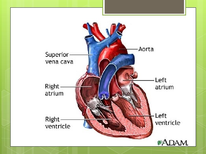
The HUMAN CIRCULATORY SYSTEM: THE HEART Slightly to the left of the middle of the chest About size of fist Walls of heart are made of a unique type of muscle called cardiac muscle Does not become fatigued In addition to pumping blood, a healthy heart ensures blood keeps flowing in one direction only AND oxygen-rich blood is separate from oxygen-poor blood
Explain to a friend the direction of blood flow starting from superior vena cava and ending with aorta
Structure of Heart Four chambers Two top chambers are the atria (singular atrium), these fill with blood returning from the body or the lungs Two bottom chambers are the ventricles receive blood from the atria and pump it out to the body or the lungs Septum: separate the atria and ventricles
The inferior vena cava collects oxygen-poor blood coming from the tissues elsewhere in the body The oxygen-poor blood flows from the right atrium into the right ventricle, and then out into the pulmonary trunk.
Four chambers of the Heart Right side of the heart receives blood from veins called the vena cavae open into the right atrium Superior vena cava receives blood that is coming back from head, chest, and arms Then pumps this blood out to the lungs
Enters left and right pulmonary arteries and then to left and right lungs for gas exchange
Left side of heart Does reverse; receives oxygen-rich blood from the left and right lungs and pumps this blood out to the body Flows from lungs through the pulmonary veins to the left atrium Left atrium pumps blood into the left ventricle, where all the blood going to the body tissues leaves through the largest vessel in the body, the aorta
Atrioventricular Valves Ensure blood flows in the correct direction Tricupsid is on the right side and has three flaps Mitral (bicuspid) valve is on the left side and it has two flaps SEMILUNAR VALVE Are the other two valves (pulmonary and aortic valve)
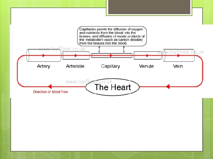
Blood vessels Arteries carry blood Away from the heart Smaller Veins carry blood toward the heart Smaller A diameter arteries are called arterioles diameter veins are called venules network of Capillaries joins the arteries and arterioles with venules and veins
The one-cell-thick capillaries are the sites where gases, nutrients, and other materials are transferred from blood to tissue cells and from tissue cells to blood
Artery An artery has highly elastic walls This elasticity allows the artery to expand as a wave of blood surges through it during the contraction of the ventricles Then to snap back during the relaxation of the ventricles Expansion and contraction of the artery walls keeps the blood flowing in the right direction and provides and additional pumping motion to help propel the blood through the blood vessels
Veins Have thinner walls than arteries Larger inner circumference Not as elastic as arteries They cannot contract to help move the blood back to the heart Also have one-way valves that prevent blood from flowing backward
Capillaries Smallest blood vessels Capillary wall is a single layer of cells
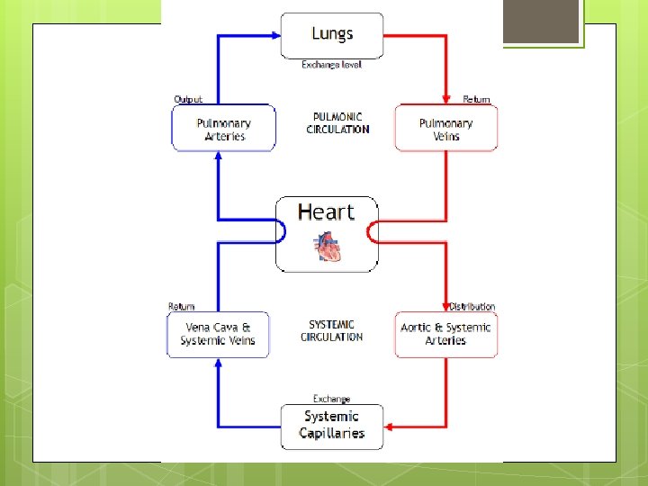
Pulmonary Circulation Movement of blood from the heart to the lungs, and then from the lungs back to the heart again Blood from the heart to the lungs carries waste carbon dioxide gas Gas exchange takes place (carbon dioxide leaves the blood and oxygen moves into the blood) Freshly oxygen-rich blood goes back to the heart
Pulmonary Circulation Flow Chart Right ______ right ___________ _______ (in the lungs) ______ ____________
Systemic circulation Takes oxygenated blood from the heart to other tissues and organs throughout the body
Systemic Circulation Flow Chart Left atrium left ___________ ____________ _________ right _____
MONITORING THE HUMAN CIRCULATORY SYSTEM
Sinoatrial Node bundle of specialized muscle tissue Stimulates the muscle cells to contract and relax rhythmically Pacemaker Wall of right atrium Generates electrical signal that spreads over the two atria and makes them contract simultaneously As atria contract, the signal reaches another node, called the……
Atrioventricular Node Transmits signal through a bundle of specialized fibres called the bundle of His These relay signal to Purkinje fibres Stimulates almost simultaneous contraction of all cells of the right and left ventricles
“Lub-Dub” “Lub” is caused by the atrioventricular valves closing Blood being pumped from the atria to ventricles “Dub” is the closing of the semilunar valves as blood is pumped from the ventricles into the arteries
Blood Pressure As blood passes through the vessels in the body, it exerts pressure against the vessel walls The maximum pressure during the ventricular contraction is called systolic pressure The ventricles contract and force blood into the pulmonary arteries and the aorta, the pressure in creases in these vessels
The lowest pressure before the ventricles contract is called the diastolic pressure The ventricles then relax and the pressure in the pulmonary arteries and the aorta drops
Blood and its components 5 L of blood moving continuously Plasma – 55% White blood cells and platelets 1% Red blood cells 44%
Plasma Clear, yellowish fluid 92% water, 7% dissolved blood proteins Strengthen immunity, helps with blood clotting
Red blood cells Erythrocytes 44% volume of blood Oxygen transport Oxygen-carrying capacity of the blood is dependent on the number of erythrocytes that are present and the amount of hemoglobin that each red blood cell contains
White Blood Cells Leukocytes Body’s response to infection 1% of total blood volume (may increase if fighting infection)
Platelets Thrombocytes Key 1. 2. 3. 4. role in blood clotting When a blood vessel is broken due to injury, it releases a chemical that attract platelets to the site of injury (do not need to copy rest of this slide) Platelets rupture producing thromoplastin If Ca ions are present, thromboplastin reacts with prothrombin to produce another enzyme called thrombin Thrombin reacts with fibrinogen to produce fibrin 1. (Fibrin is an insoluble protein that forms a fibrous mesh over the site of injury. This mesh prevents the loss of blood cells and eventually solidifies to form a clot. )
Questions (pg 481) 1. 2. 3. 4. 5. 6. What are three main functions of the circulatory system? What is the main difference between open and closed circulatory systems? Explain how hemolymph in the open circulatory system of an insect moves through its body If the left ventricle was not able to pump blood properly, what effect would this have on the lungs Describe the oxygen content (O 2 rich or O 2 -poor) of the blood in each of the four chambers of the heart. Describe the destination of the blood leaving each of the four chambers of the heart.
Questions (pg 491) 13. How does exercise help reduce high blood pressure? What is the sinoatrial node and why is it often called the pacemaker of the heart?
Using two different organ systems discussed explain how they work together to achieve a common goal.