CHRONIC PANCREATITIS A 49 yearold man was admitted


 – – – – • • older")


- Slides: 41
CHRONIC PANCREATITIS
• A 49 -year-old man was admitted with a ninemonth history of intermittent attacks of epigastric pain, jaundice and fever. • These attacks usually last up to several days associated with nausea and vomiting. • He was well in between attacks and had no loss of weight
What is your next step?
Lab Results • • AP =1017 GGT= 269 AST =103 ALT =186 • TB =2 (DB= 1. 1) • Alb= 3. 2 • Lipase =33 (up to 244 during attacks) • Amylase= 44 • Hb =12 • WBC= 5. 7 • Plts= 223 Na =141 • K =4. 2 • Ur =15 • Cr =0. 9 • Ca =8. 9 • FBS: 178
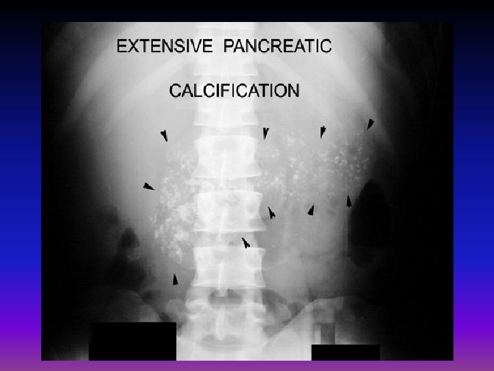
What are arrows?
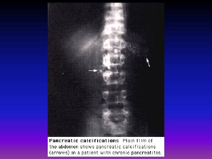
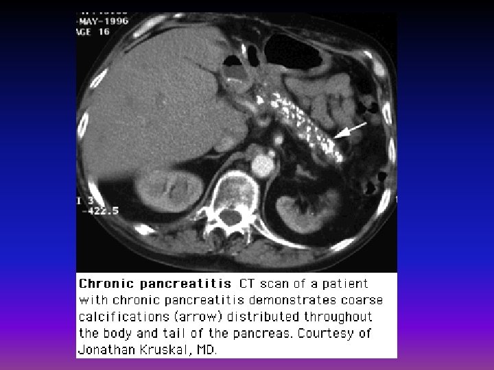
Pancreatic calcification
• Transabdominal US: No gallstones or mass in head of pancreas • CT scan: The extrahepatic bile duct was mildly dilated and "generous pancreas" was noted but there was no mass.
Endoscopic Ultrasound Diffuse hypoechoic enlargement of pancreas. Fine needle aspirate of the pancreas was negative for tumor.
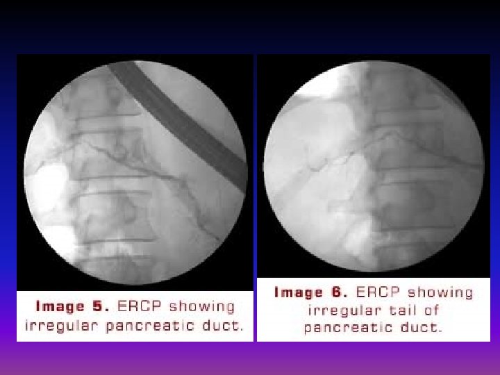
ERCP There was a long segment of extrahepatic biliary stricture. The pancreatic duct was normal in size but irregular. Brushings, biopsies and bile aspirate were negative for tumor
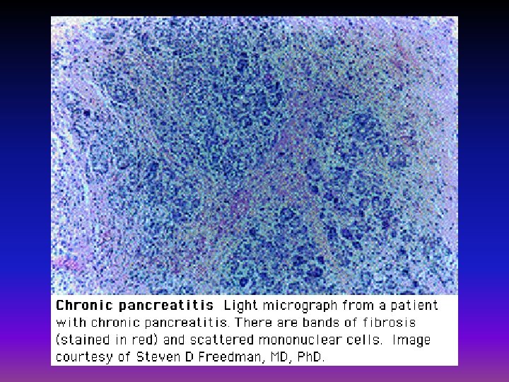
The patient underwent Whipple's operation Histology of the pancreas showed chronic pancreatitis, no malignancy
• Two presentation: – Episodes of acute inflammation in a previously injured pancreas – Chronic damage with persistent pain or malabsorption • Etiology : – same as acute pancreatitis “pancreatitis associated with gallstones predaminantly acute or relapsing-acute” – More idiopathic types
• Most common cause : – In adults: alcohol intake – In children: cystic fibrosis • Idiopathic chronic pancreatitis is the leading cause of nonalcoholic chronic pancreatitis
PATHOPHYSIOLOGY • The events that initiate an inflamatory process are still not well understood • In the alcohol-induced : suggested that the primary defect may be the precipitation of protein(inspissated enzyme ) • In fact , shown that alcohol has direct toxic effect on the pancreas
Clinical features • abdominal pain: – may be continuous, intermittent or absent – Pattern is often atypical • RUQ or LUQ of the back • Diffuse throughout upper abdomen • May be referred to the anterior chest or flank – Typical form: • Persistent , deep-seated, • Unresponsive to antacids • Worsened by alcohol intake or a heavy meal (especially fatty foods) • Often need narcotics
• Pancreatic insufficiency – Weight loss – Fat malabsorption: • Steatorrhea: 15% of patients present with steatorrhea and no pain – Pancreatic diabetes: • Like DM 1 needs insulin , but risk of hypoglycemia is more than it (because alfa cells is also affected – Fat-soluble vitamin deficiency rare
Lab data • Amylase and lipase : usually normal • CBC , electrolytes, and liver function tests are typically normal • Bilrubin and ALP may be increased • Impaired glucose intolerance and elevated fasting blood glucose • Sudan staining of feces or quantitative test for steatorrhea • fecal elastase (Among pancreatic function tests, fecal elastase measurement is the most sensitive and specific, especially in the early phases of pancreatic insufficiency)
Cont, • Classic triad “ pancreatic calcification , steatorrhea , and diabetes mellitus “usually establishes chronic pancreatitis • Classic triad : found in fewer than one-third • It is often necessary to perform secretin stimulation test (abnormal when 60% or more of pancreatic exocrine function has been lost) • A decreased serum trypsinogen (<20 ng/ml) or a fecal elastase level of <100 ug/mg of stool strongly suggests severe pancreatic insufficiency
Imaging studies • Plain films : – Pancreatic calcifications : % 30 – most common with alcoholic pancreatitis, but is also seen in the hereditary and tropical forms of the disorder; it is rare in idiopathic pancreatitis.
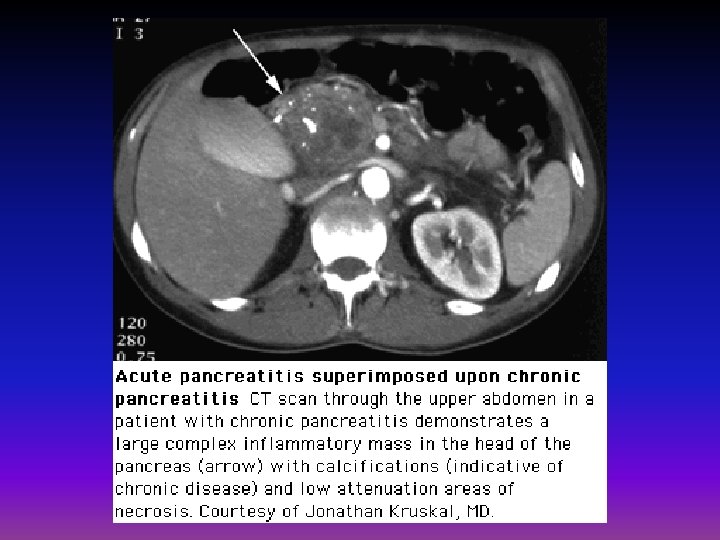
CT, MRI, US • • calcifications ductal dilatation enlargement of the pancreas fluid collections (eg, pseudocysts)
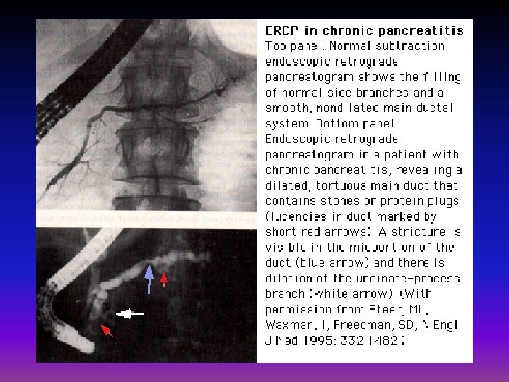
ERCP • Choice when calcifications are not present and there is no evidence of steatorrhea. • a normal study should not rule out the diagnosis of chronic pancreatitis
ERCP May provide useful information on the status of the pancreatic ductal system Abnormalities include : 1)luminal narowing 2)irregularitis in the ductal system with stenosis, dilation, saculation, and ectasia 3)blockage of the duct by calcium deposits
Endoscopic ultrasonography • The most predictive endosonographic feature is the presence of stone • Other suggestive features include: – – – – visible side branches cysts lobularity irregular main pancreatic duct, hyperechoic foci and strands dilation of the main pancreatic duct hyperechoic margins of the main pancreatic duct.
Complications • • pseudocyst formation bile duct or duodenal obstruction pancreatic ascites or pleural effusion splenic vein thrombosis Pseudoaneurysms pancreatic cancer acute attacks of pancreatitis( particularly alcoholics who continue drinking)
DIFFERENTIAL DIAGNOSIS • Pancreatic cancer (most important) – – – – • • older age absence of a history of alcohol use weight loss a protracted flare of symptoms onset of significant constitutional symptoms pancreatic duct stricture greater than 10 mm in length on ERCP Markers such as CA 19 -9 and CEA peptic ulcer disease gallstones irritable bowel syndrome Acute pancreatitis
TREATMENT
PAIN MANAGEMENT • stepwise approach : – general recommendations – pancreatic enzyme supplementation – Analgesics – invasive options
General recommendations • Establish a secure diagnosis • Cessation of alcohol intake • Small meals
Pancreatic enzyme supplements • not very effective • response may be better in young women with small duct disease. • MECHANISM: – suppression of feedback loops in the duodenum that regulate the release of cholecystokinin (CCK), the hormone that stimulates digestive enzyme secretion from the exocrine pancreas • six tablets of Viokase® which contains: – 16, 000 units of lipase – 30, 000 units of protease – 30, 000 units of amylase.
• Patients should also be treated with acid suppression (either with an H 2 receptor blocker or a proton pump inhibitor) to reduce inactivation of the enzymes from gastric acid.
Analgesics • if pancreatic enzyme therapy fails to control pain. • short course of narcotics coupled with low dose amitriptyline and a nonsteroidal antiinflammatory • Simultaneous short-term hospitalization, with the patient kept NPO to minimize pancreatic stimulation, may also be of benefit in breaking the pain cycle. • Chronic narcotic analgesia may be required in patients with persistent significant pain. Long-acting agents such as MS Contin or Fentanyl patches are generally more effective than short acting medications, which last only three or four hours.
Other medical therapies • octreotide : cannot be recommended for general use. • Antioxidant therapy : vitamin C, E, methionine and selenium
Specialized approaches • Celiac nerve blocks • Endoscopic stenting of the pancreatic duct or pancreatic sphincterotomy • Extracorporeal shock wave lithotripsy • Surgery
Maldigestion management • Pancreatic enzymes: – Steatorrhea could be abolished if 10% of the normal amount of lipase could be delivered to the duodenum at the proper time – Poor therapeutic results because of : • Lipase is inactivated by gastric acid • Food empties from the stomach faster than do the pancreatic enzymes • Batches of commercially available pancreatic extracts vary in enzyme activity
• Adjuants: – H 2 blockers – Sodium bicarbonate – PPIs