Chronic Liver Disease Rami Dwairi MD Symptoms of


 �")


- Slides: 21
Chronic Liver Disease Rami Dwairi, MD
Symptoms of chronic liver disease �Patients may be asymptomatic or complain of non- specific symptoms, particularly fatigue �Right hypochondrial pain due to liver distension �Abdominal distension due to ascites �Ankle swelling due to fluid retention �Hematemesis and melena from gastrointestinal hemorrhage �Pruritus due to cholestasis �Gynecomastia , loss of libido and amenorrhea due to endocrine dysfunction �Confusion and drowsiness due to neuropsychiatric complications (portosystemic encephalopathy)
Signs of chronic liver disease
SPIDER NEVI �Telangiectasias that consist of a central arteriole with radiating small vessels. They are found in the distribution of the superior vena cava (above the nipple line)
PALMAR ERYTHEMA �A non-specific change, indicative of a hyperdynamic circulation
DUPUYTREN’S CONTRACTURE
GYNECOMASTIA
CAPUT MEDUSA
Liver Cirrhosis results from the necrosis of liver cells followed by fibrosis and nodule formation The liver architecture is diffusely abnormal and this interferes with liver blood flow and function This derangement produces the clinical features of portal hypertension and impaired liver cell function Alcohol is the most common cause in the West, but viral infection is the most common cause world-wide
Aetiology �Alcohol �Viral hepatitis B and C �Non-alcoholic fatty liver �Drugs �Cystic fibrosis �Galactosemia disease �Glycogen storage disease �Autoimmune hepatitis �Primary Biliary Cirrhosis �Veno-occlusive disease �Hemochromatosis �Cryptogenic �Sclerosing Cholangitis �Alpha 1 antitrypsin deficiency �Hepatic venous congestion �Budd–Chiari syndrome �Wilson’s disease
Pathology �The characteristic features of cirrhosis are regenerating nodules separated by fibrous septa and loss of the normal lobular architecture within the nodules �Two types of cirrhosis have been described: �Micronodular cirrhosis. Regenerating nodules are usually less than 3 mm in size and the liver is involved uniformly. �This type is often caused by ongoing alcohol damage or biliary tract disease �Macronodular cirrhosis. The nodules are of variable size and normal acini may be seen within the larger nodules �This type is often seen following chronic viral hepatitis �A mixed picture with small and large nodules is sometimes seen
Investigations Severity assessment � Liver function: Serum albumin and prothrombin time are the best indicators of liver function � Liver biochemistry: This can be normal, depending on the severity of cirrhosis. In most cases there is at least a slight elevation in the serum ALP and serum aminotransferases. In decompensated cirrhosis all biochemistry is deranged � Serum electrolytes: A low sodium indicates severe liver disease due to a defect in free water clearance or to excess diuretic therapy � Serum creatinine: An elevated concentration > 130 μmol/ L is a marker of worse prognosis � Serum α-fetoprotein: If > 200 ng/m. L is strongly suggestive of the presence of a hepatocellular carcinoma
Investigation Cause assessment This can be determined by: �viral markers �serum autoantibodies �serum immunoglobulins �iron indices and ferritin �copper, ceruloplasmin �α 1 -antitrypsin �Serum copper and serum α 1 -antitrypsin should always be measured in young cirrhotics. �Total iron-binding capacity (TIBC) and ferritin should be measured to exclude hereditary hemochromatosis
Imaging Ultrasound examination. � This can demonstrate changes in size and shape of the liver. Fatty change and fibrosis produce a diffuse increased echogenicity � In established cirrhosis there may be marginal nodularity of the liver surface and distortion of the arterial vascular architecture. The patency of the portal and hepatic veins can be evaluated � It is useful in detecting hepatocellular carcinoma Fibroelastography CT scan � Shows hepatosplenomegaly, and dilated collaterals. Contrastenhanced scans are useful in the detection of hepatocellular carcinoma. Endoscopy MRI � Useful in the diagnosis of benign tumors
Liver Biopsy Widely replaced by the use of ultrasound and fibroelastography along with serologic testing Performed to confirm the severity and type of liver disease Special stains are required for iron and copper, and various immunocytochemical stains can identify viruses, bile ducts and angiogenic structures Chemical measurement of iron and copper is necessary to confirm diagnosis of iron overload or Wilson’s disease
Management is that of the complications seen in decompensated cirrhosis Patients should have 6 -monthly ultrasound to detect the early development of a hepatocellular carcinoma as all therapeutic strategies work best with small single tumors Treatment of the underlying cause may arrest or occasionally reverse the cirrhotic changes The only dietary restriction is to reduce salt intake Alcohol, aspirin and NSAIDs should be avoided
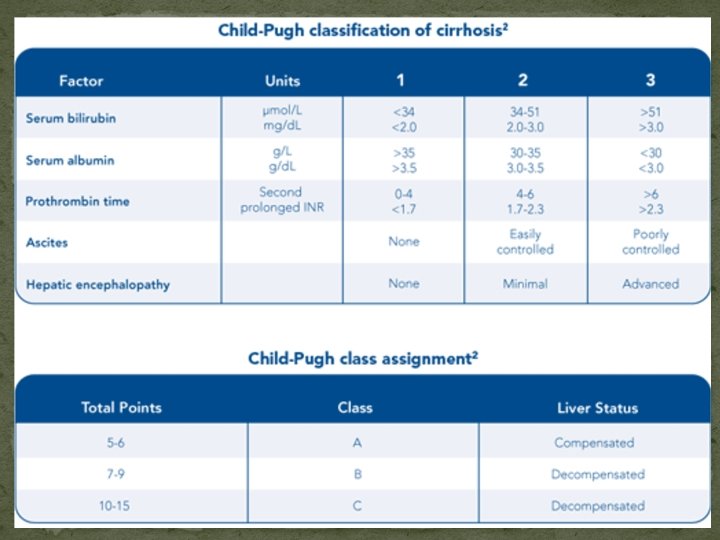
Course and Prognosis � This is extremely variable, depending on many factors, including the aetiology and the presence of complications � Development of any complication usually worsens the prognosis � In general, the 5 -year survival rate is approximately 50%, but this also varies depending on the aetiology and the stage at which the diagnosis is made � There a number of prognostic classifications based on modifications of Child’s grading (A, B and C) and the model for end-stage disease (MELD), based on serum bilirubin, creatinine and INR, which is widely used as a predictor of mortality in patients awaiting liver transplantation.
Poor Prognostic Indicators in Cirrhosis Blood tests � Low albumin (< 28 g/L) � Low serum sodium (< 125 mmol/L) � Prolonged prothrombin time > 6 seconds above normal value � Raised creatinine > 130 μmol/L Clinical � Persistent jaundice � Poor response to therapy � Ascites � Variceal hemorrhage � Neuropsychiatric complications developing with progressive liver failure � Small liver � Persistent hypotension � Aetiology (e. g. alcoholic cirrhosis, if the patient continues drinking)
Complications Portal hypertension Variceal bleeding Ascites Portosystemic encephalopathy Spontaneous bacterial peritonitis Renal failure (hepatorenal syndrome) Hepatopulmonary syndrome Primary hepatocellular carcinoma
Thank You