
Chronic inflammation 3 rd stage MSc Etab A
Chronic inflammation 3 rd stage MSc Etab A. AL-Mosawe
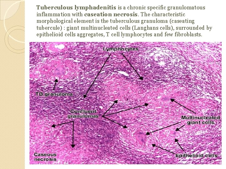
Tuberculous lymphadenitis is a chronic specific granulomatous inflammation with caseation necrosis. The characteristic morphological element is the tuberculous granuloma (caseating tubercule) : giant multinucleated cells (Langhans cells), surrounded by epithelioid cells aggregates, T cell lymphocytes and few fibroblasts.
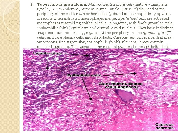
1. Tuberculous granuloma. Multinucleated giant cell (mature - Langhans type) : 50 - 100 microns, numerous small nuclei (over 20) disposed at the periphery of the cell (crown or horseshoe), abundant eosinophilic cytoplasm. It results when activated macrophages merge. Epithelioid cells are activated macrophages resembling epithelial cells : elongated, with finely granular, pale eosinophilic (pink) cytoplasm and central, ovoid nucleus. They have indistinct shape contour and form aggregates. At the periphery are the lymphocytes (T cells) and rare plasma cells and fibroblasts. Caseous necrosis is a central area, amorphous, finely granular, eosinophilic (pink). If recent, it may contain nuclear fragments. The caseum is the result of giant cells and epithelioid cells destruction.
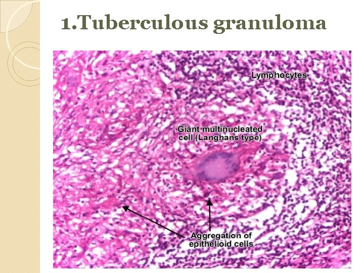
1. Tuberculous granuloma
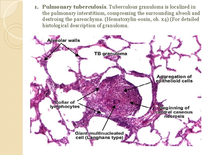
1. Pulmonary tuberculosis. Tuberculous granuloma is localized in the pulmonary interstitium, compressing the surrounding alveoli and destroing the parenchyma. (Hematoxylin-eosin, ob. x 4) (For detailed histological description of granuloma.
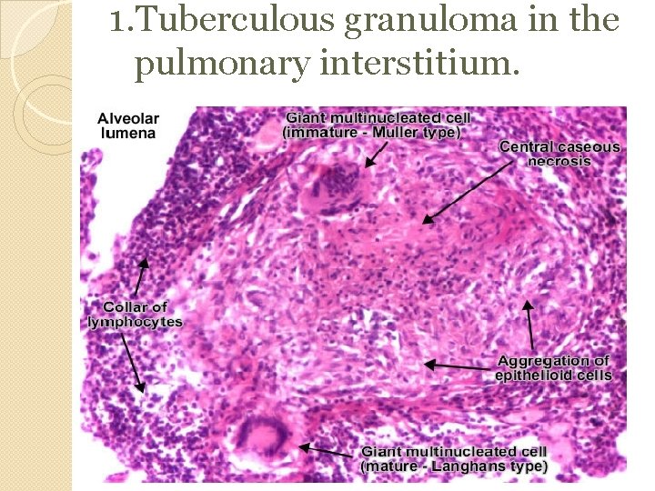
1. Tuberculous granuloma in the pulmonary interstitium.
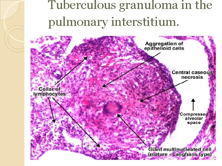
Tuberculous granuloma in the pulmonary interstitium.
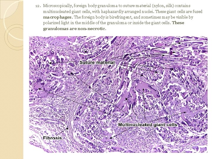
12. Microscopically, foreign body granuloma to suture material (nylon, silk) contains multinucleated giant cells, with haphazardly arranged nuclei. These giant cells are fused macrophages. The foreign body is birefringent, and sometimes may be visible by polarized light in the middle of the granuloma or inside the giant cells. These granulomas are non-necrotic.
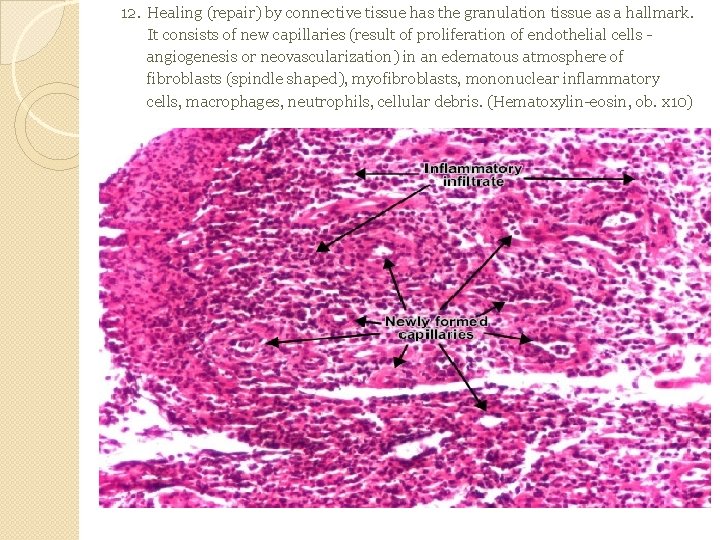
12. Healing (repair) by connective tissue has the granulation tissue as a hallmark. It consists of new capillaries (result of proliferation of endothelial cells angiogenesis or neovascularization) in an edematous atmosphere of fibroblasts (spindle shaped), myofibroblasts, mononuclear inflammatory cells, macrophages, neutrophils, cellular debris. (Hematoxylin-eosin, ob. x 10)
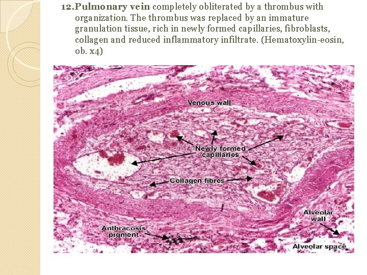
12. Pulmonary vein completely obliterated by a thrombus with organization. The thrombus was replaced by an immature granulation tissue, rich in newly formed capillaries, fibroblasts, collagen and reduced inflammatory infiltrate. (Hematoxylin-eosin, ob. x 4)
- Slides: 10