
Chromosomes Cytogenetics A subdiscipline within genetics Focuses on
Chromosomes
Cytogenetics • A subdiscipline within genetics • Focuses on chromosome variations • Abnormal number of copies of genes or chromosomes can lead to genetic abnormalities • Human genome sequence information is used to identify genes that contribute to the chromosome-related syndromes
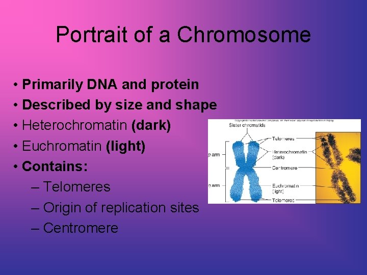
Portrait of a Chromosome • Primarily DNA and protein • Described by size and shape • Heterochromatin (dark) • Euchromatin (light) • Contains: – Telomeres – Origin of replication sites – Centromere
Chromosomes • Heterochromatin is darkly staining, contains mostly repetitive DNA • Euchromatin contains more protein encoding genes • Telomeres are chromosome tips composed of many repeats of TTAGGG and shorten with each cell division • Centromere is the largest constriction of the chromosome and where spindle fibers attach
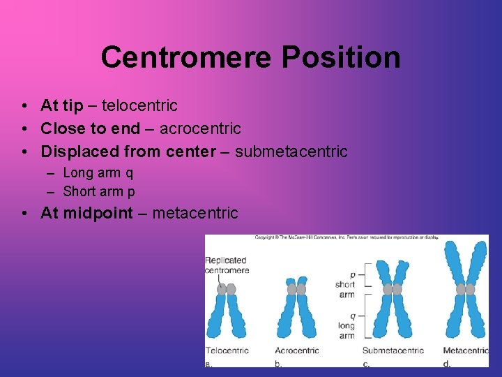
Centromere Position • At tip telocentric • Close to end acrocentric • Displaced from center submetacentric – Long arm q – Short arm p • At midpoint metacentric
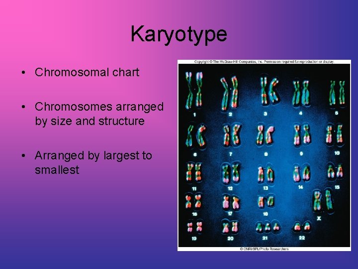
Karyotype • Chromosomal chart • Chromosomes arranged by size and structure • Arranged by largest to smallest
Visualizing Chromosomes Fetal tissue: amniocentesis chorionic villi sampling fetal cell sorting Adult tissue: blood (white blood cells) cheek swab (buccal cells) skin cells tissue biopsy
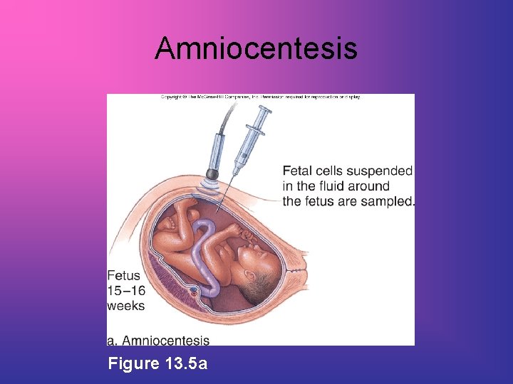
Amniocentesis Figure 13. 5 a
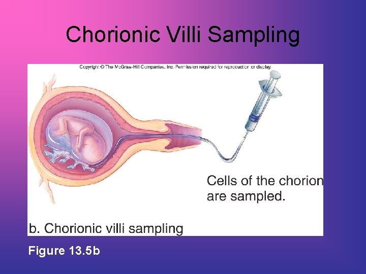
Chorionic Villi Sampling Figure 13. 5 b
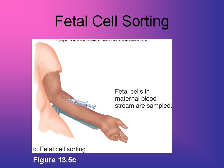
Fetal Cell Sorting Figure 13. 5 c
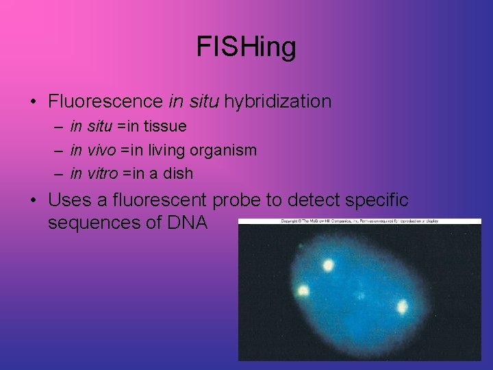
FISHing • Fluorescence in situ hybridization – in situ =in tissue – in vivo =in living organism – in vitro =in a dish • Uses a fluorescent probe to detect specific sequences of DNA
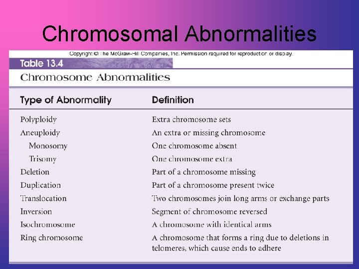
Chromosomal Abnormalities
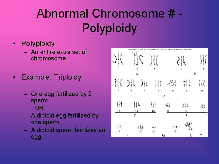
Abnormal Chromosome # Polyploidy • Polyploidy – An entire extra set of chromosome • Example: Triploidy – One egg fertilized by 2 sperm OR – A diploid egg fertilized by one sperm – A diploid sperm fertilizes an egg
Abnormal Chromosome # Aneuploidy • Missing a single chromosome (monosomy) – OR - • Having one extra chromosome (trisomy) • Euploid =“good set” • Aneuploid =“not good set” • Caused by nondisjunction during meiosis
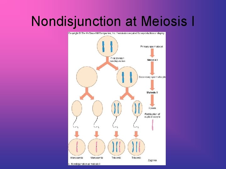
Nondisjunction at Meiosis I
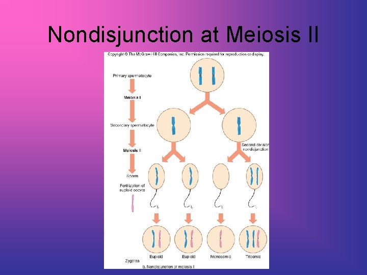
Nondisjunction at Meiosis II
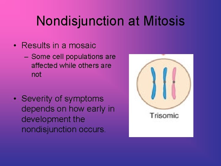
Nondisjunction at Mitosis • Results in a mosaic – Some cell populations are affected while others are not • Severity of symptoms depends on how early in development the nondisjunction occurs.
Autosomal Aneuploids • Usually lethal • Those that survive often have mental retardation • Most common for chromosomes 13, 18, and 21. Why?
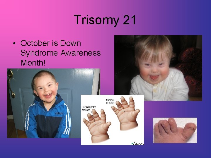
Trisomy 21 • October is Down Syndrome Awareness Month!
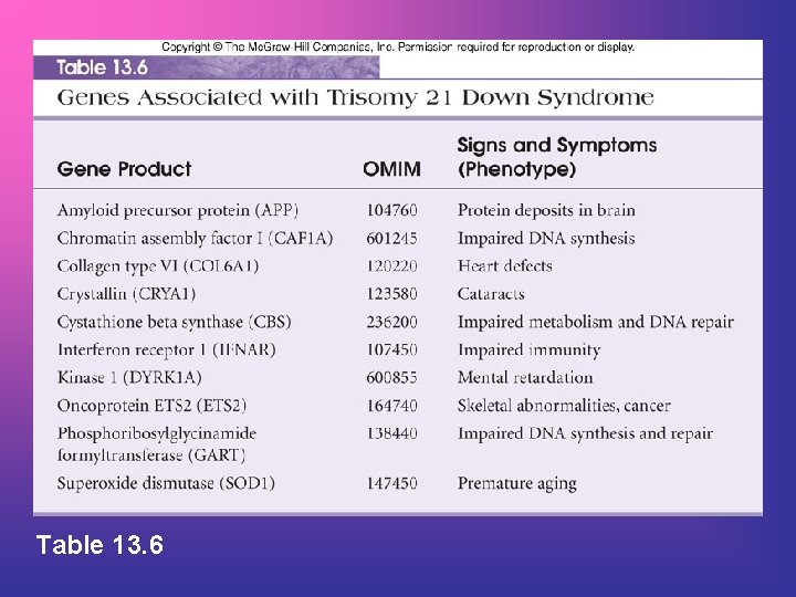
Table 13. 6
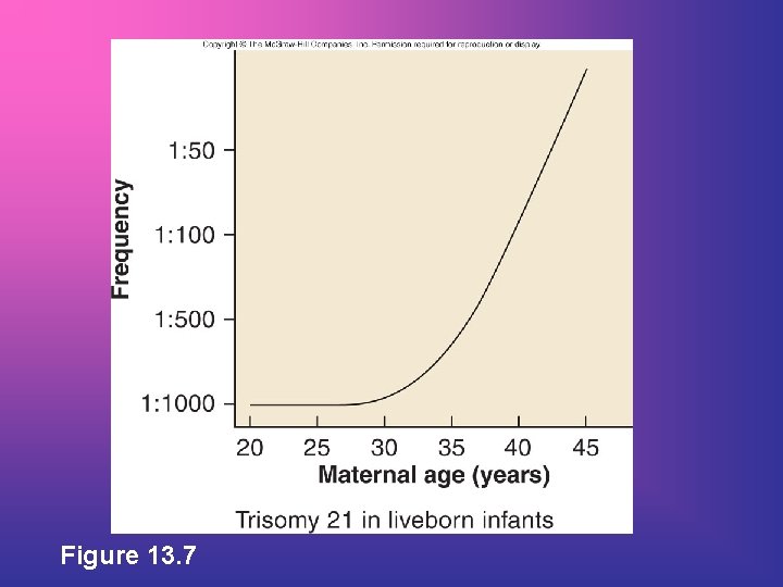
Figure 13. 7
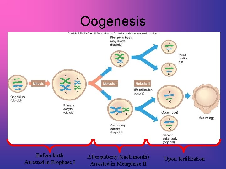
Oogenesis Before birth Arrested in Prophase I After puberty (each month) Arrested in Metaphase II Upon fertilization
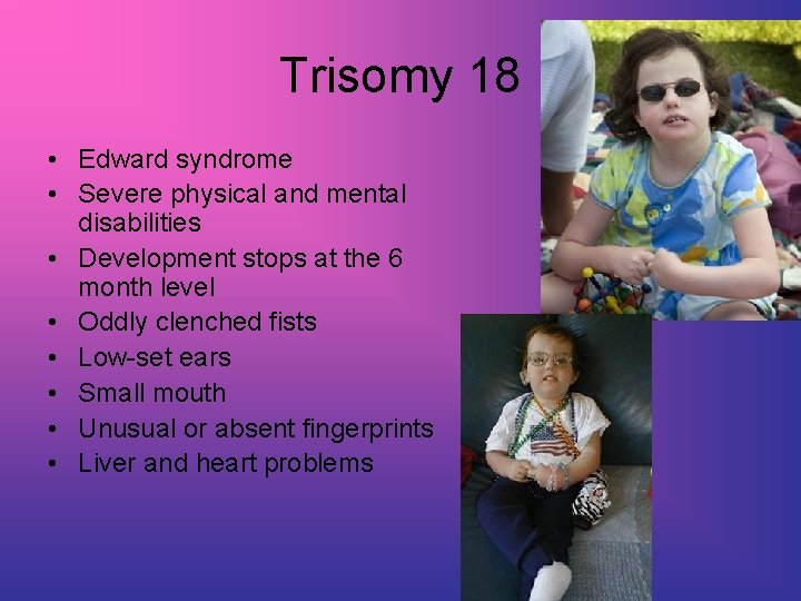
Trisomy 18 • Edward syndrome • Severe physical and mental disabilities • Development stops at the 6 month level • Oddly clenched fists • Low-set ears • Small mouth • Unusual or absent fingerprints • Liver and heart problems
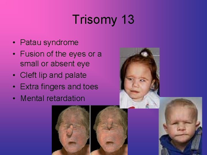
Trisomy 13 • Patau syndrome • Fusion of the eyes or a small or absent eye • Cleft lip and palate • Extra fingers and toes • Mental retardation
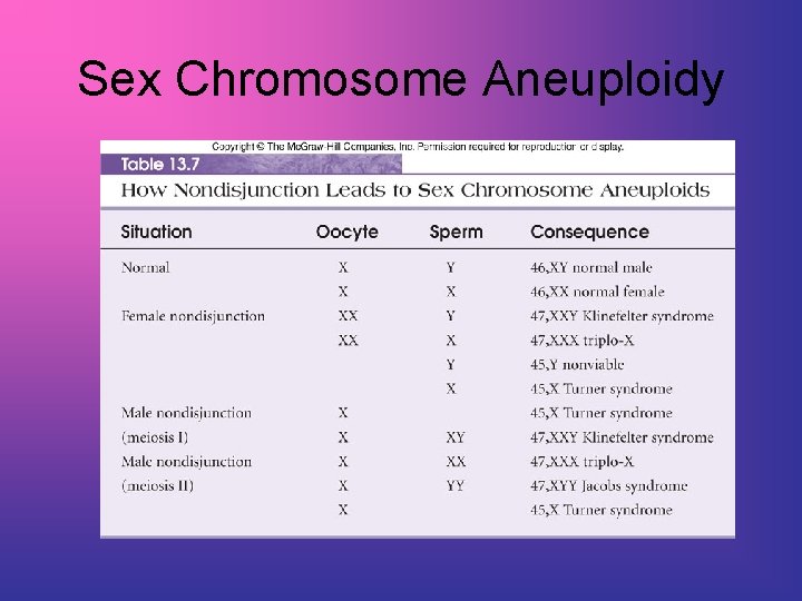
Sex Chromosome Aneuploidy
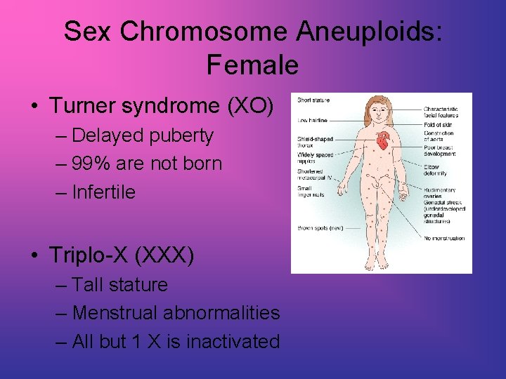
Sex Chromosome Aneuploids: Female • Turner syndrome (XO) – Delayed puberty – 99% are not born – Infertile • Triplo-X (XXX) – Tall stature – Menstrual abnormalities – All but 1 X is inactivated
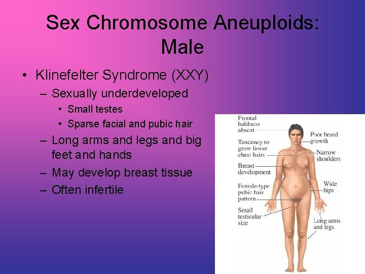
Sex Chromosome Aneuploids: Male • Klinefelter Syndrome (XXY) – Sexually underdeveloped • Small testes • Sparse facial and pubic hair – Long arms and legs and big feet and hands – May develop breast tissue – Often infertile

Sex Chromosome Aneuploids: Male • XXYY Syndrome – Slightly delayed childhood development – Behavioral problems • ADD, OCD, and learning disabilities – – – Leg ulcers due to poor circulation Sexual development is delayed Testes do not descend Infertile Abnormal (YY) sperm AND abnormal (XX) egg
Sex Chromosome Aneuploids: Male • • • Jacobs Syndrome (XYY) 1/1000 males has an extra Y 96% of XYY males are normal Tall height and acne Criminals with chromosomal abnormalities tend to have XYY
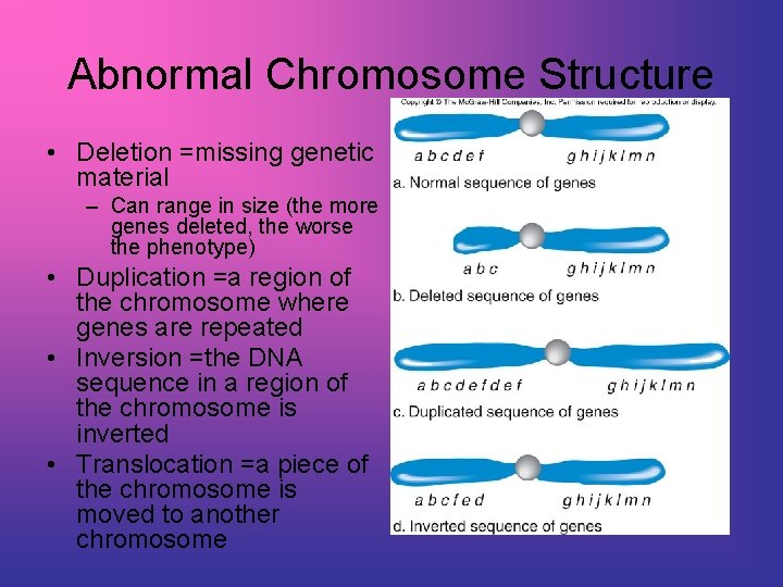
Abnormal Chromosome Structure • Deletion =missing genetic material – Can range in size (the more genes deleted, the worse the phenotype) • Duplication =a region of the chromosome where genes are repeated • Inversion =the DNA sequence in a region of the chromosome is inverted • Translocation =a piece of the chromosome is moved to another chromosome
Translocation • Nonhomologous chromosome exchange segments Two major types: • Robertsonian translocation • Two nonhomologous acrocentric chromosomes break at the centromere and long arms fuse. The short arms are often lost. • 5% of Down syndrome results from a Robertsonian translocation between chr 21 and chr 14. • Reciprocal translocation • Two nonhomologous chromosomes exchange a portion of their chromosome arms.
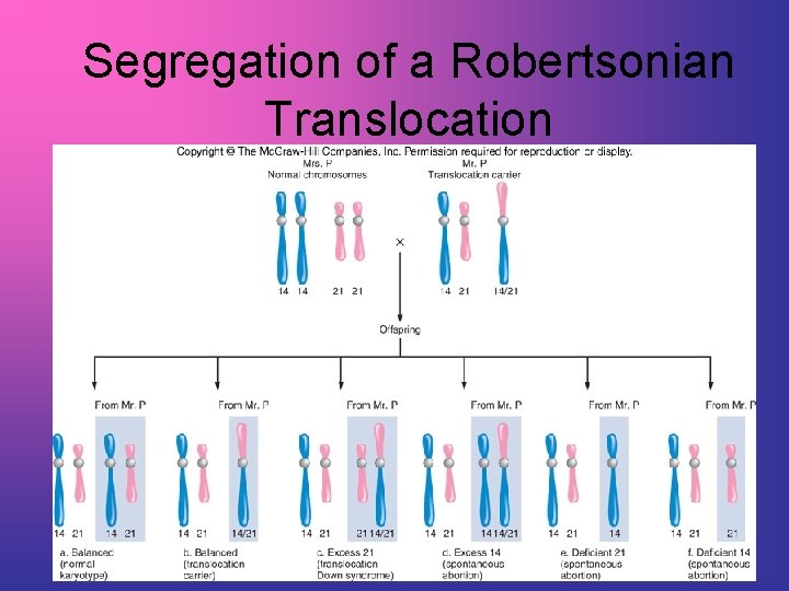
Segregation of a Robertsonian Translocation
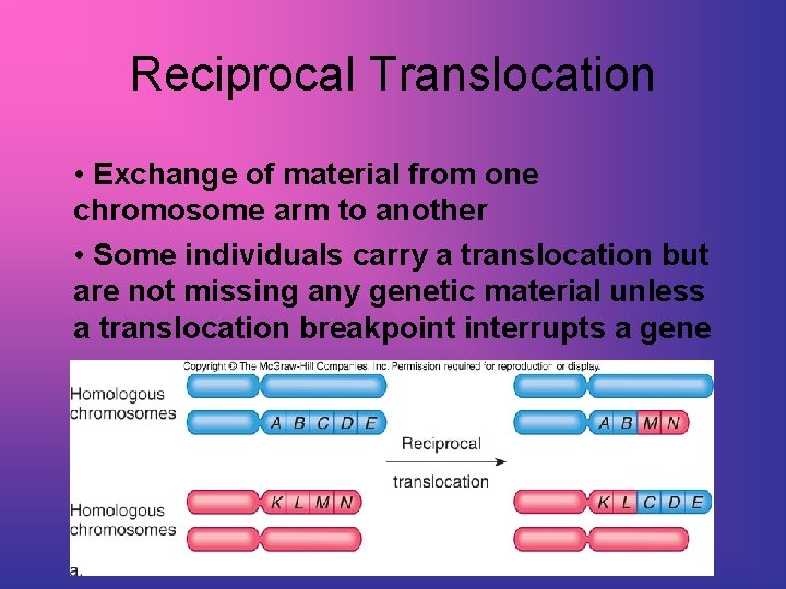
Reciprocal Translocation • Exchange of material from one chromosome arm to another • Some individuals carry a translocation but are not missing any genetic material unless a translocation breakpoint interrupts a gene
Inversions • Inverted chromosomes have a region flipped in orientation • 5 -10% cause health problems probably due to disruption of genes at the breakpoints • Inversions may impact meiotic segregation • Two types of inversions occur: • Paracentric – inverted region does NOT include centromere • Pericentric – inverted region includes centromere
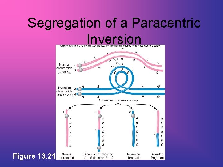
Segregation of a Paracentric Inversion Figure 13. 21
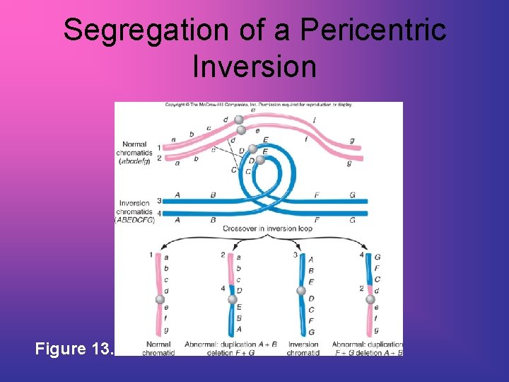
Segregation of a Pericentric Inversion Figure 13. 22
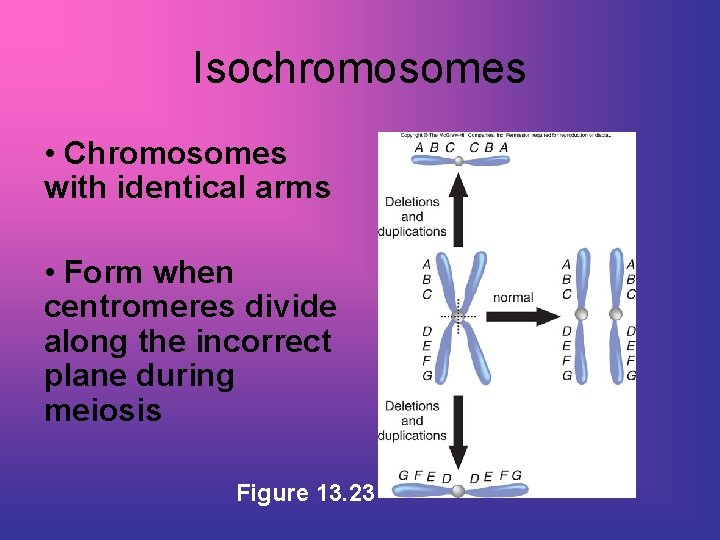
Isochromosomes • Chromosomes with identical arms • Form when centromeres divide along the incorrect plane during meiosis Figure 13. 23
Ring Chromosomes • Chromosomes shaped like a ring • Occur in 1 in 25, 000 conceptions • May arise when telomeres are lost and sticky chromosome end fuse – Radiation exposure

- Slides: 39