Chromosome 10 Contains over 1400 genes Contains over


 results from structural abnormalities in the potassium channels of the")
,")
 is a group of rare diseases caused by genetic defects")
 usually appear in the second")


 is an inherited error of metabolism caused by a deficiency in the")


 is a protein that protects the body from")

 is an uncommon inherited disorder characterized by mental retardation, decreased muscle")
 is an uncommon neurogenetic disorder characterized by mental retardation, abnormal gait,")


 occurs most commonly in people of non-Ashkenazi Jewish, Armenian, Arab,")
 is characterized by large cysts in")
 is a group of chronic disorders that causes")


 is named after its three")


 represents a group of rare, sometimes fatal, congenital disorders characterized")
 is an inherited disorder so")


 is a neurological disorder characterized by progressive degeneration of motor")


 is a rare metabolic disorder caused by")
 disease whose")
 is a rare inherited disorder characterized by the development")

 is a cancer of blood cells, characterized")


")
 is one of a group of muscular dystrophies characterized by")

 is a genetic disease in which a collagen mutation affects the")
 is a rare inherited disease that disrupts the metabolism of the")
 is a rare primary immunodeficiency characterized by the production")

 is a progressive neurodevelopmental disorder almost exclusively affecting females. With an")

 is a rare, inherited metabolic disorder that afflicts the young boy Lorenzo")

- Slides: 75
Chromosome 10 Contains over 1400 genes Contains over 130 million base pairs, of which over 95% have been determined See the diseases associated with chromosome 10 in the Map. Viewer.
Refsum disease is a rare disorder of lipid metabolism that is inherited as a recessive trait. Symptoms may include a degenerative nerve disease (peripheral neuropathy), failure of muscle coordination (ataxia), retinitis pigmentosa (a progressive vision disorder), and bone and skin changes. Refsum disease is characterized by an accumulation of phytanic acid in the plasma and tissues. is a derivative of phytol, a component of chlorophyll. In 1997 the gene for Refsum disease was identified and mapped to chromosome 10. The protein product of the gene, PAHX, is an enzyme that is required for the metabolism of phytanic acid. Refsum disease patients have impaired PAHX - phytanic acid hydrolase. It is thought that Refsum disease is a peroxisomal disorder, since human PAHX contains PTS 2 localization sequences, which target it to the peroxisome. Our bodies can not synthesize phytanic acid: we have to obtain all of it from our food. Therefore, prolonged treatment with a diet deficient in phytanic acid can be beneficial.
Gyrate atrophy of the choroid and retina People suffering from gyrate atrophy of the choroid (the thin coating of the eye) and retina face a progressive loss of vision, with total blindness usually occurring between the ages of 40 and 60. The disease is an inborn error of metabolism. The gene whose mutation causes gyrate atrophy is found on chromosome 10, and encodes an enzyme called ornithine ketoacid aminotransferase (OAT). Different inherited mutations in OAT cause differences in the severity of symptoms of the disease. OAT converts the amino acid ornithine from the urea cycle ultimately into glutamate. In gyrate atrophy, where OAT function is affected, there is an increase in plasma levels of ornithine. It is already known that reduction of the amino acid arginine in the diet has a salutary effect on most patients. Current lines of research into the disease include: (1) investigating how variant mutations of the alleles (versions of the gene inherited) interact in order to cause the differing symptoms of the disease and (2) work on mouse models of the disease is furthering our understanding, which is hoped will lead to a true cure.
Chromosome 11 Contains approximately 2000 genes Contains over 130 million base pairs, of which over 95% have been determined See the diseases associated with chromosome 11 in the Map. Viewer.
Harvey Ras oncogene Cancer occurs when the growth and differentiation of cells in a body tissue become uncontrolled and deranged. While no two cancers are genetically identical (even in the same tissue type), there are relatively few ways in which normal cell growth can go wrong. One of these is to make a gene that stimulates cell growth hyperactive; this altered gene is known as an 'oncogene'. Ras is one such oncogene product that is found on chromosome 11. It is found in normal cells, where it helps to relay signals by acting as a switch. When receptors on the cell surface are stimulated (by a hormone, for example), Ras is switched on and transduces signals that tell the cell to grow. If the cell-surface receptor is not stimulated, Ras is not activated and so the pathway that results in cell growth is not initiated. In about 30% of human cancers, Ras is mutated so that it is permanently switched on, telling the cell to grow regardless of whether receptors on the cell surface are activated or not. Usually, a single oncogene is not enough to turn a normal cell into a cancer cell, and many mutations in a number of different genes may be required to make a cell cancerous. To help unravel the intricate network of events that lead to cancer, mice are being used to model the human disease, which will further our understanding and help to identify possible targets for new drugs and therapies.
Long QT syndrome (LQTS) results from structural abnormalities in the potassium channels of the heart, which predispose affected persons to an accelerated heart rhythm (arrhythmia). This can lead to sudden loss of consciousness and may cause sudden cardiac death in teenagers and young adults who are faced with stressors ranging from exercise to loud sounds. LQTS is usually inherited as an autosomal dominant trait. In the case of LQT 1, which has been mapped to chromosome 11, mutations lead to serious structural defects in the person's cardiac potassium channels that do not allow proper transmission of the electrical impulses throughout the heart. There also appear to be other genes, tentatively located on chromosomes 3, 6 and 11 whose mutated products may contribute to, or cause, LQT syndrome. Beta blockers are used to treat the symptoms of the disease, and appear to be effective in some symptomatic patients. However, common sense therapies such as avoiding strenuous physical exercise and other stressors are also effective. Research on how the genes discussed above interact with each other should encourage the development of new treatments for long. QT syndrome
Best disease Best disease, also known as Vitelliform Macular Dystrophy type 2 (VMD 2), is a heritable disorder occurring primarily in European Caucasians. Individuals with Best disease generally show a gradual loss of visual acuity starting in their teenage years, although the frequency with which an affected individual may show symptoms and the severity of those symptoms are highly variable. Best disease is autosomal dominant; in other words, a mutation in only one copy of the VMD 2 gene located on chromosome 11 may result in development of the disease. Prior to their vision loss, individuals with Best disease accumulate a mass of fat-like material that resembles an egg yolk (vitelline is a word that means yolk-like) in the area of the retina responsible for central vision. Surprisingly, it is the breakup of this mass rather than its formation that is associated with the gradual vision loss characteristic of Best disease. Little is known about the protein product of the VMD 2 gene, although its function seems to be restricted to an area of the eye known as the retinal pigment epithelium. There is speculation that the protein encoded by VMD 2 may be involved in the removal and/or processing of photoreceptor components. Determination of the VMD 2 protein function and development of an animal model will be the next crucial steps toward a better understanding of Best disease
Multiple endocrine neoplasia (MEN) is a group of rare diseases caused by genetic defects that lead to hyperplasia (abnormal multiplication or increase in the number of normal cells in normal arrangement in a tissue) and hyperfunction (excessive functioning) of two or more components of the endocrine system. Endocrine glands are different from other organs in the body because they release hormones into the bloodstream. Hormones are powerful chemicals that travel through the blood, controlling and instructing the functions of various organs. Normally, the hormones released by endocrine glands are carefully balanced to met the body's needs. When a person has MEN, specific endocrine glands, such as the parathyroid glands, the pancreas gland, and the pituitary gland, tend to become overactive. When these glands go into overdrive, the result can be: excessive calcium in the bloodstream (resulting in kidney stones or kidney damage); fatigue; weakness; muscle or bone pain; constipation; indigestion; and thinning of bones.
Ataxia telangiectasia The first signs of ataxia telangiectasia (A-T) usually appear in the second year of life as a lack of balance and slurred speech. It is a progressive, degenerative disease characterized by cerebellar degeneration, immunodeficiency, radiosensitivity (sensitivity to radiant energy, such as x-ray), and a predisposition to cancer. Back in 1988 the gene responsible for A-T was mapped to chromosome 11. The subsequent identification of the gene proved difficult; it was 7 more years until the human ATM gene was cloned. The diverse symptoms seen in A-T reflect the main role of ATM, which is to induce several cellular responses to DNA damage. When the ATM gene is mutated, these signaling networks are impaired, and so the cell does not respond correctly to minimize the damage. Some of the ATM-dependent signaling pathways are found in yeast. Because these pathways appear to be conserved throughout evolution, they are likely to be central to the DNA damage response. Research into finding an effective therapy for A-T sufferers is likely to be helped by harnessing the power of yeast genetics, which allows more rapid and systematic study of the pathways affected by an ATM mutation.
Chromosome 12 Contains over 1600 genes Contains over 130 million base pairs, of which over 95% have been determined See the diseases associated with chromosome 12 in the Map. Viewer.
Zellweger syndrome is a rare hereditary disorder affecting infants, and usually results in death. Unusual problems in prenatal development, an enlarged liver, high levels of iron and copper in the blood, and vision disturbances are among the major manifestations of Zellweger syndrome. The PXR 1 gene has been mapped to chromosome 12; mutations in this gene cause Zellweger syndrome. The PXR 1 gene product is a receptor found on the surface of peroxisomes - microbodies found in animal cells, especially liver, kidney and brain cells. The function of peroxisomes is not fully understood, although the enzymes they contain carry out a number of metabolically important reactions. The PXR 1 receptor is vital for the import of these enzymes into the peroxisomes: without it functioning properly, the peroxisomes can not use the enzymes to carry out their important functions, such as cellular lipid metabolism and metabolic oxidations. There is a yeast homolog to human PXR 1, which should allow powerful molecular genetic techniques to be used in the investigation of the normal role of peroxisomes in cells, as well as the molecular events that occur in disease states
Phenylketonuria (PKU) is an inherited error of metabolism caused by a deficiency in the enzyme phenylalanine hydroxylase. Loss of this enzyme results in mental retardation, organ damage, unusual posture and can, in cases of maternal PKU, severely compromise pregnancy. Classical PKU is an autosomal recessive disorder, caused by mutations in both alleles of the gene for phenylalanine hydroxylase (PAH), found on chromosome 12. In the body, phenylalanine hydroxylase converts the amino acid phenylalanine to tyrosine, another amino acid. Mutations in both copies of the gene for PAH means that the enzyme is inactive or is less efficient, and the concentration of phenylalanine in the body can build up to toxic levels. In some cases, mutations in PAH will result in a phenotypically mild form of PKU called hyperphenylalanemia. Both diseases are the result of a variety of mutations in the PAH locus; in those cases where a patient is heterozygous for two mutations of PAH (ie each copy of the gene has a different mutation), the milder mutation will predominate. A form of PKU has been discovered in mice, and these model organisms are helping us to better understand the disease, and find treatments against it. With careful dietary supervision, children born with PKU can lead normal lives, and mothers who have the disease can produce healthy children
Chromosome 13 Contains approximately 800 genes Contains over 110 million base pairs, of which over 80% have been determined See the diseases associated with chromosome 13 in the Map. Viewer.
Deafness Hearing loss is extremely common and can present at any time from infancy to old age. About 1 in 1000 infants has profound hearing impairment, with half thought to be of genetic origin. Many deafness genes exist, but the most common cause of hearing loss in American and European populations is a mutation in the connexin 26 (Cx 26) gene. Cx 26 has a carrier rate of 3%, similar to that for cystic fibrosis, and it causes about 20% of childhood deafness. Mutations in Cx 26 cause congenital syndromic and nonsyndromic deafness that is, the deafness is not accompanied by other symptoms, such as blindness. Cx 26 is located on chromosome 13 q 11 -12 and codes for a gap junction protein called connexin 26. Gap junctions are plasma membrane channels that allow the movement of small molecules and ions between adjacent cells. Gap junctions of the inner ear may play a role in maintaining potassium homeostasis, which is important for inner-ear function and, thus, hearing. It has been proposed that mutations in Cx 26 may disrupt potassium circulation and result in deafness. The discovery that Cx 26 mutations are a cause of congenital hearing loss can help in
Retinoblastoma occurs in early childhood and affects about 1 child in 20, 000. The tumor develops from the immature retina - the part of the eye responsible for detecting light and color. There are both hereditary and non-hereditary forms of retinoblastoma. IN the hereditary form, multiple tumors are found in both eyes, while in the non-hereditary form only one eye is effected and by only one tumor. In the hereditary form, a gene called Rb is lost from chromosome 13. Since the absence of Rb seemed to be linked to retinoblastoma, it has been suggested that the role of Rb in normal cells is to suppress tumor formation. Rb is found in all cells of the body, where under normal conditions it acts as a brake on the cell division cycle by preventing certain regulatory proteins from triggering DNA replication. If Rb is missing, a cell can replicate itself over and over in an uncontrolled manner, resulting in tumor formation. Untreated, retinoblastoma is almost uniformly fatal, but with early diagnosis and modern methods of treatment the survival rate is over 90%. Since the Rb gene is found Retinoblastoma in all cell types, studying the molecular mechanism of tumor suppression by Rb will give insight into the progression of many types of cancer, not just retinoblastoma
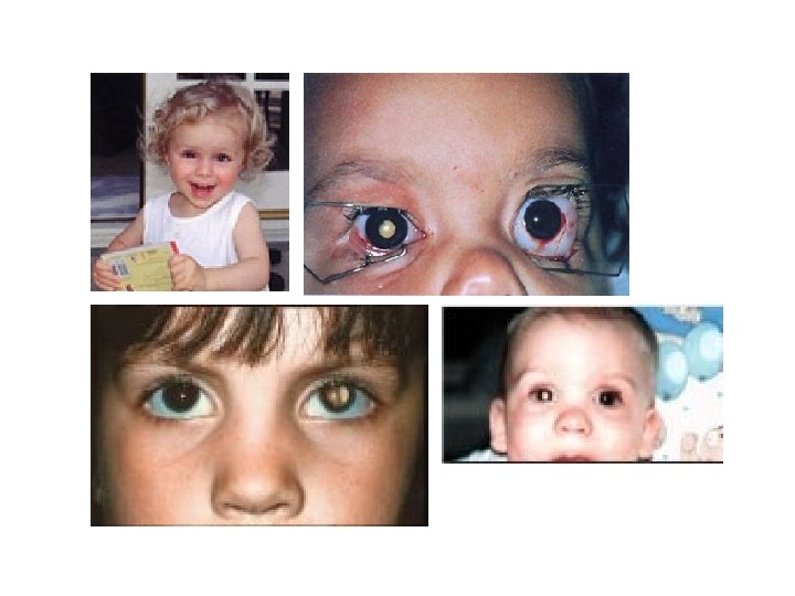
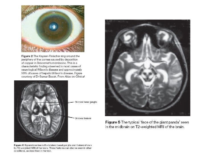
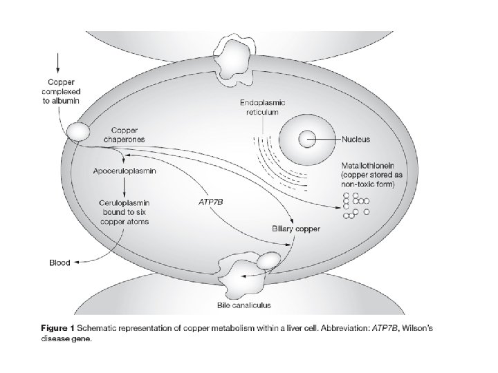
Wilson's disease Wilson's Disease is a rare autosomal recessive disorder of copper transport, resulting in copper accumulation and toxicity to the liver and brain. Liver disease is the most common symptom in children; neurological disease is most common in young adults. The cornea of the eye can also be affected: the 'Kayser-Fleischer ring' is a deep copper-colored ring at the periphery of the cornea, and is thought to represent copper deposits. The gene for Wilson's disease (ATP 7 B) was mapped to chromosome 13. The sequence of the gene was found to be similar to sections of the gene defective in Menkes disease, another disease caused by defects in copper transport. The similar sequences code for copperbinding regions, which are part of a transmembrane pump called a P-type ATPase that is very similar to the Menkes disease protein. A homolog to the human ATP 7 B gene has been mapped to mouse chromosome 8, and an authentic model of the human disease in rat is also available (called the Long. Evans Cinnamon [LEC][ rat). These systems will be useful for studying copper transport and liver pathophysiology, and should help in the development of a therapy for Wilson disease
Chromosome 14 Contains approximately 1200 genes Contains over 100 million base pairs, of which over 80% have been determined See the diseases associated with chromosome 14 in the Map. Viewer.
Alpha-1 -antitrypsin deficiency Alpha-1 -antitrypsin (AAT) is a protein that protects the body from damage by its immune cells. Deficiency of this protein leaves the lung, and occasionally the liver, vulnerable to injury. The lung is made of thin outpouchings called alveoli. These contain air, and oxygen travels across their walls into the bloodstream. White blood cells release elastase, a powerful enzyme that can fight infections. But it can also attack normal tissues. If uncontrolled elastase is released around alveoli, it would destroy their walls and surrounding tissue, leaving areas of trapped air. This abnormal accumulation of air in the lungs is called emphysema and causes shortness of breath. AAT inhibits elastase around normal tissue. Deficiency is caused by mutations in the SERPINA 1 gene, located on chromosome 14. This gene has many different versions (alleles) that produce different amounts of AAT. The M allele produces normal levels of the AAT protein, the S allele produces moderately low levels, and the Z allele produces very low levels. The alleles are expressed in a codominant manner that means that a person with MZ has levels of AAT that are between the levels of those people who have alleles MM or ZZ. Individuals who have at least one normal allele (MZ or MS) or two copies of S (SS) usually produce enough AAT to protect the lungs but do have an increased risk of lung disease. The risk is particularly high if they smoke. Individuals who inherit the Z allele from each parent (ZZ) have very low AAT and are at a higher risk of developing emphysema and liver disease. There are over 70 known mutations that occur at the SERPINA 1 gene. A common mutation that creates the Z allele involves a switch in amino acids glutamic acid is replaced by lysine at position 342 (Glu 342 Lys). The resulting AAT protein cannot fold properly. This hinders its secretion from the liver (which makes AAT) into the bloodstream (which transports AAT to the lungs). The accumulation of AAT complexes can damage the liver, whereas the lack of AAT fails to stop the destruction of lung tissue. Treatment of AAT deficiency includes the standard treatment of emphysema (bronchodilators, early use of antibiotics in infections) as well as the more experimental therapies of correcting AAT levels by replacing the protein. Gene therapy to replace the defective SERPINA 1 gene with a functional copy is currently being investigated. However, the most important part of treatment of AAT deficiency is to avoid smoking. Affected
Chromosome 15 Contains approximately 1200 genes Contains approximately 100 million base pairs, of which over 80% have been determined See the diseases associated with chromosome 15 in the Map. Viewer.
Prader-Willi syndrome (PWS) is an uncommon inherited disorder characterized by mental retardation, decreased muscle tone, short stature, emotional lability and an insatiable appetite which can lead to life-threatening obesity. The syndrome was first described in 1956 by Drs. Prader, Labhart, and Willi. PWS is caused by the absence of segment 11 -13 on the long arm of the paternally derived chromosome 15. In 70 -80% of PWS cases, the region is missing due to a deletion. Certain genes in this region are normally suppressed on the maternal chromosome, so, for normal development to occur, they must be expressed on the paternal chromosome. When these paternally derived genes are absent or disrupted, the PWS phenotype results. When this same segment is missing from the maternally derived chromosome 15, a completely different disease, Angelman syndrome, arises. This pattern of inheritance when expression of a gene depends on whether it is inherited from the mother or the father is called genomic imprinting. The mechanism of imprinting is uncertain, but, it may involve DNA methylation. Genes found in the PWS chromosomal region code for the small ribonucleoprotein N (SNRPN). SNRPN is involved in m. RNA processing, an intermediate step between DNA transcripton and protein formation. A mouse model of PWS has been developed with a large deletion which includes the SNRPN region and the PWS 'imprinting centre' (IC) and shows a phenotype similar to infants with PWS. These and other molecular biology techniques may lead to a better understanding of PWS and the mechanisms of genomic imprinting
Angelman syndrome (AS) is an uncommon neurogenetic disorder characterized by mental retardation, abnormal gait, speech impairment, seizures, and an inappropriate happy demeanor that includes frequent laughing, smiling, and excitability. The uncoordinated gait and laughter have caused some people to refer to this disorder as the "happy puppet" syndrome. The genetic basis of AS is very complex, but the majority of cases are due to a deletion of segment 15 q 11 q 13 on the maternally derived chromosome 15. When this same region is missing from the paternally derived chromosome, an entirely different disorder, Prader Willi syndrome, results. This phenomenon when the expression of genetic material depends on whether it has been inherited from the mother or the father is termed genomic imprinting. The ubiquitin ligase gene (UBE 3 A) is found in the AS chromosomal region. It codes for an enzyme that is a key part of a cellular protein degradation system. AS is thought to occur when mutations in UBE 3 A disrupt protein break down during brain development. In a mouse model of AS, affected animals had much less maternally inherited UBE 3 A than their unaffected litter mates. However, this difference in UBE 3 A levels was only found in the hippocampus
Marfan syndrome is a connective tissue disorder, so affects many structures, including the skeleton, lungs, eyes, heart and blood vessels. The disease is characterized by unusually long limbs, and is believed to have affected Abraham Lincoln. Marfan syndrome is an autosomal dominant disorder that has been linked to the FBN 1 gene on chromosome 15. FBN 1 encodes a protein called fibrillin, which is essential for the formation of elastic fibres found in connective tissue. Without the structural support provided by fibrillin, many tissues are weakened, which can have severe consequences, for example, ruptures in the walls of major arteries. Beta blockers have been used to control some of the cardiovascular symptoms of Marfan syndrome; however, they are not effective against the skeletal and ocular problems, which can also be serious. A related disease has been found in mice, and it is hoped that the study of mouse fibrillin synthesis and secretion, and connective tissue formation, will further our understanding Marfan syndrome in humans.
Tay-Sachs disease Tay-Sachs disease, a heritable metabolic disorder commonly associated with Ashkenazi Jews, has also been found in the French Canadians of Southeastern Quebec, the Cajuns of Southwest Louisiana, and other populations throughout the world. The severity of expression and the age at onset of Tay-Sachs varies from infantile and juvenile forms that exhibit paralysis, dementia, blindness and early death to a chronic adult form that exhibits neuron dysfunction and psychosis. Tay-Sachs is an autosomal recessive disease caused by mutations in both alleles of a gene (HEXA) on chromosome 15. HEXA codes for the alpha subunit of the enzyme b-hexosaminidase A. This enzyme is found in lysosomes, organelles that break down large molecules for recycling by the cell. Normally, b-hexosaminidase A helps to degrade a lipid called GM 2 ganglioside, but in Tay-Sachs individuals, the enzyme is absent or present only in very reduced amounts, allowing excessive accumulation of the GM 2 ganglioside in neurons. The progressive neurodegeneration seen in the varied forms of Tay-Sachs depends upon the speed and degree of GM 2 ganglioside accumulation, which in turn is dependent upon the level of functional b-hexosaminidase A present in the body. A mouse model has been developed for Tay-Sachs, although its usefulness is limited since Tay-Sachs mice possess a minor alternative pathway for breaking down GM 2 ganglioside. Treatment of the late onset form of Tay-Sachs with a ganglioside synthesis inhibitor shows promise. The effectiveness this and other treatments on individuals with the infantile (the most common) form of the disease is extremely limited since the extent of neurological damage prior to birth is unknown. The difficulty in reversing such damage will make it hard to develop an effective treatment for the infantile form of
Chromosome 16 Contains approximately 1300 genes ntains approximately 90 million base pairs, of which over 85% have been determined See the diseases associated with chromosome 16 in the Map. Viewer
Thalassemia is an inherited disease of faulty synthesis of hemoglobin. The name is derived from the Greek word "thalassa" meaning "the sea" because the condition was first described in populations living near the Mediterranean Sea; however, the disease is also prevalent in Africa, the Middle East, and Asia. Thalassemia consists of a group of disorders that may range from a barely detectable abnormality of blood, to severe or fatal anemia. Adult hemoglobin is composed of two alpha (a) and two beta (b) polypeptide chains. There are two copies of the hemoglobin alpha gene (HBA 1 and HBA 2), which each encode an a-chain, and both genes are located on chromosome 16. The hemoglobin beta gene (HBB) encodes the b-chain and is located on chromosome 11. In a-thalassemia, there is deficient synthesis of a-chains. The resulting excess of b-chains bind oxygen poorly, leading to a low concentration of oxygen in tissues (hypoxemia). Similarly, in bthalassemia there is a lack of b-chains. However, the excess a-chains can form insoluble aggregates inside red blood cells. These aggregates cause the death of red blood cells and their precursors, causing a very severe anemia. The spleen becomes enlarged as it removes damaged red blood cells from the circulation. Deletions of HBA 1 and/or HBA 2 tend to underlie most cases of a-thalassemia. The severity of symptoms depends on how many of these genes are lost. Loss of one or two genes is usually asymptomatic, whereas deletion of all four genes is fatal to the unborn child. In contrast, over 100 types of mutations affect HBB, and deletion mutations are rare. Splice mutations and mutations that occur in the HBB gene promoter region tend to cause a reduction, rather than a complete absence, of b-globin chains and so result in milder disease. Nonsense mutations and frameshift mutations tend to not produce any b-globin chains leading to severe disease. Currently, severe thalassemia is treated by blood transfusions, and a minority of patients are cured by bone marrow transplantation. Mouse models are proving to be useful in assessing the potential of gene therapy.
Familial Mediterranean fever (FMF) occurs most commonly in people of non-Ashkenazi Jewish, Armenian, Arab, and Turkish background. As many as 1 in 200 people in these populations have the disease, with as many as 1 in 5 acting as a disease carrier. FMF is an inherited disorder usually characterized by recurrent episodes of fever and peritonitis (inflammation of the abdominal membrane). In 1997, researchers identified the gene for FMF and found several different gene mutations that cause this inherited rheumatic disease. The gene, found on chromosome 16, codes for a protein that is found almost exclusively in granulocytes white blood cells important in the immune response. The protein is likely to normally assist in keeping inflammation under control by deactivating the immune response without this "brake, " an inappropriate fullblown inflammatory reaction occurs: an attack of FMF. Discovery of the gene mutations will allow the development of a simple diagnostic blood test for FMF. With identification of the mutant protein, it may be easier to recognize environmental triggers that lead to attacks and may lead to new treatments for not only FMF but also other inflammatory diseases.
Polycystic kidney disease Adult polycystic kidney disease (APKD) is characterized by large cysts in one or both kidneys and a gradual loss of normal kidney tissue which can lead to chronic renal failure. The role of the kidneys in the body is to filter the blood, excreting the end-products of metabolism in the form of urine and regulating the concentrations of hydrogen, sodium, potassium, phosphate and other ions in the extracellular fluid. In 1994 the European Polycystic Kidney Disease Consortium isolated a gene from chromosome 16 that was disrupted in a family with APCD. The protein encoded by the PKD 1 gene is an integral membrane protein involved in cell-cell interactions and cell-matrix interactions. The role of PKD 1 in the normal cell may be linked to microtubule-mediated functions, such as the placement of Na(+), K(+)ATPase ion pumps in the membrane. Programmed cell death, or apoptosis, may also be invoked in APKD. Further clarification of the pathogenesis of the disease await further research. The so-called 'cpk mouse' is a well known model for the human disease. Studying the molecular basis of the disease in the mouse is expected to provide a better understanding of the human disease, and is hoped to lead to more effective therapies.
Crohn's disease Inflammatory bowel disease (IBD) is a group of chronic disorders that causes inflammation or ulceration in the small and large intestines. Most often, IBD is classified either as ulcerative colitis or Crohn's disease. While ulcerative colitis affects the inner lining of the colon and rectum, Crohn's disease extends into the deeper layers of the intestinal wall. It is a chronic condition and may recur at various times over a lifetime. About 20% of cases of Crohn's disease appear to run in families. It is a "complex trait, " which means that several genes at different locations in the genome may contribute to the disease. A susceptibility locus for the disease was recently mapped to chromosome 16. Candidate genes found in this region include several involved in the inflamatory response, including: CD 19, involved in B-lymphocyte function; sialophorin, involved in leukocyte adhesion; the CD 11 integrin cluster, involved in microbacterial cell adhesion; and the interleukin-4 receptor, which is interesting, as IL-4 -mediated functions are altered in IBDs. Because some of the genetic factors involved in Crohn's disease may also contribute to ulcerative colitis susceptibility, research into Crohn's disease may assist in further understanding both types of IBD.
Chromosome 17 Contains over 1600 genes Contains approximately 80 million base pairs, of which over 95% have been determined See the diseases associated with chromosome 17 in the Map. Viewer.
The p 53 tumor suppressor protein The p 53 gene like the Rb gene, is a tumor suppressor gene, i. e. , its activity stops the formation of tumors. If a person inherits only one functional copy of the p 53 gene from their parents, they are predisposed to cancer and usually develop several independent tumors in a variety of tissues in early adulthood. This condition is rare, and is known as Li. Fraumeni syndrome. However, mutations in p 53 are found in most tumor types, and so contribute to the complex network of molecular events leading to tumor formation. The p 53 gene has been mapped to chromosome 17. In the cell, p 53 protein binds DNA, which in turn stimulates another gene to produce a protein called p 21 that interacts with a cell division-stimulating protein (cdk 2). When p 21 is complexed with cdk 2 the cell cannot pass through to the next stage of cell division. Mutant p 53 can no longer bind DNA in an effective way, and as a consequence the p 21 protein is not made available to act as the 'stop signal' for cell division. Thus cells divide uncontrollably, and form tumors. Help with unraveling the molecular mechanisms of cancerous growth has come from the use of mice as models for human cancer, in which powerful 'gene knockout' techniques can be used. The amount of information that exists on all aspects of p 53 normal function and mutant expression in human cancers is now vast, reflecting its key role in the pathogenesis of human cancers. It is clear that p 53 is just one component of a network of events
Breast and ovarian cancer Breast cancer is the second major cause of cancer death in American women, with an estimated 44, 190 lives lost (290 men and 43, 900 women) in the United States in 1997. Although ovarian cancer accounts for fewer deaths than breast cancer, ovarian cancer still represents 4% of all female cancers. For some of the cases of both types of cancer, there is also a clear genetic link. In 1994, two breast cancer susceptibility genes were identified: BRCA 1 on chromosome 17 and BRCA 2 on chromosome 13. When individuals carry a mutation in either BRCA 1 or BRCA 2, they are at an increased risk of being diagnosed with breast or ovarian cancer at some point in their lives. Until recently, it was not clear what the function of these genes was, until studies on a related protein in yeast revealed their normal role: they participate in repairing radiation-induced breaks in double-stranded DNA. It is thought that mutations in BRCA 1 or BRCA 2 might disable this mechanism, leading to more errors in DNA replication and ultimately to cancerous growth. Thus far, the best opportunity to reduce mortality is through early detection (general screening of the population for BRCA 1 and BRCA 2 is not yet recommended). However, new strategies to find anticancer drugs are constantly being developed. The latest strategy, called "synthetic lethal screening", looks for new drug targets in organisms such as yeast and fruit flies. In the same way that studies in yeast recently helped to identify the functions of BRCA 1 and BRCA 2, it is thought that drugs that work in more primitive organisms will also be applicable to humans.
Charcot Marie Tooth syndrome Charcot Marie Tooth disease (CMT) is named after its three discoverers, who first noted the disease around the turn of the century. It is the most common inherited peripheral neuropathy in the world, characterized by a slowly progressive degeneration of the muscles in the foot, lower leg, hand, and forearm and a mild loss of sensation in the limbs, fingers, and toes. Full expression of CMT's clinical symptoms generally occurs by age 30. CMT is not a fatal disease, however, and the disorder does not affect normal life expectancy. CMT is a genetically heterogeneous disorder in which mutations in different genes can produce the same clinical symptoms. In CMT there are not only different genes but different patterns of inheritance. One of the most common forms of CMT is Type 1 A. The gene for Type 1 A CMT maps to chromosome 17 and is thought to code for a protein (PMP 22) involved in coating peripheral nerves with myelin, a fatty sheath that is important for their conductance. Other types of CMT include Type 1 B, autosomal-recessive, and X-linked. The same proteins involved in the Type 1 A and Type 1 B CMT are also involved in a disease called Dejerine Sottas Syndrome (DSS), in which similar clinical symptoms are presented, but they are more severe. Research into understanding the pathogenesis of CMT, through the use of animal models for the disease, should also give insight into DSS and may lead to therapies for both diseases
Chromosome 18 Contains over 600 genes Contains over 70 million base pairs, of which over 95% have been determined See the diseases associated with chromosome 18 in the Map. Viewer
Niemann Pick disease In 1914, German pediatrician Albert Niemann described a young child with brain and nervous system impairment. Later, in the 1920's, Luddwick Pick studied tissues after the death of such children and provided evidence of a new disorder, distinct from those storage disorders previously described. Today, there are three separate diseases that carry the name Niemann Pick: Type A is the acute infantile form, Type B is a less common, chronic, non-neurological form, while Type C is a biochemically and genetically distinct form of the disease. Recently, the major locus responsible for Niemann Pick type C (NP-C) was cloned from chromosome 18, and found to be similar to proteins that play a role in cholesterol homeostasis. Usually, cellular cholesterol is imported into lysosomes 'bags of enzymes' in the cell for processing, after which it is released. Cells taken from NP-C patients have been shown to be defective in releasing cholesterol from lysosomes. This leads to an excessive build-up of cholesterol inside lysosomes, causing processing errors. NPC 1 was found to have known sterol-sensing regions similar to those in other proteins, which suggests it plays a role in regulating cholesterol traffic.
Pancreatic cancer The pancreas is responsible for producing the hormone insulin, along with other substances. It also plays a key role in the digestion of protein. There were an estimated 27, 000 new cases of pancreatic cancer in the US in 1997, with 28, 100 deaths from the disease. About 90% of human pancreatic carcinomas show a loss of part of chromosome 18. In 1996, a possible tumor suppressor gene, DPC 4 (Smad 4), was discovered from the section that is lost in pancreatic cancer, so may play a role in pancreatic cancer. There is a whole family of Smad proteins in vertebrates, all involved in signal transduction of transforming growth factor b (TGFb) related pathways. Other tumor suppressor genes include p 53 and Rb, which, if mutated or absent from the genome can contribute to cancerous growth in a variety of tissues. DPC 4 (Smad 4) homologs exist in the worm (Caenorhabditis elegans), mouse and the fly (Drosophila). In Drosophila, when the gene is not present, there a number of developmental defects. Likewise, homozygous Smad 4 mutant mouse embryos die before embryonic day 7. 5, and have reduced size because of reduced cell proliferation. Research on these model organisms should help elucidate the role of
Chromosome 19 Contains over 1700 genes Contains over 60 million base pairs, of which over 85% have been determined See the diseases associated with chromosome 19 in the Map. Viewer.
Severe combined immunodeficiency (SCID) represents a group of rare, sometimes fatal, congenital disorders characterized by little or no immune response. The defining feature of SCID, commonly known as "bubble boy" disease, is a defect in the specialized white blood cells (B- and T-lymphocytes) that defend us from infection by viruses, bacteria and fungi. Without a functional immune system, SCID patients are susceptible to recurrent infections such as pneumonia, meningitis and chicken pox, and can die before the first year of life. Though invasive, new treatments such as bone marrow and stem-cell transplantation save as many as 80% of SCID patients. All forms of SCID are inherited, with as many as half of SCID cases linked to the X chromosome, passed on by the mother. X-linked SCID results from a mutation in the interleukin 2 receptor gamma (IL 2 RG) gene which produces the common gamma chain subunit, a component of several IL receptors. IL 2 RG activates an important signalling molecule, JAK 3. A mutation in JAK 3, located on chromosome 19, can also result in SCID. Defective IL receptors and IL receptor pathways prevent the proper development of Tlymphocytes that play a key role in identifying invading agents as well as activating and regulating other cells of the immune system. In another form of SCID, there is a lack of the enzyme adenosine deaminase (ADA), coded for by a gene on chromosome 20. This means that the substrates for this enzyme accumulate in cells. Immature lymphoid cells of the immune system are particularly sensitive to the toxic effects of these unused substrates, so fail to reach maturity. As a result, the immune system of the afflicted individual is severely compromised or completely lacking. Some of the most promising developments in the search for new therapies for SCID center on 'SCID mice', which can be bred deficient in various genes including ADA, JAK 3, and IL 2 RG. It is now possible to reconstitute the impaired mouse immune system by using human components, so these animals provide a very useful model for studying both normal and pathological immune systems in biomedical research.
Maple syrup urine disease Maple Syrup Urine Disease (MSUD) is an inherited disorder so named because one of its first signs is urine that has an odor reminiscent of maple syrup. The underlying defect disrupts the metabolism of certain amino acids. These are amino acids that have a branched side chain. Because they cannot be fully broken down, they accumulate in the urine, along with their metabolites (alpha-ketoacids) to give the distinctive smell. Left untreated, there is progressive neurodegeneration leading to death within the first months of life. Three amino acids have branched side chains: valine, leucine, and isoleucine. They are an essential element in the diet and are broken down by the body to yield energy. One step in this breakdown involves the branched-chain alpha-ketoacid dehydrogenase (BCKDH) complex, which consists of three catalytic components and two regulatory enzymes. In total, six gene loci encode for the BCKDH, and mutations in different loci are responsible for the genetic variety seen in MSUD. The Mennonite community of Lancaster County, Pennsylvania is particularly afflicted by MSUD, with over 1 of 176 individuals affected. This is due to a high carrier rate of a mutation in the E 1 alpha-subunit of the BCKDH complex. By contrast, the disease is rare in the general population. Currently treatment consists of restricting the dietary intake of branched-chain amino acids to the absolute minimum that is needed for growth. However, studies
Myotonic dystrophy is an inherited disorder in which the muscles contract but have decreasing power to relax. With this condition, the muscles also become weak and waste away. Myotonic dystrophy can cause mental deficiency, hair loss and cataracts. Onset of this rare disorder commonly occurs during young adulthood. However, it can occur at any age and is extremely variable in degree of severity. The myotonic dystrophy gene, found on chromosome 19, codes for a protein kinase that is found in skeletal muscle, where it likely plays a regulatory role. An unusual feature of this illness is that its symptoms usually become more severe with each successive generation. This is because mistakes in the faithful copying of the gene from one generation to the next result in the amplification of a genomic 'AGC/CTG triplet repeat', similar to that found in Huntington disease. Unaffected individuals have between 5 and 27 copies of AGC/CTG, myotonic dystrophy patients who are minimally affected have at least 50 repeats, while more severely affected patients have an expansion of up to several kilobase pairs.
Atherosclerosis is a disease that can affect people at any age, although it usually doesn't pose a threat until people reach their forties or fifties. It is characterized by a narrowing of the arteries caused by cholesterol-rich plaques of immune system cells. Key risk factors for atherosclerosis, which can be genetic and/or environmental, include: elevated levels of cholesterol and triglyceride in the blood, high blood pressure, and cigarette smoke. A protein called apolipoprotein E, which can exist in several different forms, is coded for by a gene found on chromosome 19. It is important for removing excess cholesterol from the blood, and does so by carrying cholesterol to receptors on the surface of liver cells. Defects in apolipoprotein E sometimes result in its inability to bind to the receptors, which leads to an increase a person's blood cholesterol and consequently their risk of atherosclerosis. Currently, a debate is raging over how the various mutated forms of apolipoprotein E effect the body. As a result, many of the treatments proposed remain in their experimental phase. While mice are proving useful for modeling the human disease, a great deal of research is still required before we can fully understand the mechanisms that regulate the levels of lipoproteins like apolipoprotein E in the blood.
Chromosome 20 Contains over 900 genes Contains over 60 million base pairs, of which over 90% have been determined See the diseases associated with chromosome 20 in the Map. Viewer.
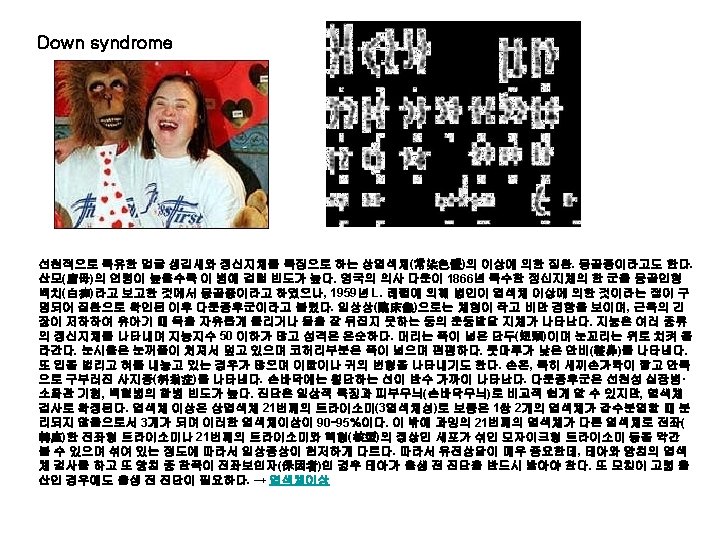
Chromosome 21 Contains over 400 genes Contains over 40 million base pairs, of which over 70% have been determined See the diseases associated with chromosome 21 in the Map. Viewer.
Autoimmune polyglandular syndrome The endocrine system is responsible for the release of hormones into the blood or lymph. Deficiencies in the endocrine system can be caused by infection, infarction, or a tumor destroying all or a large part of the gland. However, the activity of an endocrine organ is most often depressed as a result of an autoimmune reaction that ultimately results in partial or complete destruction of the gland. Autoimmune disease affecting one organ is frequently followed by the impairment of other glands, resulting in multiple endocrine failure. Autoimmune polyglandular syndrome type I (APS 1, also called APECED) is a rare autosomal recessive disorder that maps to human chromosome 21. At the end of 1997, researchers reported that they isolated a novel gene, which they called AIRE (autoimmune regulator). Database searches revealed that the protein product of this gene is a transcription factor a protein that plays a role in the regulation of gene expression. The researchers showed that mutations in this gene are responsible for the pathogenesis of APS 1. The identification of the gene defective in APS 1 is the first step toward developing tests that will be able to genetically diagnose the disease. Further investigations of the gene and its function should also facilitate finding a potential treatment for the disease as well as increasing our general understanding of the mechanisms underlying other autoimmune diseases
Amyotrophic lateral sclerosis (ALS) is a neurological disorder characterized by progressive degeneration of motor neuron cells in the spinal cord and brain, which ultimately results in paralysis and death. The disease takes its less-scientific name from Lou Gehrig, a baseball player with the New York Yankees in the late 1920 s and 1930 s, who was forced to retire in 1939 as a result of the loss of motor control caused by the disease. In 1991, a team of researchers linked familial ALS to chromosome 21. Two years later, the SOD 1 gene was identified as being associated with many cases of familial ALS. The enzyme coded for by SOD 1 carries out a very important function in cells: it removes dangerous superoxide radicals by converting them into nonharmful substances. Defects in the action of this enzyme mean that the superoxide radicals attack cells from the inside, causing their death. Several different mutations in this enzyme all result in ALS, making the exact molecular cause of the disease difficult to ascertain. Recent research has suggested that treatment with drugs called antioxidants may benefit ALS patients. However, since the molecular genetics of the disease are still unclear, a significant amount of research is still required to design other promising treatments for ALS.
Chromosome 22 Contains over 800 genes Contains over 40 million base pairs, of which approximately 70% have been determined See the diseases associated with chromosome 22 in the Map. Viewer
Glucose galactose malabsorption Glucose Galactose Malabsorption (GGM) is a rare metabolic disorder caused by a defect in glucose and galactose transport across the intestinal lining. GGM is characterized by severe diarrhea and dehydration as early as the first day of life and can result in rapid death if lactose (milk sugar), sucrose (table sugar), glucose, and galactose are not removed from the diet. Half of the 200 severe GGM cases found worldwide result from familial intermarriage. At least 10% of the general population has glucose intolerance, however, and it is possible that these people may have milder forms of the disease. GGM is an autosomal recessive disorder in which affected individuals inherit two defective copies of the SGLT 1 gene, located on chromosome 22. Normally within the space enclosed by the small intestine (called the lumen), lactose is broken down into glucose and galactose by an enzyme called lactase, while sucrose is broken down into glucose and fructose by an enzyme called sucrase. The protein product of SGLT 1 then moves the glucose and the galactose from the lumen of the small intestine into intestinal cells. Usually the mutations carried by GGM individuals result in nonfunctional truncated SGLT 1 proteins or in the improper placement of the proteins such that they can not transport glucose and galactose out of the intestinal lumen. The glucose and galactose, if left untransported, draw water out of the body into the intestinal lumen, resulting in diarrhea. Although no cure exists for GGM, patients can control their symptoms (diarrhea) by removing lactose, sucrose, and glucose from their diets. Infants showing a prenatal diagnosis of GGM will thrive on a fructose-based replacement formula and will later continue their "normal" physical development on a fructose-
Di. George syndrome is a rare congenital (i. e. present at birth) disease whose symptoms vary greatly between individuals but commonly include a history of recurrent infection, heart defects, and characteristic facial features. Di. George syndrome is caused by a large deletion from chromosome 22, produced by an error in recombination at meiosis (the process that creates germ cells and ensures genetic variation in the offspring). This deletion means that several genes from this region are not present in Di. George syndrome patients. It appears that the variation in the symptoms of the disease is related to the amount of genetic material lost in the chromosomal deletion. Although researchers now know that the DGS gene is required for the normal development of the thymus and related glands, counteracting the loss of DGS is difficult. Some effects, for example the cardiac problems and some of the speech impairments, can be treated either surgically or therapeutically, but the loss of immune system T-cells (produced by the thymus) is more challenging and requires further research on recombination and immune function
Neurofibromatosis Neurofibromatosis, type 2, (NF-2) is a rare inherited disorder characterized by the development of benign tumors on both auditory nerves (acoustic neuromas). The disease is also characterized by the development of malignant central nervous system tumors as well. The NF 2 gene has been mapped to chromosome 22 and is thought to be a so-called 'tumor- suppressor gene'. Like other tumor suppressor genes (such as p 53 and Rb), the normal function of NF 2 is to act as a brake on cell growth and division, ensuring that cells do not divide uncontrollably, as they do in tumors. A mutation in NF 2 impairs its function, and accounts for the clinical symptoms observed in neurofibromatosis sufferers. NF-2 is an autosomal dominant genetic trait, meaning it affects both genders equally and that each child of an affected parent has a 50% chance of inheriting the gene. We are learning more about the function of the NF 2 gene through studies of families with neurofibromatosis type 2 and through work in model organisms, particularly mice. The exact molecular function of NF 2 in the cell is still unknown, although the protein is similar to the ERM family of cytoskeleton-membrane linker proteins. Further work on the binding partners of NF 2 would help to identify potential specific targets for future drug therapies.
Nf 1 knock out –embryonic lethal at mid-gastration
Leukemia, chronic myeloid Chronic myeloid leukemia (CML) is a cancer of blood cells, characterized by replacement of the bone marrow with malignant, leukemic cells. Many of these leukemic cells can be found circulating in the blood and can cause enlargement of the spleen, liver, and other organs. CML is usually diagnosed by finding a specific chromosomal abnormality called the Philadelphia (Ph) chromosome (see figure), named after the city where it was first recorded. The Ph chromosome is the result of a translocation or exchange of genetic material between the long arms of chromosomes 9 and 22. This exchange brings together two genes: the BCR (breakpoint cluster region) gene on chromosome 22 and the proto-oncogene ABL (Ableson leukemia virus) on chromosome 9. The resulting hybrid gene BCR-ABL codes for a fusion protein with tyrosine kinase activity, which activates signal transduction pathways, leading to uncontrolled cell growth. A mouse model has been created that develops a CML-like disease when given bone marrow cells infected with a virus containing the BCR-ABL gene. In other animal models, the fusion proteins have been shown to transform normal blood precursor cells to malignant cells. To research the human disease, antisense oligomers (short DNA segments) that block BCR-ABL were developed that specifically
Chromosome X Contains over 1400 genes Contains over 150 million base pairs, of which approximately 95% have been determined See the diseases associated with chromosome X in the Map. Viewer.
Paroxysmal nocturnal hemoglobinuria The distinct and rather peculiar characteristics of paroxysmal nocturnal hemoglobinuria (PNH) have puzzled hematologists for more than a century. PNH is characterized by a decreased number of red blood cells (anemia), and the presence of blood in the urine (hemoglobinuria) and plasma (hemoglobinemia), which is evident after sleeping. PNH is associated with a high risk of major thrombotic events, most commonly thrombosis of large intra-abdominal veins. Most patients who die of their disease die of thrombosis. PNH blood cells are deficient in an enzyme known as PIG-A, which is required for the biosynthesis of cellular anchors. Proteins that are partly on the outside of cells are often attached to the cell membrane by a glycosylphosphatidylinositol (GPI) anchor, and PIG-A is required for the synthesis of a key anchor component. If PIG-A is defective, surface proteins that protect the cell from destructive components in the blood (complement) are not anchored and therefore absent, so the blood cells are broken down. The PIG-A gene is found on the X chromosome. Although not an inherited disease, PNH is a genetic disorder, known as an acquired genetic disorder. The affected blood cell clone passes the altered PIG-A to all its descendants red cells, leukocytes (including lymphocytes), and platelets. The proportion of abnormal red blood cells in the blood determines the severity of the disease
Duchenne muscular dystrophy (DMD) is one of a group of muscular dystrophies characterized by the enlargement of muscles. DMD is one of the most prevalent types of muscular dystrophy and is characterized by rapid progression of muscle degeneration that occurs early in life. All are Xlinked and affect mainly males an estimated 1 in 3500 boys worldwide. The gene for DMD, found on the X chromosome, encodes a large protein dystrophin. Dystrophin is required inside muscle cells for structural support; it is thought to strengthen muscle cells by anchoring elements of the internal cytoskeleton to the surface membrane. Without it, the cell membrane becomes permeable, so that extracellular components enter the cell, increasing the internal pressure until the muscle cell "explodes" and dies. The subsequent immune response can add to the damage. A mouse model for DMD exists and is proving useful for furthering our understanding on both the normal function of dystrophin and the pathology of the disease. In particular, initial experiments that increase the production of utrophin, a dystrophin relative, in order to compensate for the loss of dystrophin in the mouse are promising and may lead to the development of effective therapies for this devastating disease.
Menkes syndrome is an inborn error of metabolism that markedly decreases the cells' ability to absorb copper. The disorder causes severe cerebral degeneration and arterial changes, resulting in death in infancy. The disease can often be diagnosed by looking at a victim's hair, which appears to be both whitish and kinked when viewed under a microscope. Menkes' disease is transmitted as an X-linked recessive trait. Sufferers can not transport copper, which is needed by enzymes involved in making bone, nerve and other structures. A number of other diseases, including type IX Ehlers-Danlos syndrome, may be the result of allelic mutations (i. e. mutations in the same gene, but having slightly different symptoms) and it is hoped that research into these diseases may prove useful in fighting Menkes' disease. If administered within the first few months of life, copper histidinate appears to be effective in increasing the life expectancy of some patients. However, this treatment only increases life expectancy from three to thirteen years of age, so can only be considered a palliative. A similar condition to Menkes' disease exists in mice; working with these model organisms will help give insight into human copper transport mechanisms, so helping to develop effective treatments for Menkes' sufferers.
Alport syndrome (AS) is a genetic disease in which a collagen mutation affects the kidneys, the ears, and the eyes. The syndrome was named for Dr. Alport who in 1927 described a British family in which many members developed renal disease as well as deafness. He noted that affected men in the family died as a result of their kidney problems, whereas females were less affected and lived until old age. It is now known that most cases of AS are caused by a mutation in the collagen gene COL 4 A 5. This gene encodes for the alpha-5 chain of collagen type IV and is located on the X chromosome. Because women have two X chromosomes (XX), affected women usually have one normal copy and one abnormal copy of the gene. Men only have one copy of the X chromosome (XY). If they inherit the COL 4 A 5 mutation, this abnormal copy of the gene is the only copy they have and the effects are more severe. Type IV collagen is found in basement membranes (BM), which are selective barriers between cells. In the kidney, the glomerular BM filters waste products into the urine while keeping useful molecules within the blood stream. In AS, the abnormal collagen disrupts this filter, leading to the loss of proteins and red blood cells into the urine. Blood in the urine (hematuria) is a sign common to all types of AS. In the ear, abnormal collagen in the cochlea results in a progressive deafness in which the ability to hear high tones is lost first. Abnormal collagen can also affect the lens of the eye. Currently, renal failure due to AS is treated by dialysis or, for some, renal transplantation. However, gene therapy may one
Lesch-Nyhan syndrome (LNS) is a rare inherited disease that disrupts the metabolism of the raw material of genes. These raw materials are called purines, and they are an essential part of DNA and RNA. The body can either make purines (de novo synthesis) or recycle them (the resalvage pathway). Many enzymes are involved in these pathways. When one of these enzymes is missing, a wide range of problems can occur. In LNS, there is a mutation in the HPRT 1 gene located on the X chromosome. The product of the normal gene is the enzyme hypoxanthine-guanine phosphoribosyltransferase, which speeds up the recycling of purines from broken down DNA and RNA. Many different types of mutations affect this gene, and the result is a very low level of the enzyme. The mutation is inherited in an X-linked fashion. Females who inherit one copy of the mutation are not affected because they have two copies of the X chromosome (XX). Males are severely affected because they only have one X chromosome (XY), and therefore their only copy of the HPRT 1 gene is mutated. Mutations of the HPRT 1 gene cause three main problems. First is the accumulation of uric acid that normally would have been recycled into purines. Excess uric acid forms painful deposits in the skin (gout) and in the kidney and bladder (urate stones). The second problem is selfmutilation. Affected individuals have to be restrained from biting their fingers and tongues. Finally, there is mental retardation and severe muscle weakness. In the year 2000 it was shown that the genetic deficiency in LNS could be corrected in vitro. A virus was used to insert a normal copy of the HPRT 1 gene into deficient human cells. Such techniques used in gene therapy may one day provide a cure for this disease. For now, medications are
Immunodeficiency with hyper-Ig. M (HIM) is a rare primary immunodeficiency characterized by the production of normal to increased amounts of Ig. M antibody of questionable quality and an inability to produce sufficient quantities of Ig. G and Ig. A. Individuals with HIM are susceptible to recurrent bacterial infections and are at an increased risk of autoimmune disorders and cancer at an early age. In a normal immune response to a new antigen, B cells first produce Ig. M antibody. Later, the B cells switch to produce Ig. G, Ig. A and Ig. E, antibodies that protect tissues and mucosal surfaces more effectively. In the most common form of HIM there is a defect in the gene TNFSF 5, found on chromosome X at q 26. This gene normally produces a CD 40 antigen ligand (CD 154), a protein on T cells which binds to the CD 40 receptor on B and other immune cells. Without CD 154, B cells are unable to receive signals from T cells, and thus fail to switch antibody production to Ig. A and Ig. G. The absence of CD 40 signals between other immune cells makes individuals with HIM susceptible to infections by opportunistic organisms such as Pneumocystis and Cryptosporidium species. Treatment of HIM mainly consists of regular IV replacement of the missing Ig. G antibodies and prompt treatment of infections. Long lasting immunity, however, cannot be maintained without a bone marrow transplant, which is done when a suitable donor is available.
Fragile X syndrome is the most common inherited form of mental retardation currently known. Fragile X syndrome is a defect in the X chromosome and its effects are seen more frequently, and with greater severity, in males than females. In normal individuals, the FMR 1 gene is transmitted stably from parent to child. However, in Fragile X individuals, there is a mutation in one end of the gene (the 5' untranslated region), consisting of an amplification of a CGG repeat. Patients with fragile X syndrome have 200 or more copies of the CGG motif. The huge expansion of this repeat means that the FMR 1 gene is not expressed, so no FMR 1 protein is made. Although the exact function of FMR 1 protein in the cell is unclear, it is known that it binds RNA. A similar nucleotide repeat expansion is seen in other diseases, such as Huntington disease. Research in mice has proven helpful in elucidating some of the mechanisms that cause the instability of this gene. Our methods for identifying carriers of Fragile X syndrome have also improved, and further research will help people carrying "premutations" to avoid having children who have a larger expansion (i. e. more CGG repeats) in FMR 1, and therefore suffer from Fragile X syndrome.
Rett syndrome (RTT) is a progressive neurodevelopmental disorder almost exclusively affecting females. With an incidence of about 1 in 10, 000 births, it is a common cause of profound mental impairment in girls. Typically, babies with RTT develop normally until the age of 6 to 18 months, when their developmental milestones regress. They lose purposeful use of their hands and are seriously disabled for life, with reduced muscle tone and seizures. A temporary "autistic-like" phase often occurs at the onset of the disorder, and older children are known for their social engagement through intense eye gaze. RTT is caused by mutations in the gene Me. CP 2, found on the X chromosome. Me. CP 2 is called a "transcriptional repressor" because it codes for a protein that controls the expression of other genes. Depending on what part of the gene contains the mutation, partial loss of this protein changes the environment experienced by developmentally important proteins which, in turn, leads to the RTT phenotype. Me. CP 2 mutations seem to be more common in the X chromosome from sperm cells, which may explain why RTT is rare in boys, who do not inherit an X chromosome from their father. Girls, however, inherit one copy of the X chromosome from each parent. Girls with RTT have one functional copy of Me. CP 2 to counteract the mutated Me. CP 2 copy. But, in a normal process called X inactivation, one X chromosome is randomly inactivated in every cell. This results in the normal copy of Me. CP 2 being inactivated in some cells but not in others. X-inactivation coupled with the specific Me. CP 2 mutation type causes the range of symptoms seen in RTT. By studying the relationships between the Me. CP 2
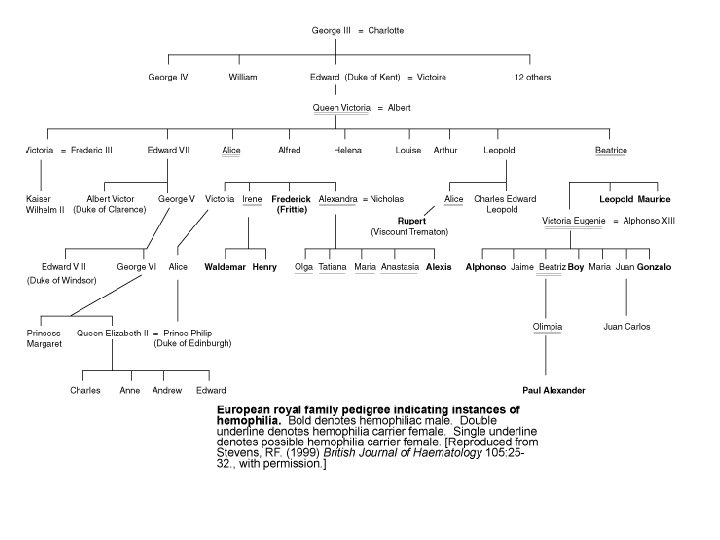
Hemophilia A is a hereditary blood disorder, primarily affecting males, characterized by a deficiency of the blood clotting protein known as Factor VIII that results in abnormal bleeding. Babylonian Jews first described hemophilia more than 1700 years ago; the disease first drew widespread public attention when Queen Victoria transmitted it to several European royal families. Mutation of the HEMA gene on the X chromosome causes Hemophilia A. Normally, females have two X chromosomes, whereas males have one X and one Y chromosome. Since males have only a single copy of any gene located on the X chromosome, they cannot offset damage to that gene with an additional copy as can females. Consequently, X-linked disorders such as Hemophilia A are far more common in males. The HEMA gene codes for Factor VIII, which is synthesized mainly in the liver, and is one of many factors involved in blood coagulation; its loss alone is enough to cause Hemophilia A even if all the other coagulation factors are still present. Treatment of Hemophilia A has progressed rapidly since the middle of the last century when patients were infused with plasma or processed plasma products to replace Factor VIII. HIV contamination of human blood supplies and the consequent HIV infection of most hemophiliacs in the mid-1980 s forced the development of alternate Factor VIII sources for replacement therapy, including monoclonal antibody purified Factor VIII and recombinant Factor VIII, both of which are used in replacement therapies today. Development of a gene replacement therapy for
Adrenoleukodystrophy (ALD) is a rare, inherited metabolic disorder that afflicts the young boy Lorenzo Odone, whose story is told in the 1993 film "Lorenzo's oil. " In this disease, the fatty covering (myelin sheath) on nerve fibers in the brain is lost, and the adrenal gland degenerates, leading to progressive neurological disability and death. People with ALD accumulate high levels of saturated, very long chain fatty acids in their brain and adrenal cortex because the fatty acids are not broken down by an enzyme in the normal manner. So, when the ALD gene was discovered in 1993, it was a surprise that the corresponding protein was in fact a member of a family of transporter proteins, not an enzyme. It is still a mystery as to how the transporter affects the function the fatty acid enzyme and, for that matter, how high levels of very long chain fatty acids cause the loss of myelin on nerve fibers. More recently, all the transporters related to ALD protein have been found in the yeast Saccharomyces cerevisiae, and a mouse model for the human disease has been developed. These and other molecular biology approaches should further our understanding of ALD and hasten our progress toward effective therapies.
Chromosome Y Contains over 200 genes • Contains over 50 million base pairs, of which approximately 50% have been determined • See the diseases associated with chromosome Y in the Map. Viewer. SRY: Sex determination We have come a long way in our understanding of sexual dimorphism since 355 BC. In those days, Aristotle suggested that the difference between the two sexes was due to the heat of semen at the time of copulation: hot semen generated males, whereas cold semen made females. Thankfully, we now know a little more about the molecular events of sex determination. Usually, a woman has two X chromosomes (XX) and a man one X and one Y (XY). However, both male and female characteristics can sometimes be found in one individual, and it is possible to have XY women and XX men. Analysis of such individuals has revealed some of the molecules involved in sex determination, including one called SRY, which is important for testis formation. SRY (which stands for sex-determining region Y gene) is found on the Y chromosome. In the cell, it binds to other DNA and in doing so distorts it dramatically out of shape. This alters the properties of the DNA and likely alters the expression of a number of genes, leading to testis formation. Most XX men who lack a Y chromosome do still have a copy of the SRY region on one of their X chromosomes. This copy accounts for their maleness. However, because the remainder of the Y chromosome is missing they frequently do not develop secondary sexual characteristics in the usual way. Since human SRY is similar to SRY of mice, a model of SRY function