
Choosing the Proper Power for the IOL Brannon
Choosing the Proper Power for the IOL Brannon Aden, MD Miles H. Friedlander, MD, FACS
Goal’s of Surgery Have Changed. b In past the goal was good visual outcome b Now an equal goal is a good refractive outcome • Central to that is an accurate calculation of the correct IOL power • Next came a variety of formulas aimed at achieving that accuracy
Possible Sources of Error in IOL Calculation b Systematic error-weakness in formula or weakness in a measurement technique • Example of technique is altering the axial length of the eye by using a contact type probe b Random error • Not common but tend to produce larger errors – Example is presence of a staphyloma
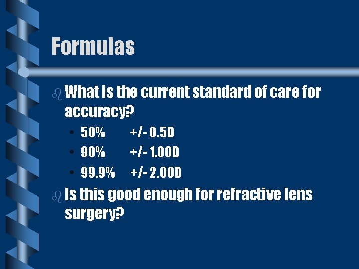
Formulas b What is the current standard of care for accuracy? • • • 50% +/- 0. 5 D 90% +/- 1. 00 D 99. 9% +/- 2. 00 D b Is this good enough for refractive lens surgery?
Factors Needed to Calculate IOL Power b Axial length of globe (distance from anterior corneal vertex to fovea) b Corneal power b Location of lens in eye (related to anterior chamber depth)

Axial Length b Most important anatomical variable b Greater deviation away from 22. 5 the greater the IOL power calculated especially with short eyes
Axial Length Measurement b Contact • Very personal dependent • Average error +/-. 2 mm (. 50 D) b Immersion • Technician unfriendly • Accurate +/-. 1 mm
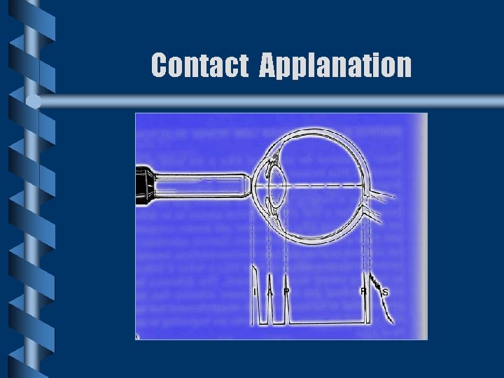
Contact Applanation

Immersion Scan
Measurement Continued b Buzard “Touch and Go” • Table mounted A-scan • Flood eye with tears • Advance probe toward eye until retinal spike produced on oscilloscope • Requires skilled and experienced examiner
IOL Master (Humphrey and Zeiss) b Uses optical interference (Partial Coherence Interferometry) to measure axial length b Keratometry also performed by machine
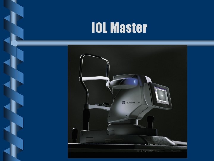
IOL Master
Corneal Curvature b Error of 0. 1 mm = 1 Diopter error b Sources of error • • Contact lens ware Refractive surgery
Anterior Chamber Depth b Now refers to final position of IOL or the distance from the posterior vertex of the cornea to the anterior surface of the IOL b ACD shallows 0. 1 mm per decade because of lens growth b In myopia deepens 0. 06 mm per 1 D b Of less importance than past
Early Formulas (First Generation) b Anterior chamber depth (ACD) was constant value b Early lenses were iris supported which produced small variations in Post Op ACD b Later with the introduction of PC IOL’s formula was less accurate • Difference of in the bag vs. sulcus was 1 mm therefore 1 D
Next First Generation Regression Formula (SRK 1) b Used multiple regression analysis b Eliminated ACD variable and replaced it with A-constant • Given by manufacturer and is based on expected position in eye, haptic and optic design, and refractive index of IOL material
Problems With SRK 1 Formula b Formula assumes 2. 5 D refractive change for each 1 mm of axial length regardless the axial length of the globe b Tended to under estimate IOL power in globes 25 to 29 mm long
Second Generation Regression Formulas b SRK II recognized the non linear relationship between axial length and IOL power b Binkhorst II, Holladay, Donzis also addressed same problems
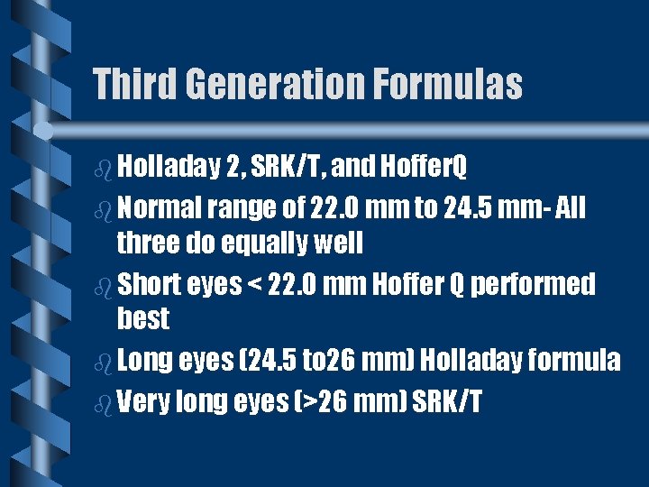
Third Generation Formulas b Holladay 2, SRK/T, and Hoffer. Q b Normal range of 22. 0 mm to 24. 5 mm- All three do equally well b Short eyes < 22. 0 mm Hoffer Q performed best b Long eyes (24. 5 to 26 mm) Holladay formula b Very long eyes (>26 mm) SRK/T
IOL Design and Materials b Majority of lenses are convex-plano, biconvex, or plano-convex b Vitreous pressure, haptic flexibility, and final position of ccc by contraction of the lens capsule effect final refractive error
Choice of Lens Materials b b In normal, non allergic, disease free eye either PMMA , silicone, or acrylic ok Eyes with silicone oil or anticipated vitro-retinal surgery need heparin surface-modified 100% PMMA -tend to retard adhesion of silicone oil to lens Uveitis- use heparin surface-modified lenses Posterior capsule opacification - Prevent? with acrylic lenses (stick to pc and stop proliferation of epithelial cells)
Lens Position b Plus lens- need more power as approach the retina b Minus lens- need less power as approach the retina b. Anterior iris plane, sulcus, capsule bag. • For every 1 mm of displacement- 1 D of corrective change • Example If a capsular bag lens is placed in the sulcus then the power is reduced by 1 D
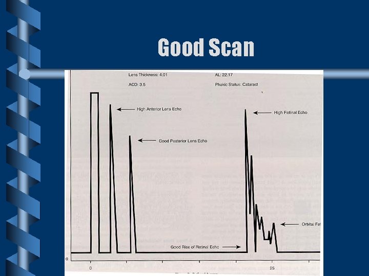
Good Scan
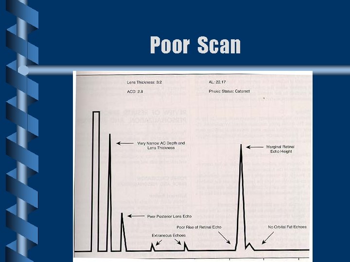
Poor Scan
- Slides: 24