
Charge Drift in partiallydepleted epitaxial Ga As detectors

- Slides: 28
Charge Drift in partially-depleted epitaxial Ga. As detectors P. J. Sellin, H. El-Abbassi, S. Rath Department of Physics University of Surrey, Guildford, UK J. C. Bourgoin LMDH, Université Pierre et Marie Curie, Paris, France Paul Sellin, Radiation Imaging Group
Overview r Chemical reaction growth of thick epitaxial Ga. As layers r Depletion thickness and residual impurity concentration r Performance of partially depleted detectors r C-V measurements of impurity concentration at low temperature r Optical probing of charge transport using a focussed laser Paul Sellin, Radiation Imaging Group
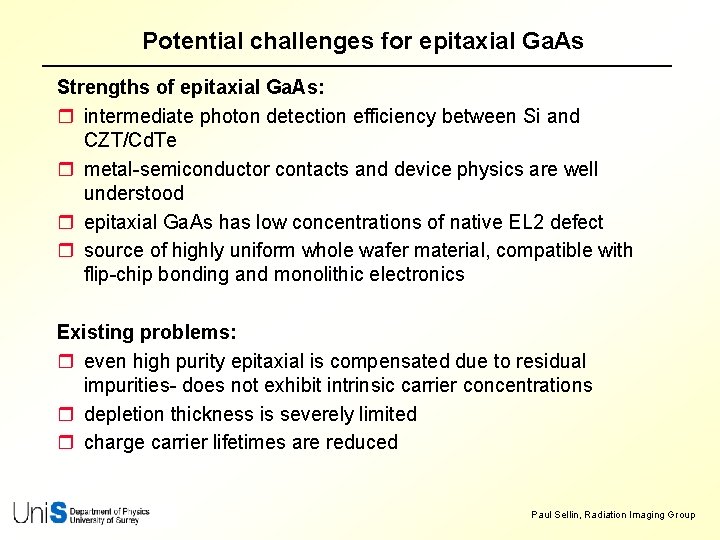
Potential challenges for epitaxial Ga. As Strengths of epitaxial Ga. As: r intermediate photon detection efficiency between Si and CZT/Cd. Te r metal-semiconductor contacts and device physics are well understood r epitaxial Ga. As has low concentrations of native EL 2 defect r source of highly uniform whole wafer material, compatible with flip-chip bonding and monolithic electronics Existing problems: r even high purity epitaxial is compensated due to residual impurities- does not exhibit intrinsic carrier concentrations r depletion thickness is severely limited r charge carrier lifetimes are reduced Paul Sellin, Radiation Imaging Group
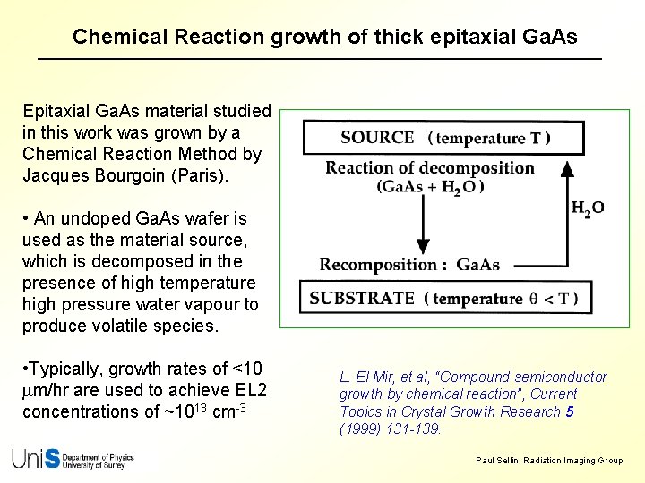
Chemical Reaction growth of thick epitaxial Ga. As Epitaxial Ga. As material studied in this work was grown by a Chemical Reaction Method by Jacques Bourgoin (Paris). • An undoped Ga. As wafer is used as the material source, which is decomposed in the presence of high temperature high pressure water vapour to produce volatile species. • Typically, growth rates of <10 mm/hr are used to achieve EL 2 concentrations of ~1013 cm-3 L. El Mir, et al, “Compound semiconductor growth by chemical reaction”, Current Topics in Crystal Growth Research 5 (1999) 131 -139. Paul Sellin, Radiation Imaging Group
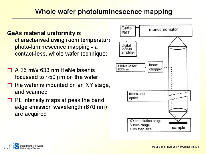
Whole wafer photoluminescence mapping Ga. As material uniformity is characterised using room temperature photo-luminescence mapping - a contact-less, whole wafer technique: r A 25 m. W 633 nm He. Ne laser is focussed to ~50 mm on the wafer r the wafer is mounted on an XY stage, and scanned r PL intensity maps at peak the band edge emission wavelength (870 nm) are acquired Paul Sellin, Radiation Imaging Group
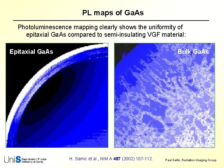
PL maps of Ga. As Photoluminescence mapping clearly shows the uniformity of epitaxial Ga. As compared to semi-insulating VGF material: Epitaxial Ga. As Bulk Ga. As H. Samic et al. , NIM A 487 (2002) 107 -112. Paul Sellin, Radiation Imaging Group
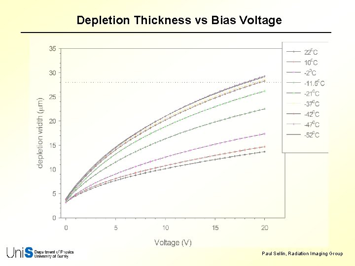
Calculated depletion thickness This material is nominally 1 -5 x 1014 cm-3 - corresponds to a 10 -20 mm depletion thickness @ 30 V, and 15 -30 mm @ 80 V Paul Sellin, Radiation Imaging Group
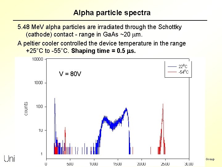
Alpha particle spectra 5. 48 Me. V alpha particles are irradiated through the Schottky (cathode) contact - range in Ga. As ~20 mm. A peltier cooler controlled the device temperature in the range +25°C to -55°C. Shaping time = 0. 5 ms. V =V 30 V = 80 V Paul Sellin, Radiation Imaging Group
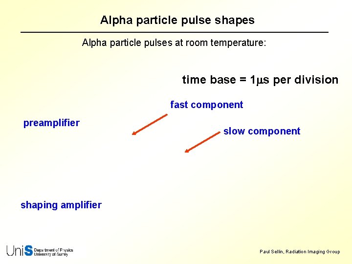
Alpha particle pulse shapes Alpha particle pulses at room temperature: time base = 1 ms per division fast component preamplifier slow component shaping amplifier Paul Sellin, Radiation Imaging Group
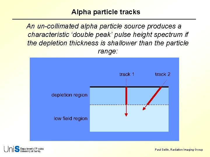
Alpha particle tracks An un-collimated alpha particle source produces a characteristic ‘double peak’ pulse height spectrum if the depletion thickness is shallower than the particle range: Paul Sellin, Radiation Imaging Group
59. 5 ke. V gamma spectra Depth-dependent CCE produces poorly resolved gamma spectra: T = -50°C Paul Sellin, Radiation Imaging Group
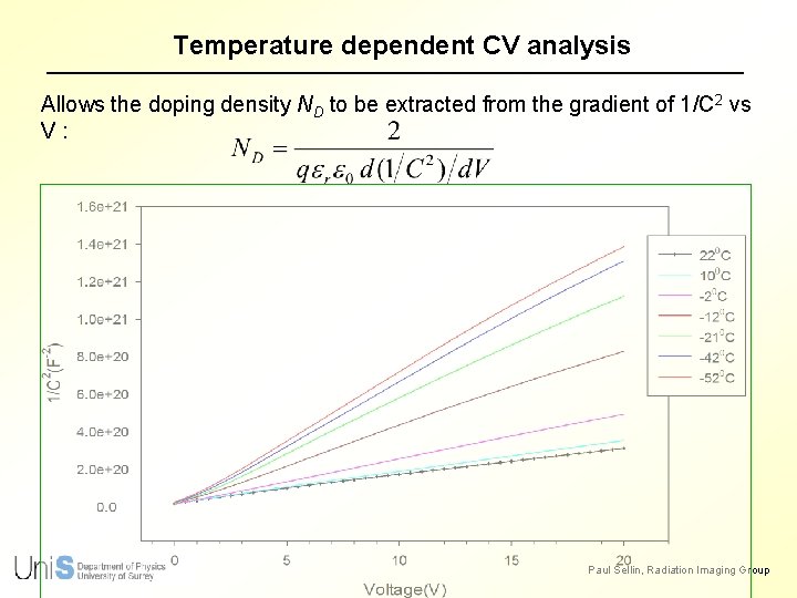
Temperature dependent CV analysis Allows the doping density ND to be extracted from the gradient of 1/C 2 vs V: Paul Sellin, Radiation Imaging Group
Depletion Thickness vs Bias Voltage Paul Sellin, Radiation Imaging Group
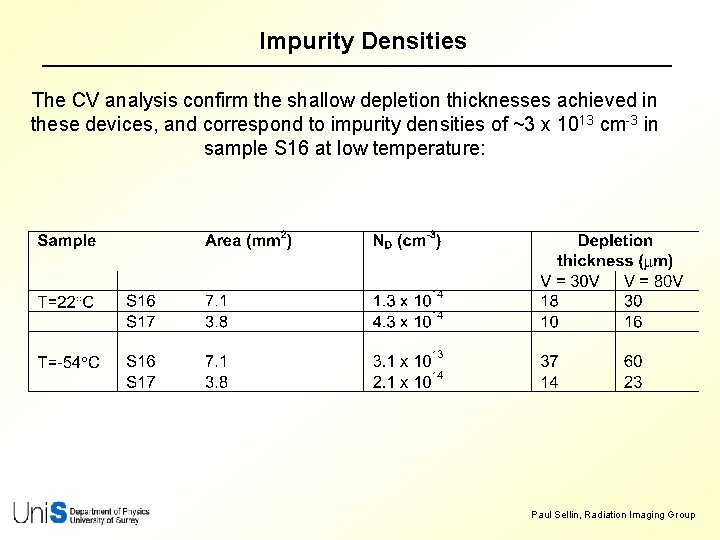
Impurity Densities The CV analysis confirm the shallow depletion thicknesses achieved in these devices, and correspond to impurity densities of ~3 x 1013 cm-3 in sample S 16 at low temperature: Paul Sellin, Radiation Imaging Group
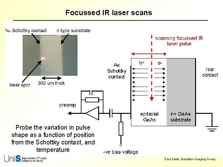
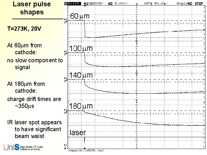
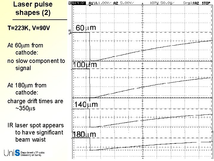
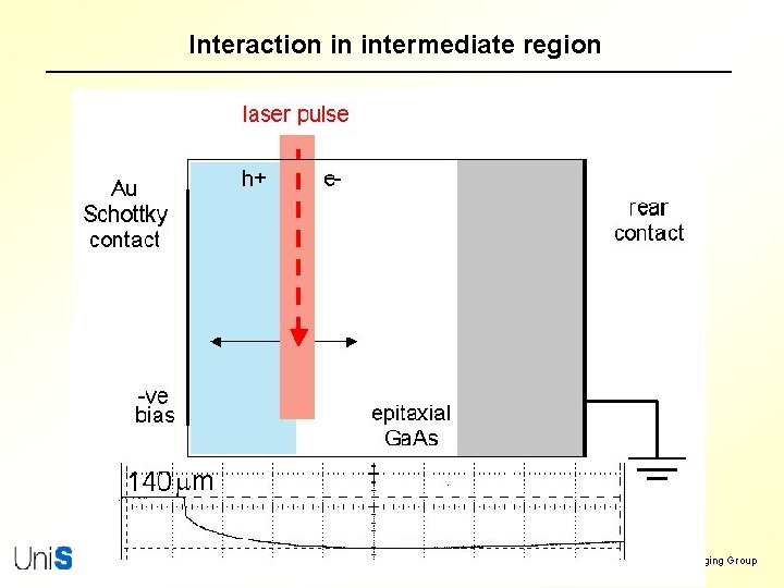
Focussed IR laser scans Probe the variation in pulse shape as a function of position from the Schottky contact, and temperature Paul Sellin, Radiation Imaging Group
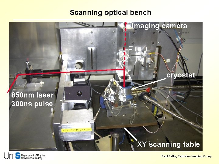
Scanning optical bench imaging camera cryostat 850 nm laser 300 ns pulse XY scanning table Paul Sellin, Radiation Imaging Group
Laser pulse shapes T=273 K, 20 V At 60 mm from cathode: no slow component to signal At 180 mm from cathode: charge drift times are ~350 ms IR laser spot appears to have significant beam waist Paul Sellin, Radiation Imaging Group
Laser pulse shapes (2) T=223 K, V=90 V At 60 mm from cathode: no slow component to signal At 180 mm from cathode: charge drift times are ~350 ms IR laser spot appears to have significant beam waist Paul Sellin, Radiation Imaging Group
Pulse risetime and amplitude vs bias Paul Sellin, Radiation Imaging Group
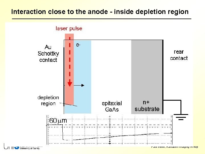
Interaction close to the anode - inside depletion region Paul Sellin, Radiation Imaging Group
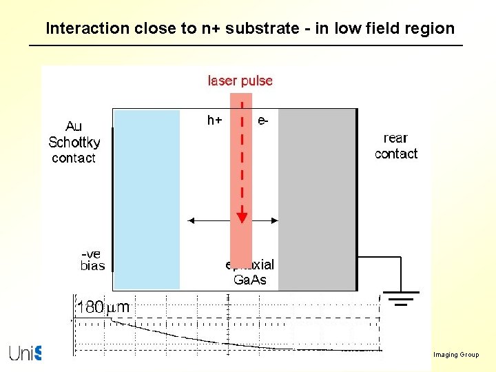
Interaction close to n+ substrate - in low field region Paul Sellin, Radiation Imaging Group
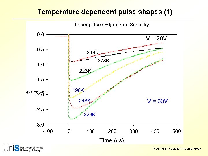
Temperature dependent pulse shapes (1) Paul Sellin, Radiation Imaging Group
Temperature dependent pulse shapes (2) Paul Sellin, Radiation Imaging Group
Conclusions r The epitaxial Ga. As layers studied showed excellent uniformity, and a residual impurity concentration of 1 -5 x 1014 cm-3 r Long electron lifetimes > 300 ms were observed in the low field regions - confirms the very low EL 2 concentration Lateral laser scans show: r good charge transport in the shallow depleted region r long-lived components to the pulse shapes when irradiated close to n+ substrate - consistent with slow electron diffusion towards the substrate r significant penetration of the depletion region when cooled to -50°C Future work: r further lateral scanning is required with focussed lasers and high resolution proton microbeams to quantify these phenomena r further modest reductions in impurity concentration will produce significant performance improvements Paul Sellin, Radiation Imaging Group
Acknowledgements This work was partially funded by the UK’s Engineering and Physics Science Research Council Paul Sellin, Radiation Imaging Group
Alpha particle spectra Epitaxial Ga. As pad detectors were irradiated with an uncollimated 241 Am alpha particle source. The detector was mounted in a vacuum cryostat, attached to a peltier cooler to allow operation in the temperature range of +25°C to -55°C. Pulse height spectra (figure 3) were acquired using a conventional charge integrating preamplifier and spectroscopy amplifier (shaping time = 0. 5 ms). Figure 3: a spectra at room temperature, as a function of bias. Paul Sellin, Radiation Imaging Group
Interaction in intermediate region Paul Sellin, Radiation Imaging Group
Temperature dependent pulse shapes (2) Paul Sellin, Radiation Imaging Group