Charge Coupled Device CCD Disease Profiling Through Medical
: Disease Profiling Through Medical Imaging • MUHAMMAD • ASRA TAYYAB")


- Slides: 14
Charge Coupled Device (CCD): Disease Profiling Through Medical Imaging • MUHAMMAD • ASRA TAYYAB NAZIR IMTIAZ (INSTITUTE OF SPACE TECHNOLOGY, ISLAMABAD , PAKISTAN)
CONTENTS • What is CCD? • How does CCD operate? • Usage of CCD in Space Telescopes • Proposal of idea • Background of invention • Description of idea • Claims • References • Acknowledgement
WHAT IS CCD? A light sensitive integrated circuit which: • was invented in 1969 by Williard Boyle & George E Smith from AT&T Bell Labs • stores and displays data for an image; each pixel is converted into an electric signal • Used in digital cameras, Photocopiers, Telescopes as a CCD sensor
HOW DOES CCD WORK? • It's basic principle is photoelectric effect • It is divided up into a large number of pixels which can be used to build up an image of the scene of interest
WORKING PRINCIPLES
Usage of CCD in Space Telescopes Conversion of a distant star’s light directly into digital images NASA has developed a software that traces, categorizes and procures images of deep sky objects allowing a user to control computer driven telescopes
BACKGROUND OF INVENTION: • Since the evolution of medicine and technology, mankind has faced difficulties in detection of damaged tissues of human brain etc. • For the diagnosis of disease, body temperature has always been helpful parameter • Early discovery of cancer depended on visual investigation of the skin and specifically on detection of skin patches • Human body temperature has been recorded with different devices like ultrasound , MRI etc. • Ultrasound Doppler techniques are used to gauge blood flow in blood vessels • However, the Doppler procedures are not delicate to blood stream in smaller vessels. • MRI has high temporal and spatial resolution • But, MRIs clinical utilization is constraint because of its high cost, poor versatility and its capacity structure has poor sensitivity.
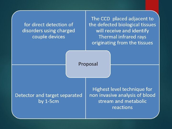
DESCRIPTION OF IDEA • Proposed idea uses CCD which collects the photons emitted from the source and sense • it to the CCD controller through the line which further applies some data analysis and processing • The energy radiation is directly proportional to T^4 according to Stephen Boltzmann Law • After completion of irradiation phenomenon the count on the cells are save and used for • generating the image for its visual representation on the basis of energy distribution over the charged couple device
Graphical description of idea
CLAIMS: non-invasive method of measuring biological tissue sample a light sensor that consists of of multi-spectral light sensitivity measures a segment of tissue being at least one centimeter beneath the skin measures the extent of energy content being collected by the light detector more specifically determines the type of disease by auto co-relation of temperature energy content
References Reference: BIOMEDICAL IMAGING SYSTEM WITH NON-INVASIVE METHOD FOR DIRECT DETECTION OF PERIPHERAL ARTERIAL DISORDERS AND CANCER DISEASES USING DEEP DEPLETION CHARGE COUPLED DEVICE • Muhammad Irfan Saeed (Institute of Space Technology) • Zahoor Sarwar Sheikh (Institute of Space Technology)
Acknowledgement We express our gratitude to two people in particular. Firstly, Mr. Irfan Saeed , space scientist at the Institute of Space Technology for his assistance throughout our work. Secondly Mr. Waqas Basheer , also at the Institute of Space Technology for his unceasing advice and encouragement.
THANK YOU