Characterization of Nanoparticles UltravioletVisible UVVis Spectroscopy nanoparticles present

Characterization of Nanoparticles ﻡ ﺩ ﻧﻴﺮﺍﻥ ﻋﻠﻲ ﺛﺎﻣﺮ
 Spectroscopy nanoparticles present distinct optical and physical properties, which are dependent upon")
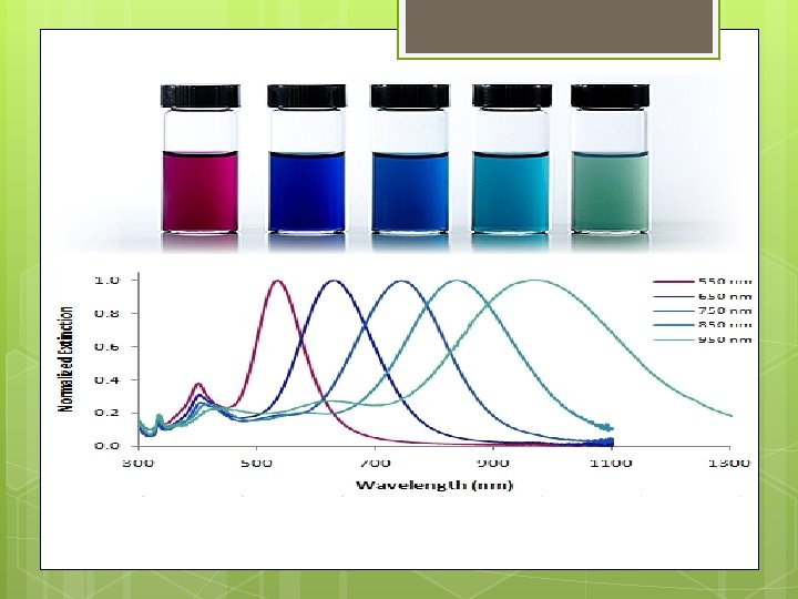
Ultraviolet-Visible (UV-Vis) Spectroscopy nanoparticles present distinct optical and physical properties, which are dependent upon their size (diameter), shape, surface structure and agglomeration state. optical feature commonly referred to as localized surface plasmon resonance (LSPR), that is, the collective oscillation of electrons in the conduction band of nanoparticles in resonance with a specific wavelength of incident light
 of gold nanoparticles due to collective oscillation")
Basics of localized surface plasmon resonance (LSPR) of gold nanoparticles due to collective oscillation of surface electrons with incident light at a specific wavelength.

Figure 1. Gold nanoparticle size dependant surface plasmon resonance. Note the red-shift of the absorption maximum as the gold nanoparticle size increases.

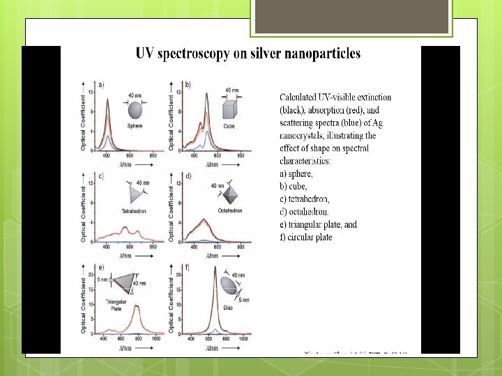
Gold nanoparticle shape dependant localized surface plasmon resonance as indicated by the visual appearance and UV-VIS spectra of spherical (A), and urchin-shaped (B) gold nanoparticles ("spiky gold").
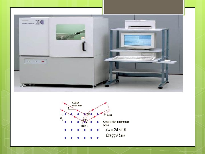

Debye–Scherrer formula, D = kλ/βcosθ,

Debye–Scherrer formula, D = kλ/βcosθ, where D is particle diameter size, k is a constant equals 1, λ is wavelength of X-ray source (0. 1541 nm), β is the full width at half maximum (FWHM)=0. 23, θ is the diffraction angle corresponding to the lattice plane (111).
 Dynamic light scattering (DLS) is an analytical technique used for")
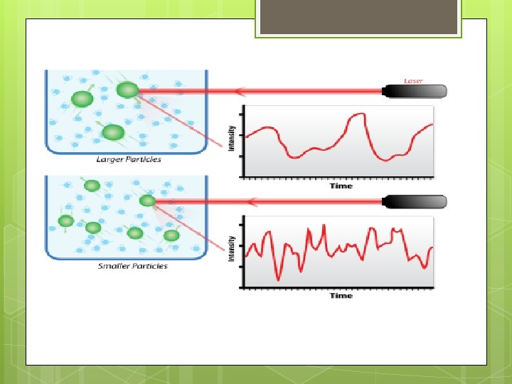
Dynamic Light Scattering (DLS) Dynamic light scattering (DLS) is an analytical technique used for measuring the size and size distribution of submicron-sized particles. In the measurement, a particle suspension is illuminated by a laser beam, and the fluctuation of scattered light is monitored analyzed, to acquire the velocity of the particles’ Brownian motion and thereby inferring their size.

62. 38 22. 3 -25

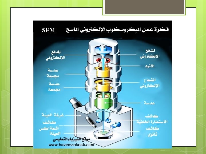
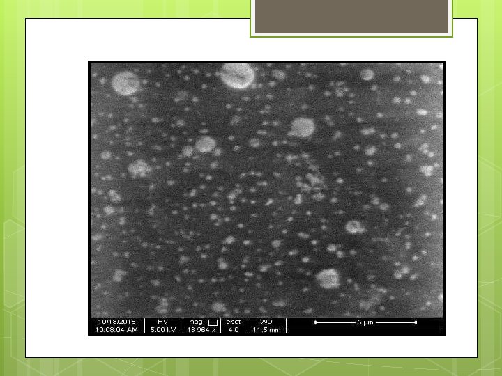
Scanning Electron Microscope SEM

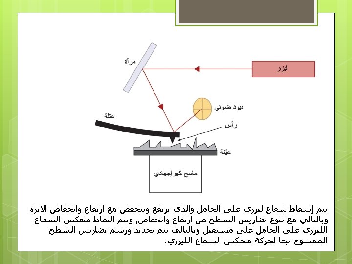
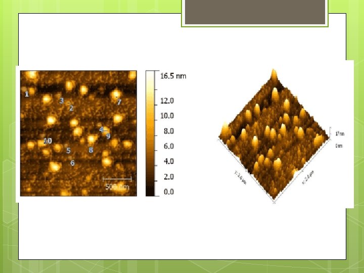
Atomic Force Microscope AFM

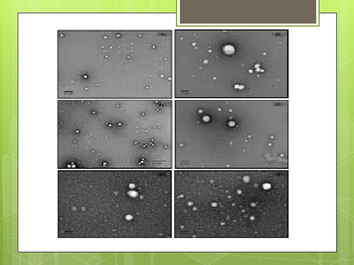
Transmission Electron Microscope TEM

It is a microscopy technique whereby a beam of electrons is transmitted through an ultra thin specimen, interacting with the specimen as it passes through. An image is formed from the interaction of electrons transmitted through the specimen; the image is magnified and focuses onto an imaging device, such as a fluorescent screen, on a layer of photographic film, or to be detected by a sensor such as a CCD camera.

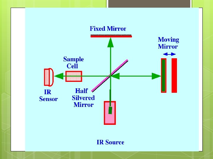
Fourier Transform Infrared Spectroscopy FTIR

- Slides: 25