Characteristics of life include 1 Movement internal or
 2. Responsiveness (reaction")


, a supportive cytoskeleton,")
 is rough ER, and functions in protein")


- Slides: 80
• Characteristics of life include: 1. Movement (internal or gross) 2. Responsiveness (reaction to internal or external change) 3. Growth (increase in size without change in shape) 4. Reproduction (new organisms or new cells) 5. Respiration (use of oxygen; removal of CO 2) 6. Digestion (breakdown of food into simpler forms) 7. Absorption (movement of substances through membranes and into fluids) 8. Circulation (movement within body fluids) 9. Assimilation (changing nutrients into chemically different forms) 10. Excretion (removal of metabolic wastes) The total of all the chemical reactions that are continuously at work to maintain these characteristics constitutes metabolism.
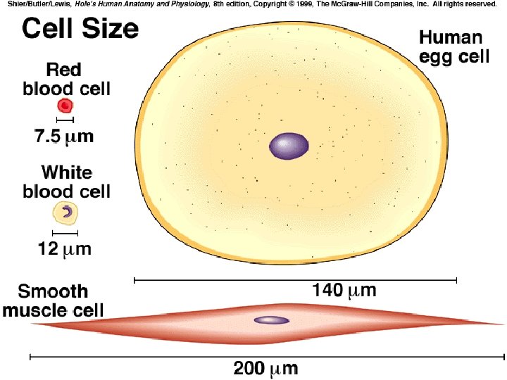
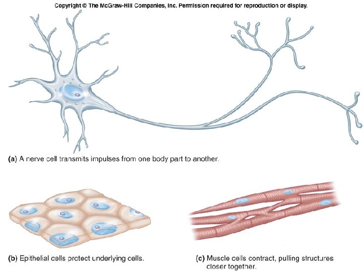
v. Introduction: A. The human body consists of 75 trillion cells that vary considerably in shape and size yet have much in common. B. Differences in cell shape make different functions possible.
v. A Composite Cell: A. A composite cell includes many different cell structures. B. A cell consists of three main parts---the nucleus, the cytoplasm, and the cell membrane. C. Within the cytoplasm are specialized organelles that perform specific functions for the cell.
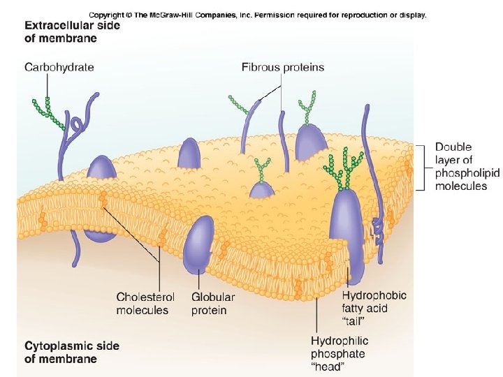
2. General Characteristics of Cell Membrane a. b. The cell membrane is extremely thin and selectively permeable. It has a complex surface with adaptations to increase surface area.
3. Cell Membrane Structure: a. The basic framework of the cell membrane consists of a double layer of phospholipids, with fatty acid tails turned inward. b. Molecules that are soluble in lipids (gases, steroid hormones) can pass through the lipid bilayer.
c. d. Embedded cholesterol molecules strengthen the membrane and help make the membrane less permeable to water-soluble substances. Many types of proteins are found in the cell membrane, including transmembrane proteins and peripheral membrane proteins.
e. f. g. Membrane proteins perform a variety of functions and vary in shape. Some proteins function as receptors on the cell surface Other proteins aid the passage of molecules and ions.
h. i. Proteins protruding into the cell anchor supportive rods and tubules. Still other proteins have carbohydrates attached; these complexes are used in cell identification. Membrane proteins called cellular adhesion molecules (CAMs) help determine one cell’s interactions with others.
The Structure of the Cell Membrane Section 7 -3 Outside of cell 4 3 Cell membrane Inside of cell (cytoplasm) 1 2
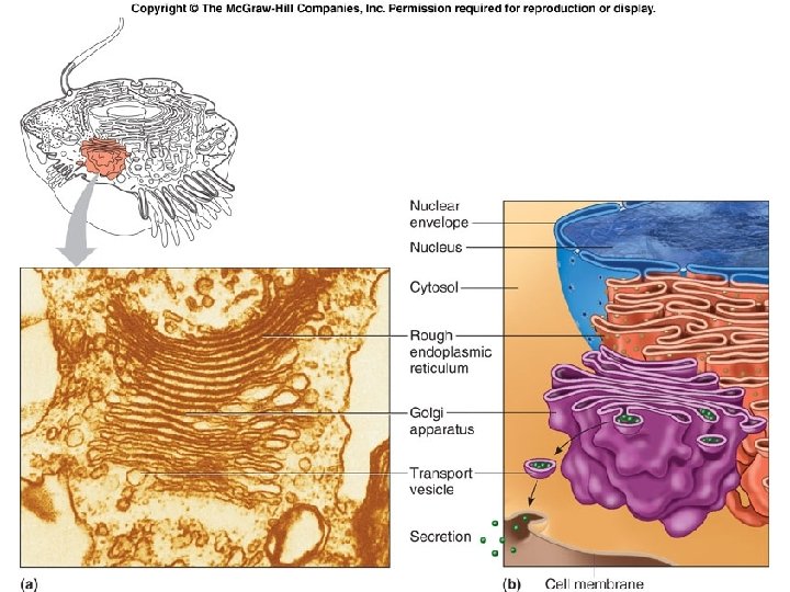
E. Cytoplasm: 1. The cytoplasm consists of a clear liquid (cytosol), a supportive cytoskeleton, and networks of membranes and organelles. a. Endoplasmic reticulum is made up of membranes, flattened sacs, and vesicles, and provides a tubular transport system inside the cell.
i. ii. With ribosomes, endoplasmic reticulum (ER) is rough ER, and functions in protein synthesis. Without ribosomes, it is smooth ER, and functions in lipid synthesis.
b. Ribosomes are found with ER and are scattered throughout the cytoplasm. They are composed of protein and RNA and provide a structural support for the RNA molecules that come together in protein synthesis.
Fig. 3. 04
c. The Golgi apparatus is composed of flattened sacs, and refines, packages, modifies, and delivers proteins. i. Vesicles form a “delivery service”, carrying chemicals throughout the cell.
d. Mitochondria are the powerhouses of the cell and contain enzymes needed for aerobic respiration. i. The inner membrane of the mitochondrion is folded into cristae which hold the enzymes needed in energy transformations to make ATP. ii. Very active cells contain thousands of mitochondria.
Fig. 3. 06
e. f. Lysosomes are the "garbage disposals" of the cell and contain digestive enzymes to break up old cell components and bacteria. Peroxisomes contain enzymes that function in the synthesis of bile acids, breakdown of lipids, degradation of rare biochemicals, and detoxification of alcohol.
g. Microfilaments and microtubules are thin, threadlike structures that serve as the cytoskeleton of the cell. i. Microfilaments, made of actin, cause various cellular movements. ii. Microtubules, made of the globular protein tubulin, are arranged in a 9 + 2 pattern of tubules.
Fig. 3. 07
h. A centrosome is made up of two hollow cylinders called centrioles that function in the separation of chromosomes during cell division.
Fig. 3. 08
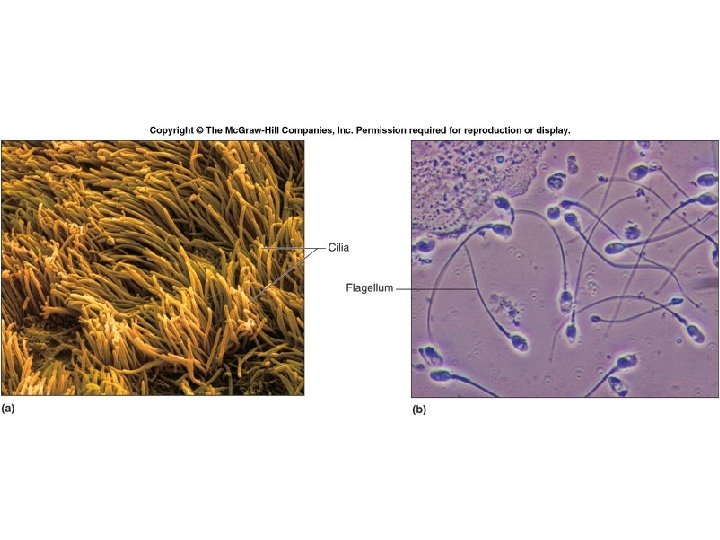
i. Cilia and flagella are motile extensions from the cell; shorter cilia are abundant on the free surfaces of certain epithelial cells (respiratory linings, for example), and a lengthy flagellum can be found on sperm cells.
j. Vesicles form from part of the cell membrane, or the Golgi apparatus, and store materials.
F. Cell Nucleus: 1. The fairly large nucleus is bounded by a double-layered nuclear membrane containing relatively large nuclear pores that allow the passage of certain substances.
Fig. 3. 10
a. b. The nucleolus is composed of RNA and protein and is the site of ribosome production. Chromatin consists of loosely coiled fibers of protein and DNA.
Fig. 3. 02
Pg 59
Section 7 -2 5 4 6 3 7 2 8 9 1 10 11 Animal Cell
v Movements Through Cell Membranes A. B. The cell membrane controls what passes through it. Mechanisms of movement across the membrane may be passive, requiring no energy from the cell (diffusion, facilitated diffusion, osmosis, and filtration) or active mechanisms, requiring cellular energy (active transport, endocytosis, and exocytosis).
DIFFUSION
Diffusion Slide number: 2 High O 2 Low O 2 High CO 2 Low CO 2 Copyright © The Mc. Graw-Hill Companies, Inc. Permission required for reproduction or display.
Diffusion Slide number: 3 High O 2 Low O 2 High CO 2 Low CO 2 Copyright © The Mc. Graw-Hill Companies, Inc. Permission required for reproduction or display.
Fig. 3. 16 a
Fig. 3. 16 b
Osmosis Selectively permeable membrane A Slide number: 2 Protein molecule Water molecule B 1 Copyright © The Mc. Graw-Hill Companies, Inc. Permission required for reproduction or display.
Osmosis Selectively permeable membrane A Slide number: 3 Protein molecule Water molecule B 1 Copyright © The Mc. Graw-Hill Companies, Inc. Permission required for reproduction or display.
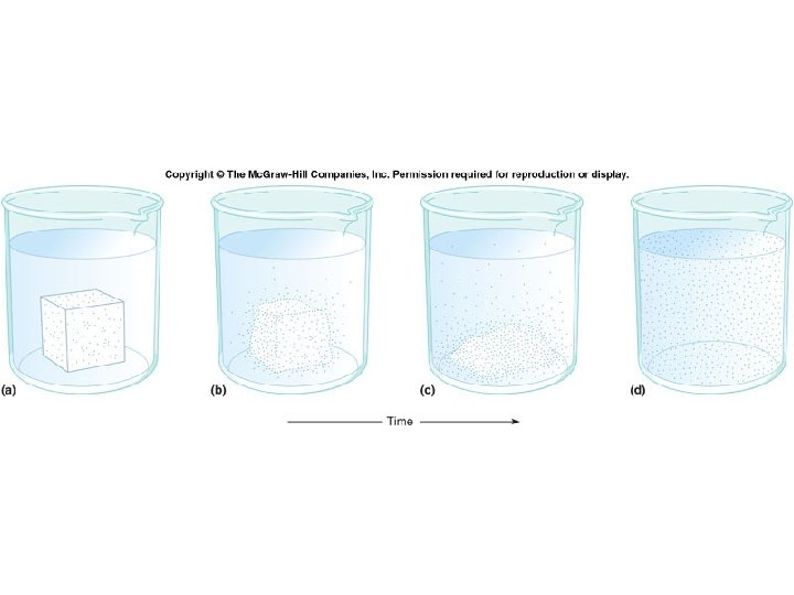
Osmosis Slide number: 4 A B Time 2 Copyright © The Mc. Graw-Hill Companies, Inc. Permission required for reproduction or display.
Osmosis Slide number: 5 Protein molecule Selectively permeable membrane Water molecule A B 2 Copyright © The Mc. Graw-Hill Companies, Inc. Permission required for reproduction or display.
Osmosis Selectively permeable membrane A Slide number: 1 Protein molecule Water molecule A B 1 B Time 2 Copyright © The Mc. Graw-Hill Companies, Inc. Permission required for reproduction or display.
Facilitated diffusion Slide number: 2 Region of higher concentration Region of lower concentration Protein carrier molecule Cell membrane Copyright © The Mc. Graw-Hill Companies, Inc. Permission required for reproduction or display.
Facilitated diffusion Slide number: 3 Region of higher concentration Transported substance Region of lower concentration Protein carrier molecule Copyright © The Mc. Graw-Hill Companies, Inc. Permission required for reproduction or display.
Filtration example Slide number: 2 Capillary wall Tissue fluid Larger molecules Smaller molecules Copyright © The Mc. Graw-Hill Companies, Inc. Permission required for reproduction or display.
Filtration example EDEMA Slide number: 3 Capillary wall Tissue fluid Blood pressure Larger molecules Smaller molecules Copyright © The Mc. Graw-Hill Companies, Inc. Permission required for reproduction or display.
Active transport Slide number: 2 Cell membrane Carrier protein Binding site Region of higher concentration Phospholipid molecules Region of lower concentration Copyright © The Mc. Graw-Hill Companies, Inc. Permission required for reproduction or display.
Active transport Slide number: 3 Carrier protein Binding site Region of higher concentration Region of lower concentration Transported particle Copyright © The Mc. Graw-Hill Companies, Inc. Permission required for reproduction or display.
Active transport Slide number: 4 Carrier protein with altered shape Cellular energy Region of higher concentration Region of lower concentration Copyright © The Mc. Graw-Hill Companies, Inc. Permission required for reproduction or display.
Sodium-Potassium Pump
Endocytosis and Exocytosis
Endocytosis and Exocytosis
Receptor-mediated endocytosis Slide number: 2 Molecules outside cell Receptor protein Cell membrane Cytoplasm Copyright © The Mc. Graw-Hill Companies, Inc. Permission required for reproduction or display.
Receptor-mediated endocytosis Slide number: 3 Receptor-ligand combination Copyright © The Mc. Graw-Hill Companies, Inc. Permission required for reproduction or display.
Receptor-mediated endocytosis Slide number: 4 Cell membrane indenting Copyright © The Mc. Graw-Hill Companies, Inc. Permission required for reproduction or display.
Receptor-mediated endocytosis Slide number: 5 Vesicle Cytoplasm Copyright © The Mc. Graw-Hill Companies, Inc. Permission required for reproduction or display.
Receptor-mediated endocytosis Slide number: 1 Molecules outside cell Receptor-ligand combination Vesicle Receptor protein Cell membrane Cytoplasm Cell membrane indenting Copyright © The Mc. Graw-Hill Companies, Inc. Permission required for reproduction or display.
Pg 65 Table. 3. 02
Why Cells have to divide
q. Most cells only divide every 40 – 60 times, called the Hayflick Limit. q. Some cells divide the maximum number of times – cells that line the small intestine q. Some cells normally don’t divide at all – nerve cells q. Telomeres – At the chromosome tips. They shorten with each mitosis until they get to a certain length when the cell will stop dividing
q. There are 260 specialized types of cells. The process of specialization from one egg cell is called differentiation.
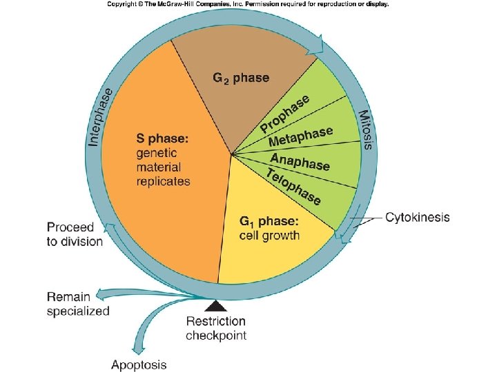
Mitosis and cytokinesis Slide number: 2 Early interphase of daughter cells— a time of normal cell A Restriction checkpoint growth and function. Copyright © The Mc. Graw-Hill Companies, Inc. Permission required for reproduction or display. Mitosis Cytokinesis G 1 phase S phase Interphase G 2 phase
Mitosis and cytokinesis Slide number: 3 Restriction checkpoint Nuclear envelope Chromatin fibers Centrioles Late Interphase Cell has passed the restriction checkpoint and completed DNA replication, as well as replication of centrioles and mitochondria, and synthesis of extra membrane. Copyright © The Mc. Graw-Hill Companies, Inc. Permission required for reproduction or display. Mitosis Cytokinesis G 1 phase S phase Interphase G 2 phase
Mitosis and cytokinesis Slide number: 4 Mitosis Cytokinesis G 1 phase S phase Interphase G 2 phase Prophase Aster Microtubules B Chromosomes condense and become visible. Nuclear envelope and nucleolus disperse. Spindle apparatus forms. Copyright © The Mc. Graw-Hill Companies, Inc. Permission required for reproduction or display.
Mitosis and cytokinesis Slide number: 5 Mitosis Cytokinesis G 1 phase S phase Interphase G 2 phase B Centromere Spindle fiber Late prophase Sister chromatids Copyright © The Mc. Graw-Hill Companies, Inc. Permission required for reproduction or display.
Mitosis and cytokinesis Slide number: 6 Mitosis Cytokinesis G 1 phase S phase Interphase G 2 phase Metaphase Chromosomes align along equator, or metaphase plate of cell. C Copyright © The Mc. Graw-Hill Companies, Inc. Permission required for reproduction or display.
Mitosis and cytokinesis Slide number: 7 Mitosis Cytokinesis G 1 phase S phase Interphase G 2 phase Anaphase Sister chromatids separate to opposite D poles of cell. Copyright © The Mc. Graw-Hill Companies, Inc. Permission required for reproduction or display.
Mitosis and cytokinesis Slide number: 8 Mitosis Cytokinesis G 1 phase S phase Interphase G 2 phase Telophase and cytokinesis Cleavage furrow Nuclear envelopes begin to reassemble around two daughter nuclei. Chromosomes decondense. Spindle disappears. Division of the cytoplasm into two cells. E Chromosomes Nuclear envelopes Copyright © The Mc. Graw-Hill Companies, Inc. Permission required for reproduction or display.
Mitosis and cytokinesis Slide number: 9 Early interphase of daughter cells— a time of normal cell A Restriction checkpoint growth and function. Copyright © The Mc. Graw-Hill Companies, Inc. Permission required for reproduction or display. Mitosis Cytokinesis G 1 phase S phase Interphase G 2 phase
Mitosis and cytokinesis Early interphase Late interphase Restriction A checkpoint Nuclear envelope Chromatin fibers Centrioles Cleavage furrow Aster Microtubules E Slide number: 1 Mitosis Cytokinesis G 1 phase S phase Interphase G 2 phase Prophase B Centromere Spindle fiber Telophase and cytokinesis Late prophase Chromosomes Nuclear envelopes D Anaphase Sister chromatids C Metaphase Copyright © The Mc. Graw-Hill Companies, Inc. Permission required for reproduction or display.
Fig. 3. 22
Fig. 3. 23
Chapter 3 Organelles