CHAPTER V Immunohistochemistry Acknowledgements Addisa Ababa University Jimma

CHAPTER V Immunohistochemistry

Acknowledgements • • Addisa Ababa University Jimma University Hawassa University Haramaya University of Gondar American Society for Clinical Pathology Center for Disease Control and Prevention-Ethiopia

Learning objectives • Upon the completion of this chapter, the student should be able to: 1. Explain the difference between immunohistochemistry and immunocytochemistry. 2. Define polyclonal and monoclonal antibodies. 3. Discuss methods of antigen retrieval. 4. Explain methods of antibody staining. 5. Explain methods of antibody detection

5. 1 Introduction • Immunocytochemistry is a method used to detect a specific antigen in the tissue to identify the type of disease. • In these techniques, an antibody is used to link a cellular antigen specifically to a stain that can be more readily seen with a microscope. • Detection of antigens in tissues is known as Immunohistochemistry, while detection in cultured cells is generally

• termed immunocytochemistry. For both, there is a wide range of specimen source, antigenavailability, antigen antibody affinity, antibody type, and detection enhancement methods. • Thus optimal conditions for immunohistochemical or immunocytochemical detection must be determined for each individual situation, dependent on the above variables.

Antigen • Antigen may be protein, carbohydrate, and lipid or a combination. • On the antigen molecule there are specific sites for attachment of antibody molecules. • The antibody attaching sites on antigens are known as antigenic determinants or epitopes. Antibodies • Antibodies are classes of immunoglobulins. There are five classes of antibodies: namely Ig. G, Ig. A, Ig. M, Ig. E, and Ig. D. Immunogloblin G (Ig. G) is the most common and most frequently used antibody for immunocytochemistry.

• Each immunoglobulin is composed of light chain and two heavy polypeptide chains linked by a disulphide bond to form a Y shaped structure. • The terminal region of each arm varies in their amino acid sequence and is known as variable domain. • The variability in amino acid sequence is responsible for a particular epitope. • The particular epitope enables the antibody to bind specifically to the particular antigen for which the antibody is raised.

• The amino acid chain on the variable domain forms a cavity. The cavity is geometrically and chemically complementary to a single type of antigen epitope. The complementary antigen • and antibody are held together by a combination of hydrogen bonds, electrostatic forces and. Van-deer Walls forces.

Polyclonal antibodies • The antibodies react major with corresponding antigen and also react minor with similar antigen. Monoclonal antibody • The antibody reacts specifically with its corresponding antigen and does not react with other antibody even with antibody having minor viabilities.

5. 2 Production of Polyclonal antibodies • An antigen of interest is introduced in to the animal. • The animal will mount an immune response to produce antibody. • The B-cells will proliferate to produce numerous clones of plasma cells. • Each clone of cells produces an antibody with slightly different specificity to a variety of epitope present on the antigen. • Some of these antibodies may cross react with other molecules and therefore they have to be removed by absorption with appropriate antigen.

N. B: Animals may have many other antibodies present in the serum. Theses antibodies may lead to the problem of cross-reactivity or non-specific staining. However, if the required • antibodies are present in high concentration, many unwanted reactions can be eliminated by dilution.

5. 3 Production of monoclonal antibody • Monoclonal antibodies are produced by hybridoma technique. • When antigen is introduced in to the body; B-cells proliferate to produce plasma cells. • Plasma cells produce antibodies against specific antigens. By fusion of normal plasma cells and myeloma cells (cancer cells) a hybrid cell can be produced.

• The hybrid plasma cell is an immortal cell and produces specific antibodies. • The hybrid plasma cell can be grown and multiplied in cell culture or ascitic fluid and it can produce an unlimited number of antibodies. By careful screening, hybrids producing antibodies of interest, i. e. , of single epitope can be produced that do not cross-react with other molecules.

• Fixation strengths and times must be optimized so that antigens and cellular structures can be retained and epitope masking is minimal. • Requirements for fixation can vary widely between tissues. For example, when using antibodies to probe for neurotransmitter substances, most tissues must be either immersion fixed with a mixture of glutaraldehyde and paraformaldehyde, or with paraformaldehyde alone. • Both acetone and methanol (precooled to -20 0 C) have been used successfully as fixatives for frozen tissue in other instances.

• The next consideration for immunological staining is the type of section to use. For immunohistochemistry, the common options are fixed or unfixed cryostat (frozen) sections, fixed “wet” or vibritome sections, or fixed, paraffin-embedded sections. • The choice of section is determined by a number of issues, including the time and skill of the investigator. • Because of the ease of use, fixed frozen sections are often quickest and easiest to use. • However, because of their superior fidelity, clarity, and preservation properties, fixed paraffin-embedded tissues have become the ultimate standard of immunohistochemistry in histology and pathology, and anytime where archiving of immunohistochemical information is required.

Today, there are two types of Cryostat sections: 1. Fresh, or unfixed sections where quickly frozen (snap frozen) tissues are first cut, theneither air-dried or fixed prior to staining and 2. Fixed frozen tissue, where the tissue is first fixed then cryo-protected with sucrose or other stabilizer (to stabilize the tissue cell structure) prior to freezing and sectioning.

• The advantages of frozen sections are that they allow excellent antigen preservation, they are typically faster to perform, and they offer flexibility, since any fixative can be used, thereby facilitating the optimization of fixative for each antigen. • However, frozen sections give less morphological detail and resolution than other methods
 Tissue Sections: 1. Snap-freeze")
Sample protocols for Cryostat sections - Fresh Frozen (then fixed) Tissue Sections: 1. Snap-freeze small tissue blocks (5 x 5 x 3 mm) in liquid nitrogen. 2. Transfer to cryostat and cut thin (5 -30 μm) sections. 3. Collect specimens on clean poly-L-lysine-coated glass slides and dry at room temperature overnight (if you want to stain the same day let air-dry for 1 -2 hours. until completely dry). Thorough drying is essential for good adhesion to the slides.

4. Fix sections in acetone or absolute ethanol at 4 0 C for 15 minutes. Use fresh ethanol or acetone for every 1015 slides for best results. The organic solvents absorb moisture from the air and tissue, as they do so, they lose their ability to fix the tissue effectively. 5. Thoroughly air-dry at room temperature or on mild heat (30 -370 C). It is during this stage that much of the chemical fixation is being finalized. Improper air-drying will lead to “soft” sections and likely loss of proper reactivity. 6. Proceed with immunostaining or freeze.

Fixed, frozen tissue sections 1. Fix tissue either by perfusion with fixative or by immersion in fixative for a set time period. Most commonly, 4% Paraformaldehyde (PFA) solutions are used. 2. Fixed tissue is then prepared for cryoprotection by submerging the target tissue in a hydro stabilizing solution. • The cryoprotection is complete when the target tissue no longer floats in the stabilizing solution. Because it works well and is relatively inexpensive, phosphate buffer saline (PBS) + sucrose solutions ranging from 10% w/v (less protection), to 30% (w/v) sucrose (greater

3. Once stabilized, tissues can be removed from the protectant solution and frozen at -70 0 C until sectioned. 4. Via cryostat (5 -40 μm), where sections can be collected directly onto slide, or floated onto slides via a PBS/water bath. • Usually up to three sections per slide can be placed. Each spaced well apart. The spacing prevents reagent mixing between samples. • Individual skill and tissue type will determine thickness of the sections. Sections between 10 -15 μm provide the best results for clarity and integrity. Sections between 6 -9 μm tend to tear during cutting,

5. Sections on slides are thoroughly air/heat dried on a slide warmer, usually overnight or at least 2 -3 hours at 4050 0 C. 6. Prepared slides can be stored dry at -70 0 C until stained. Equilibrate to room temperature and briefly re-dry prior to rehydration and staining. • The largest proportions of samples used in immunostaining are embedded in paraffin because it provides excellent morphological detail and resolution. Modern “paraffin” is typically a mixture of paraffin wax

• It is an excellent embedding medium because it can be heated to liquid state, and dissolved by xylene for infiltrating the tissue, and then relatively quickly turned to a solid state again for maximum structural support during sectioning. • Typically, small blocks of tissue (10 x 3 mm) are fixed for up to 24 hours.

• The most common fixatives used in paraffin sections are formalin-based. These fixatives are well tolerated by the tissues and achieve good penetration. • The blocks are then infiltrated and embedded with paraffin and 5 -10 μm sections are cut in ribbons and mounted on slides. • Once mounted, the slides can be stored indefinitely until immunostaining is required, then the paraffin must be removed from the tissue to allow the waterbased buffers and antibodies to penetrate.

Sample Protocol for Paraffin-embedded Sections • A. Conventional deparaffinization and dehydration sequence: 1. Incubate sections in Xylene: 2 to 3 changes, 5 minutes each. 2. 100% absolute ethanol: 2 changes, 3 minutes each. 3. 95% ethanol: 2 changes, 3 minutes each. 4. 80% ethanol: 3 minutes. 5. 50% ethanol: 3 minutes. 6. Rinse with distilled water, PBS, or Tris buffer: 2 changes, 3 minutes. each. Note: Once sections have been rehydrated, do not allow them to dry.
 0. 1% trypsin in PBS for")
B. Place slides in pre-warmed (37 0 C) 0. 1% trypsin in PBS for 5 -60 min. or 0. 4% pepsin in 0. 01 N HCl for 30 minutes to one hour. Follow by rinsing with distilled water. C. If peroxidase conjugate is used; endogenous peroxidase should be blocked at this stage. • Peroxidase activity results in the decomposition of hydrogen peroxide (H 2 O 2). It is a common property of all hem-proteins such as hemoglobin, myoglobin, cytochrome and catalase.

• Suppression of endogenous peroxidase activity in formalin-fixed tissue entails the incubation of sections in 3% H 2 O 2 for 8 -10 minutes. Methanolic H 2 O 2 treatment (1 part 3% H 2 O 2 plus 4 parts absolute methanol) for 20 minutes. can also used, but it is not recommended for specimens where cell surface markers are to be stained. • Methanolic treatment may also detach frozen sections from their carrier glass. D. Wash twice with PBS. E. Proceed with immunostaining procedure (see section 5. 5)

5. 4 Antigen Retrieval can be achieved through: – Enzyme digestion; – Microwave; – Autoclaving or pressure-cooking. • To facilitate the immunological reaction of antibodies with antigens in fixed tissue, it may benecessary to unmask or “retrieve” the antigens through pretreatment of the specimens. There are many forms of antigen retrieval (sometimes called antigen recovery), and different antigens and different antibodies will require different antigen retrieval methods.

• Antigenretrieval has been shown to increase reactivity of the majority of antigens in tissues. • The use of antigen retrieval in immunocytochemistry is less common. However, depending upon the particular antibody/antigen combination it can be performed on cell preparations, although the length of time and intensity is typically much less than for tissue. Antigen retrieval includes a variety of methods by which the availability of the antigen for interaction with aspecific antibody is maximized. • The most common techniques are enzymatic digestion or heat induced epitope retrieval (HIER) through microwave irradiation, autoclaving or pressure-cooking.

5. 4. 1 Enzymatic Digestion • This technique involves dewaxing, rehydrating, and rinsing the specimen in running water. • The specimen is then equilibrated with the appropriate buffer and incubated with aproteolytic enzyme at 37 0 C, or at room temperature. Enzymes used include pronase (0. 05% (w/v) in PBS), trypsin (0. 05% (w/v) in PBS with 0. 1% Ca. Cl 2) and pepsin (0. 05% (w/v) in 2 NHCl).

• The conditions of concentration, time and temperature must be controlled, so that the enzymes can break some of the bonds formed during fixation, uncovering antigenic sites, but the antigen should not be digested completely. • The enzymatic activity is stopped by placing the specimen in cold buffer (4°C) prior to processing with antibody. These methods should be considered for some antigens/tissues. However, proteolytic enzymes can abolish the reactivity of some antigens.

5. 4. 2 Microwave irradiation • Microwave irradiation of formalin-fixed, paraffinembedded specimens in buffer has been found to markedly enhance the retrieval of antigens. • During this procedure the energy provided helps break some of the bonds formed during fixation, thus increasing the number of positive cells available, and the intensity of reactions although the exact mechanism is unclear. • It is important to monitor the sections during the micro waving process, to prevent damage and drying.

• Consistency of conditions between experiments including buffer volumes, irradiation times, and microwave unit used, will result in less variability in staining results. • The number of samples that can be treated by microwave irradiation at one time is limited. Typically, specimens in some buffer are heated either at full or partial power for a few minutes. • Periodically, the heating is stopped and liquid is replenished. After a set time, the solution containing the slides is allowed to cool to room temperature slowly, then the slides are rinsed in PBS and used for staining.

5. 4. 3 Autoclaving • In order to standardize the procedure, it is important to start with standard volumes of preheated solutions. • After adding the specimens to the boiling retrieval solution, the autoclave or pressure cooker should be brought to full pressure as quickly as possible and the heating times measured exactly from this point. • At the end of the heating time (usually 1 to 2 minutes) the pressure should be released. As soon as possible the hot buffer should be flushed out with cold water. (Sections should not be allowed to dry. ) The specimens should then be washed in buffer.

• Although the most critical feature of both microwaving and autoclaving is probably the heating of the tissues, the p. H and composition of the solutions used are also important in the unmasking of antigenic sites. • Studies have found no significant difference between microwave and autoclave treatment, but there are significant differences based on the solutions used. Some of the buffer solutions commonly used are 0. 01 M citrate buffer (p. H 6. 0), 0. 1 M Tris-HCl (p. H 8. 0) and 1 m. M EDTA (p. H 8. 0), with citrate buffer used most commonly. • It should be noted that many more specimens could be treated at any one timeusing an autoclave or pressure cooker than using a microwave oven.

• However, preservation of the cytological detail may be slightly inferior in sections that undergo pressurecooking. • A more mild procedure that can be used on many tissues is a simple incubation in citric acid buffer, p. H 3. 0 (2. 1 grams Citric Acid added to 400 m. L of add. H 20. • Adjust to p. H 3. 0 with acetic acid if above 3. 0, or Na. OH if below 3. 0, make up to one liter final volume with distilled H 20) for 30 minutes, at 37 0 C after blocking but prior to primary antibody addition. • Rinse slide in PBS or TBS p. H 7. 4 prior to staining.

5. 5 Antibody Staining • Primary antibody may be directly labeled with an enzyme (such as horseradish peroxidase or alkaline phosphatase) or fluorophore (such as FITC or rhodamine), or unlabeled, with detection by a labeled secondary antibody or more complex detection system. • If a secondary antibody is used, it must be generated against the immunoglobulins of the primary antibody source, e. g. , if the primary antibody is raised in rabbit, then the secondary antibody could be goat anti-rabbit. • The optimal titer of both the primary and secondary antibody should be determined for each batch.

• For staining of tissue sections, it is customary to incubate with 25 -50 μl of diluted antibody. • The volume used must be sufficient to completely cover the tissue, and to ensure that the tissue will not dry out during incubation. Incubation times may range from 3090 minutes at 370 C, from one to six hours at room temperature, or overnight at 40 C. • Incubation times should be optimized empirically for each antibody/antigen combination.

General Protocol for Immunohistochemical Staining with Polyclonal Rabbit or Monoclonal Mouse Primary Antibody: • The following general protocol is intended for use as a guideline in developing antibodyspecific procedures. Different antibodies and tissues may require changes to this procedure. • Review of individual product datasheets and relevant literature references may be helpful in customizing this procedure for specific applications. 1. Gently rinse slide containing sections with distilled water or buffer from a wash bottle. Place slide at room temperature in a buffer bath for 5 minutes to rehydrate sections. 2. Gently remove excess liquid from around the specimen. Avoid touching the tissue directly 3. Apply 4 -6 drops of normal serum, (normal serum from the host of the secondary antibody) diluted 1: 5– 1: 30 (final concentration. 3%– 20%). Incubate for 20– 30 minutes at 37 0 C.

4. Tilt off serum and wipe away excess. Do not rinse. 5. Perform any antigen retrieval if necessary. 6. Apply 25 -50 μL of rabbit (mouse) primary antibody diluted appropriately per tissue section. Antibody should cover sections completely. Incubate for desired time (see above for suggested parameters and temperatures). • If optimal antibody dilution is unknown, perform a series of antibody dilutions in the range of 1: 20– 1: 1, 000 to obtain initial results. Note: Antibody diluent is often very important for consistent reactivity. Simple solutions are easier to troubleshoot than complex ones, thus antibodies diluted only with simple buffers (Tris buffer saline (TBS) or PBS) are usually recommended.

5. 6 Methods of antibody detection – Enzyme mediated; – Fluorescence; – Signal amplification. • Two of the most commonly used detection methods are fluorescence and colorimetric (enzyme mediated) detection. • With the advent of electron microscopy, detection of antigens by antibodies that contain large gold particles is often used and these may also be visualized at the light microscopic level as well, but their use is quite rare today, outside of electron microscopy. • Described below are the common antibody detection methods for light microscopy.

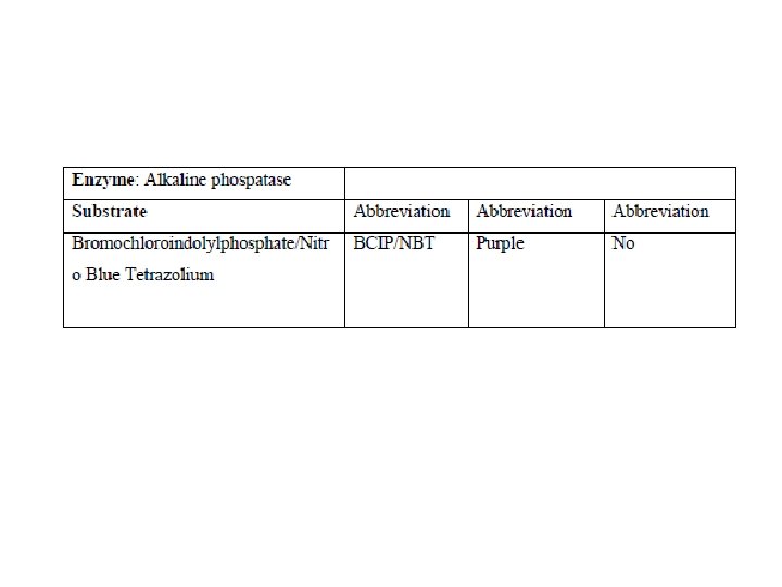
5. 6. 1 Enzyme-Mediated Detection • When choosing a substrate for conversion by an enzyme, one should select a substrate, which yields a precipitating product. • Examples of commonly used substrates are listed below Comments: 4 -chloro-1 -naphthol (CN) precipitates as a blue end product. Since CN is soluble in alcohol and other organic solvents, the slides must not be dehydrated, exposed to alcoholic counter stains, or cover slipped with mounting media containing organic solvents. Unlike DAB, CN tends to diffuse from the site of precipitation, thus it is not usually recommended for Immunohistochemistry but can be used for Western blotting.

Comments: Napthol AS acts as the substrate for alkaline phosphatase, and the Fast Red chromogen precipitates at the enzymatic sites producing a vibrant red/pink color. Precipitate is soluble in alcohol, thus aqueous counter stain and mounting medium should be used.

5. 6. 2 Fluorescence • A molecule that fluoresces can be attached to the antibody for detection using UV light. • Examples are Fluorescein, Rhodamine, Texas Red®, Cy 3 and Cy 5. In selecting fluorescence, one is limited by the available microscope filter sets. Most filter sets are best matched with rhodamine or fluorescein. Texas Red® may also be used with a rhodamine filter set. • Many mounting media contain “anti-fading” solutions, such as DABCO, which will prolong the viewing time of the sample. Chemicon offers a variety of fluorescence mounting fluids and counterstain solutions including our basic fluorescent mounting fluid and enhanced counterstaining fluid containing nuclear stains such as DAPI and PI.

5. 6. 3 Signal Amplification • Signal amplification techniques greatly enhance the sensitivity of immunohistochemical and immunocytochemical methods. • The signal amplification methods may be used in conjugation with either of the above detection techniques. • Signals may be amplified by using polyconjugated secondary antibodies, or Avidin-Biotin interactions or other commercially available amplifiers (i. e. , tyramide catalyzed systems), which increase the signal to antibody ratio. • When signal amplification is used to amplify the specific signal, however, one should be aware that non-specific

5. 7 Troubleshooting • Trouble shooting procedures should be performed to check for: – No staining; – Weak staining; – Background staining. • When tissue staining has not given the expected results, the experiment should be examined in a systematic way, wherein only single experimental variables are altered at one time. • Proper immunohistochemical troubleshooting requires one to determine whether difficulties are related to specimen, antibodies, technique, environment, or slide interpretation

- Slides: 48