Chapter five Amniotic Fluid Acknowledgements Addisa Ababa University

Chapter five Amniotic Fluid

Acknowledgements • • Addisa Ababa University Jimma University Hawassa University Haramaya University of Gondar American Society for Clinical Pathology Center for Disease Control and Prevention-Ethiopia

Chapter outline • Introduction to Cerebrospinal fluid • Routine laboratory assays • Collection of sample • Chemical analysis • Morphological Examination • Microbiological Examination • Serological Examination

Learning Objectives Upon completion of this chapter the student will be able to: 1 State the functions of amniotic fluid. 2 Describe the formation and composition of amniotic fluid. 3 State indications for performing an amniocentesis. 4 Describe the specimen-handling and processing procedures for testing amniotic fluid for bilirubin, fetal lung maturity (FLM), and cytogenetic analysis. 5 Discuss the principle of the spectrophotometric analysis for evaluation of hemolytic disease of the newborn. 6 Interpret a Liley graph.

Learning Objectives cont’d 7 Describe the analysis of amniotic fluid for the detection of neural tube disorders. 8 Explain the physiologic significance of the lecithinsphingomyelin (L/S) ratio. 9 State the relationship of phosphatidyl glycerol to FLM. 10 Discuss the principle of and sources of error for the L/S ratio, Amniostat-FLM, Foam Stability Index, and microviscosity tests for FLM. 11 Describe the relationship of lamellar bodies to FLM and the laboratory tests performed

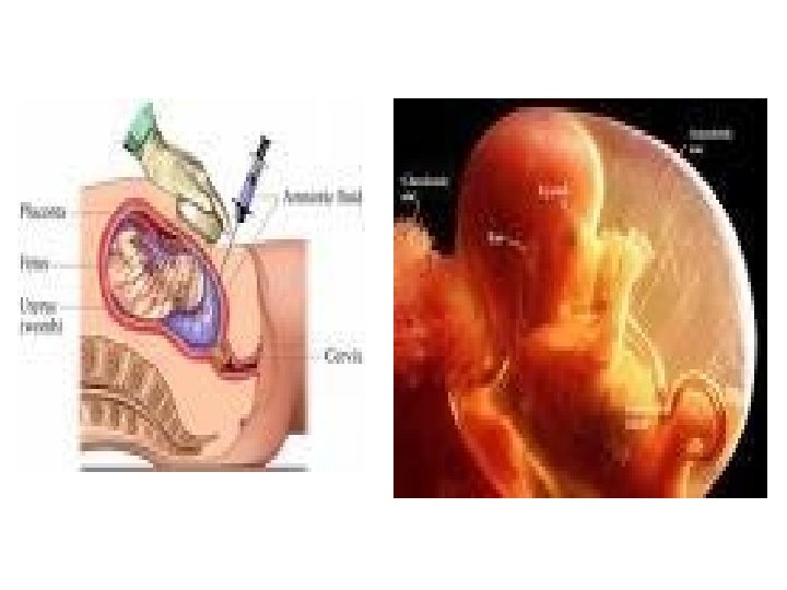
Amniotic Fluid • It is liquid that surrounds the fetus in the amniotic cavity – The primary function is to provide protective cushion for the fetus, allow for movement, and regulate temperature. • Amniocentesis is the fluid collection procedure in which a sample of the amniotic fluid surrounding a fetus is removed by means of a fine needle inserted through the abdomen and into the uterus of the pregnant woman.

Function of Amniotic Fluid • Amniotic fluid is present in the amnion, a membranous sac • that surrounds the fetus. • The primary functions of the fluid are to provide a protective cushion for the fetus, allow fetal movement, stabilize the temperature to protect the fetus from extreme temperature changes, and to permit proper lung development. • Exchanges of water and chemicals also take place between the fluid, the fetus, and the maternal circulation

Amniotic Fluid cont’d • testing of amniotic fluid is frequently associated with cytogenetic analysis, the clinical laboratory also performs several significant tests on amniotic fluid. • Because amniotic fluid is a product of fetal metabolism, the constituents that are present in the fluid provide information about the metabolic processes taking place during—as well as the progress of—fetal maturation. • When conditions that adversely affect the fetus arise, the danger to the fetus must be measured • The tests used to determine the extent of fetal distress and fetal maturity

Volume of Amniotic Fluid • regulated by a balance b/n the production of fetal urine and lung fluid and the absorption from fetal swallowing and intramembranous flow. • The amount of amniotic fluid increases throughout pregnancy, reaching a peak of 1 L during the third trimester, and then decreases prior to delivery. During the first trimester, the approximately 35 m. L (from the maternal circulation).

Volume of Amniotic Fluid cont’d • During each episode of fetal breathing movement, secreted lung liquid enters the amniotic fluid, as evidenced by lung surfactants that serve as an index of fetal lung maturity. • After the first trimester, fetal urine is the major contributor to the amniotic fluid volume. • At the time that fetal urine production occurs, fetal swallowing of the amniotic fluid begins and regulates the increase in fluid from the fetal urine.

Volume of Amniotic Fluid cont’d • Failure of the fetus to begin results in excessive accumulation of amniotic fluid (polyhydramnios) and is an indication of fetal distress, often associated with neural tube disorders. • Polyhydramnios may be secondarily associated with fetal structural anomalies, cardiac arrhythmias, congenital infections, or chromosomal abnormalities. • Increased fetal swallowing, urinary tract deformities, and membrane leakage are possible causes of decreased amniotic fluid (oligohydramnios).

Fetus in amniotic sac

Amniotic Fluid cont’d… • Sample collection – Collected by experienced professional – A maximum of 30 m. L of fluid can be collected. – The first 2 or 3 m. L should be discarded because they may contain maternal blood, tissue fluid, and maternal cells. – Sterile plastic syringes and conical centrifuge tubes should be used for the collection and transportation of the amniotic fluid. – Specimens for cytogenetic studies are maintained at 25 - 37 0 C incubation prior to analysis to prolong the life of the cells needed for analysis. – Do not freeze, refrigerate, or centrifuge.

Amniotic Fluid specimen – Specimens for fetal lung maturity tests should be placed in ice for delivery to the laboratory and refrigerated prior to testing. – The specimen will be centrifuged or filtered before analysis. – Specimens for bilirubin analysis in cases of Hemolytic Disease of the Newborn, must be protected from light. – The sample should be collected in an amber-colored tube. – The appearance of the amniotic fluid can indicate presence of certain chemicals. For example, degree of redness indicates presence of hemoglobin.

Color and Appearance • Normal amniotic fluid is colorless and may exhibit slight to moderate turbidity particularly in later stages of fetal development. • The presence of bilirubin gives the fluid a yellow color and is indicative of red blood cell destruction resulting from HDN. • Meconium, which is usually defined as a newborn’s first bowel movement, may be present in the amniotic fluid as the result of fetal intestinal secretions. It produces a dark green color. • Fetal aspiration of meconium during fetal swallowing is a concern when increased amounts are present in the fluid. A very dark red-brown fluid is associated with fetal death

Color

Gross examination of amniotic fluids color COLOR SIGNIFICANCE Colorless Normal Blood-streaked Traumatic tap, abdominal trauma, intra-amniotic hemorrhage Yellow Hemolytic Disease of the Newborn (HDN), Bilirubin Dark green Meconium (first bowel movement) Dark red-brown Fetal Death

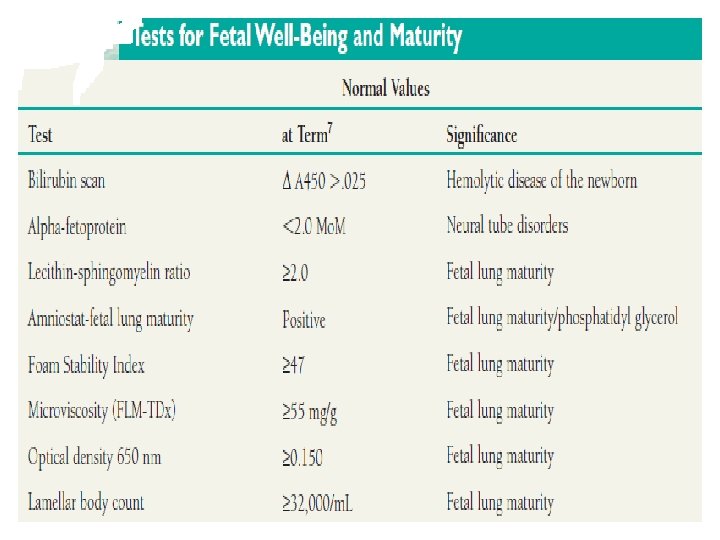
Amniotic Fluid testing • Laboratory testing of amniotic fluid involves analysis of bilirubin, alpha-fetoprotein and a variety of tests for fetal lung maturity (FLM). • During the third trimester of pregnancy but less than 35 to 36 weeks gestation, fluid collected from the amniocentesis procedure is analyzed to evaluate fetal lung maturity. • Limitations: – Contamination of the amniotic fluid specimen by blood or meconium invalidates the FLM results.

Chemical Composition • The placenta is the ultimate source of amniotic fluid water • and solutes. Amniotic fluid has a composition similar to that of the maternal plasma and contains a small amount of sloughed fetal cells from the skin, digestive system, and urinary tract. These cells provide the basis for cytogenetic analysis. • The fluid also contains biochemical substances that are produced by the fetus, such as bilirubin, lipids, enzymes, electrolytes, nitrogenous compounds, and proteins that can be tested to determine the health or maturity of the fetus.

Amniotic Fluid testing • Bilirubin Scan in Amniotic Fluid • Hemolysis and bilirubin assessed by optical density of amniotic fluid at 450 nm spectrophotometricaly • Change in Absorbance due to bilirubin • Clinical significance – evaluate fetal hemolysis in hemolytic disease of newborn

Hemolytic Disease of the Newborn • amniotic fluid evaluates the severity of the fetal anemia produced by HDN. • antibodies against other red cell antigens are also capable of producing HDN, and immunization of Rh-negative mothers may not be effective or even performed in all cases. • Initial exposure to foreign red cell antigens occurs during gestation and delivery of the placenta when fetal red blood cells enter into the maternal circulation and stimulate the mother to produce antibodies to the antigen. • When these antibodies present in the maternal circulation cross the placenta into the fetal circulation and bind to the antigen on the fetal cells, the cells are destroyed.

Hemolytic Disease of the Newborn • The destruction of fetal red blood cells results in the appearance of the red blood cell degradation product, unconjugated bilirubin, in the amniotic fluid. By measuring the amount of bilirubin in the fluid, the extent of hemolysis taking place may be determined, and the danger this anemia presents to the fetus may be assessed

Rh antibodies crossing the placenta

Summary for Amniotic Fluid • Mother and unborn child testing – Health care provider makes decisions about care and treatment – Assess chemical changes in mother and fetus – Understanding the stage of the fetus – An understanding of the developing physiology of the fetus is used to predict outcome by assessing chemical changes in the mother and fetus

Exercises 1. What may cause yellow color in amniotic fluids 2. Mention the limitation of chemical tests in amniotic fluid for specimen transport 3. Describe the principle of bilirubin test in amniotic fluid 4. Describe the clinical significance of amniotic fluid tests 5. describe the function of amniotic 6. What is the primary cause of the normal increase in amniotic fluid as a pregnancy progresses? 7. What is the reason for decreased amounts of amniotic fluid?

References: • Urinalysis and body fluids / Susan King Strasinger, 5 th ed. 2008 • District laboratory practice in tropical countries. 2 nd ed. Part I. Monica • • Cheesbrough, 2005 Text book of urinalysis and body fluids. Doris LR, Ann EN, 1983 Urinalysis and body fluids: A color text and atlas. Karen MR, Jean JL. 1995 Clinical chemistry: Principles, procedures, correlation. 3 rd ed. Michael L. Bishop et al. 1996 Tietz Text book of clinical chemistry. 3 rd ed. Carl AB, Edward RA, 1999 Clinical chemistry: Theory, analysis, correlation 4 th ed. Lawrence AK. 2003 ASCP Document Urinalysis lecture note. Mistire W. , Dawite Y. 28
- Slides: 28