Chapter 9 The Senses CLASSIFICATION OF SENSE ORGANS

 – Often exist")


 • Distribution is widespread; single-cell receptors are common")
 – Layers of eyeball •")
 – Eye fluids • Aqueous humor—in")

- Slides: 11
Chapter 9 The Senses
CLASSIFICATION OF SENSE ORGANS • General sense organs (Table 9 -1) – Often exist as individual cells or receptor units – Widely distributed throughout the body • Special sense organs (Table 9 -2) – Large and complex organs – Localized grouping of specialized receptors
CLASSIFICATION OF SENSE ORGANS • Classification by presence or absence of covering capsule – Encapsulated – Unencapsulated (“free” or “naked”) • Classification by type of stimuli required to activate receptors – – – Photoreceptors (light) Chemoreceptors (chemicals) Pain receptors (injury) Thermoreceptors (temperature change) Mechanoreceptors (movement or deforming of capsule) – Proprioceptors (position of body parts or changes in muscle length or tension)
CONVERTING A STIMULUS INTO A SENSATION • All sense organs have common functional characteristics – All are able to detect a particular stimulus – A stimulus is converted into a nerve impulse – A nerve impulse is perceived as a sensation in the central nervous system
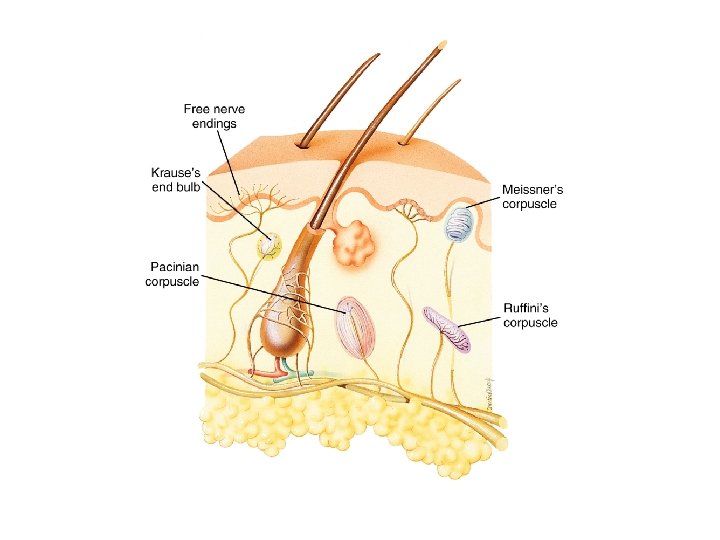
GENERAL SENSE ORGANS (Table 9 -1) • Distribution is widespread; single-cell receptors are common • Examples (Figure 9 -1, Table 9 -1) – Free nerve endings—pain, temperature, and crude touch – Meissner’s corpuscles—fine touch and vibration – Ruffini’s corpuscles—touch and pressure – Pacinian corpuscles—pressure and vibration
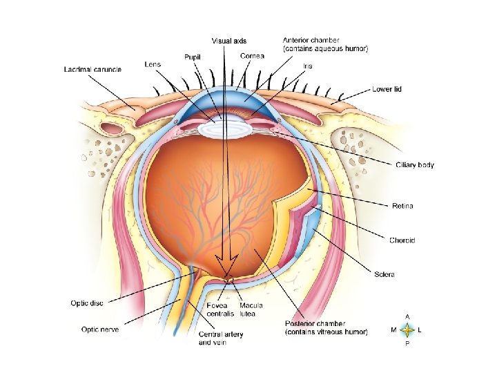
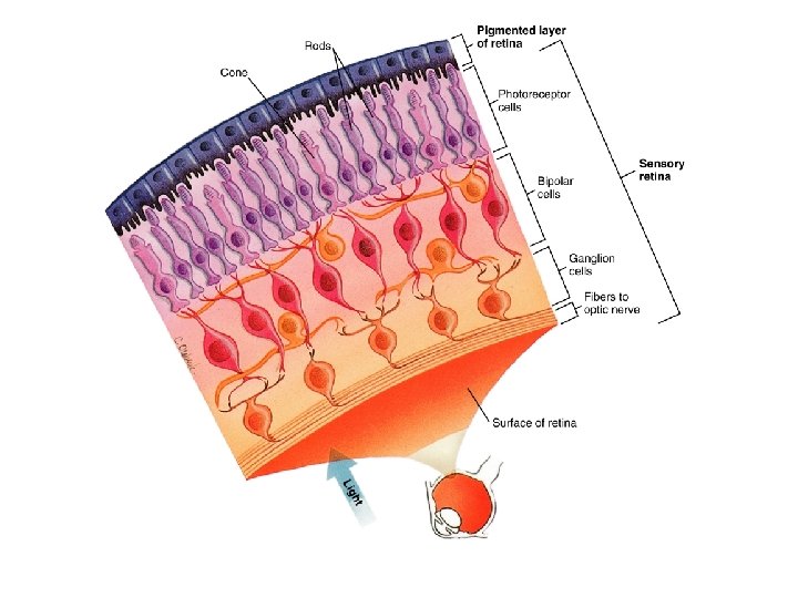
SPECIAL SENSE ORGANS • The eye (Figure 9 -2) – Layers of eyeball • Sclera—tough outer coat; “white” of eye; cornea is transparent part of sclera over iris • Choroid—pigmented vascular layer prevents scattering of light; front part of this layer made of ciliary muscle and iris, the colored part of the eye; the pupil is the hole in the center of the iris; contraction of iris muscle dilates or constricts pupil • Retina (Figure 9 -4)—innermost layer of the eye; contains rods (receptors for night vision) and cones (receptors for day vision and color vision) – Conjunctiva—mucous membrane covering the front surface of the sclera and also lines the eyelid; kept moist by tears found in the lacrimal gland – Lens—transparent body behind the pupil; focuses light rays on the retina
SPECIAL SENSE ORGANS • The eye (cont. ) – Eye fluids • Aqueous humor—in the anterior chamber in front of the lens • Vitreous humor—in the posterior chamber behind the lens – Visual pathway • Innermost layer of retina contains rods and cones • Impulse travels from the rods and cones through the bipolar and ganglionic layers of retina (Figure 9 -4) • Nerve impulse leaves the eye through the
SPECIAL SENSE ORGANS • The ear – The ear functions in hearing and in equilibrium and balance—receptors called mechanoreceptors – Divisions of the ear (Figure 9 -5) • External ear – Auricle (pinna) – External auditory canal » Curving canal 2. 5 cm (1 inch) in length » Contains ceruminous glands » Ends at the tympanic membrane