Chapter 8 The Cellular Basis of Reproduction and

Chapter 8 The Cellular Basis of Reproduction and Inheritance Power. Point Lectures for Campbell Biology: Concepts & Connections, Seventh Edition Reece, Taylor, Simon, and Dickey © 2012 Pearson Education, Inc. Lecture by Edward J. Zalisko

CONNECTIONS BETWEEN CELL DIVISION AND REPRODUCTION Organisms can reproduce sexually or asexually Reproduction 1. Sexual reproduction : the reproduction process that involves the union of a sperm and an egg 2. Asexual reproduction : the production of offsprings by a single parent without the participation of sperm and egg Copyright © 2003 Pearson Education, Inc. publishing as Benjamin Cummings

8. 1 Cell division plays many important roles in the lives of organisms • Some organisms make exact copies of themselves, asexual reproduction Copyright © 2003 Pearson Education, Inc. publishing as Benjamin Cummings

• Other organisms make similar copies of themselves in a more complex process, sexual reproduction Adults Sperm Egg fertilization Zygote (fertilized egg) development Offspring Copyright © 2003 Pearson Education, Inc. publishing as Benjamin Cummings

8. 2 Cells arise only from preexisting cells • All cells come from cells • Cellular reproduction is called cell division – Cell division allows an embryo to develop into an adult – It also ensures the continuity of life from one generation to the next Copyright © 2003 Pearson Education, Inc. publishing as Benjamin Cummings
 divide asexually")
8. 3 Prokaryotes reproduce by binary fission • Prokaryotic cells (bacteria, archaea) divide asexually – These cells possess a single chromosome, containing genes – The chromosome is replicated – The cell then divides into two cells, a process called binary fission Prokaryotic chromosomes Figure 8. 3 B Copyright © 2003 Pearson Education, Inc. publishing as Benjamin Cummings

• Binary fission of a prokaryotic cell Plasma membrane Prokaryotic chromosome Cell wall Duplication of chromosome and separation of copies Continued elongation of the cell and movement of copies Division into two daughter cells Copyright © 2003 Pearson Education, Inc. publishing as Benjamin Cummings

Figure 11 -8 STEPS IN BACTERIAL CELL DIVISION https: //www. youtube. com/watch? v=7 Lh-M-r. X 86 Q 1. Chromosome is located midcell. 2. Chromosome replicates. 3. Chromosomes 4. Fts. Z ring pull apart; ring of Fts. Z protein forms. Copyright © 2003 Pearson Education, Inc. publishing as Benjamin Cummings constricts. Membrane and cell wall infold. 5. Fission complete.

THE EUKARYOTIC CELL CYCLE AND MITOSIS • Eukaryotic cells have multiple chromosomes, while prokaryotic cells have usually one chromosome • A chromosome consists of one molecule of DNA and a number of proteins • The eukaryotic chromosome consists of a linear DNA, while prokaryotic one consists of a circular DNA • The number of chromosomes in a eukaryotic cell depends on the species Copyright © 2003 Pearson Education, Inc. publishing as Benjamin Cummings

Chromatin Chromosome Copyright © 2003 Pearson Education, Inc. publishing as Benjamin Cummings

Copyright © 2003 Pearson Education, Inc. publishing as Benjamin Cummings

• Chromosomes contain a very long DNA molecule with thousands of genes – Individual chromosomes are only visible during cell division – They are packaged as chromatin Copyright © 2003 Pearson Education, Inc. publishing as Benjamin Cummings

• Before a cell starts dividing, the chromosomes are duplicated – This process produces sister chromatids Sister chromatids Centromere Figure 8. 4 B Copyright © 2003 Pearson Education, Inc. publishing as Benjamin Cummings

• When the cell divides, the sister chromatids separate – Two daughter cells are produced – Each has a complete and identical set of chromosomes Copyright © 2003 Pearson Education, Inc. publishing as Benjamin Cummings

Figure 8. 3 B Chromosomes DNA molecules Sister chromatids Chromosome duplication Centromere Sister chromatids Chromosome distribution to the daughter cells

8. 5 The cell cycle multiplies cells • The cell cycle consists of two major phases: – Interphase, where chromosomes duplicate and cell parts are made – The mitotic phase, when cell division occurs Copyright © 2003 Pearson Education, Inc. publishing as Benjamin Cummings

8. 6 Cell division is a continuum of dynamic changes • Eukaryotic cell division consists of two stages: – Mitosis : one nucleus two nuclei – Cytokinesis : the cytoplasm is divided into two Mitosis Copyright © 2003 Pearson Education, Inc. publishing as Benjamin Cummings Cytokinesis

Mitosis : the equal division of one nucleus into two genetically identical daughter nuclei 1. Prophase 2. Metaphase 3. Anaphase 4. Telophase Interphase (G 1, S, G 2 phases) Chromosomal DNA is present as a form of chromatin DNA duplication in S-phase Copyright © 2003 Pearson Education, Inc. publishing as Benjamin Cummings

Prophase 1. Chromatin chromosome structure compaction 2. Nucleoli disappear 3. The nuclear envelope disappears 4. Formation of the mitotic spindle 5. Spindle microtubules attach to the kinetochores of chromosomes At the centromere region, each sister chromatid has a protein structure called a “kinetochore” Copyright © 2003 Pearson Education, Inc. publishing as Benjamin Cummings
 Centrioles Nuclear envelope Chromatin Early mitotic spindle")
MITOSIS INTERPHASE Prophase Centrosomes (with centriole pairs) Centrioles Nuclear envelope Chromatin Early mitotic spindle Prometaphase Centrosome Fragments of the nuclear envelope Kinetochore Plasma membrane Centromere Chromosome, consisting of two sister chromatids Copyright © 2003 Pearson Education, Inc. publishing as Benjamin Cummings Spindle microtubules

Metaphase Arrangement of the chromosomes on the metaphase plate Anaphase Separation of the sister chromatids toward the poles Telophase Daughter nuclei reappear Chromosome chromatin Nucleoli reappear Mitotic spindles disappear Cytokinesis와 telophase는 동시에 진행 Copyright © 2003 Pearson Education, Inc. publishing as Benjamin Cummings

MITOSIS Anaphase Metaphase plate Mitotic spindle Daughter chromosomes Copyright © 2003 Pearson Education, Inc. publishing as Benjamin Cummings Telophase and Cytokinesis Cleavage furrow Nuclear envelope forming

Microtubules – Fibers containing motor proteins Kinetochore plates + + Chromosome Copyright © 2003 Pearson Education, Inc. publishing as Benjamin Cummings Chromosome movement Tubulin subunits

8. 7 Cytokinesis differs for plant and animal cells • In animals, cytokinesis occurs by cleavage – This process pinches the cell apart Cytokinesis occurs with telophase, although it may actually begin in late anaphase Cleavage furrow SEM 140 Cleavage furrow Contracting ring of microfilaments Daughter cells Figure 8. 7 A Copyright © 2003 Pearson Education, Inc. publishing as Benjamin Cummings

• In plants, a membranous cell plate splits the cell in two New cell wall Cytokinesis Cell wall of the parent cell Cell wall Plasma membrane Daughter nucleus Cell plate forming Copyright © 2003 Pearson Education, Inc. publishing as Benjamin Cummings Vesicles containing cell wall material Cell plate Daughter cells

8. 8 Anchorage, cell density, and chemical growth factors affect cell division • Most animal cells divide only when stimulated, and others not at all • In laboratory cultures, most normal cells divide only when attached to a surface – They are anchorage dependent – Most animal cells are normally anchored to an extracellular matrix or to other cells of the same tissue Copyright © 2003 Pearson Education, Inc. publishing as Benjamin Cummings

• Cells continue dividing until they touch one another – This is called density-dependent inhibition Figure 8. 8 A Copyright © 2003 Pearson Education, Inc. publishing as Benjamin Cummings

Cultured cells suspended in liquid The addition of growth factor

Anchorage Single layer of cells Removal of cells Restoration of single layer by cell division Copyright © 2003 Pearson Education, Inc. publishing as Benjamin Cummings

8. 9 Growth factors signal the cell cycle control system • Proteins within the cell control the cell cycle – Signals affecting critical checkpoints determine whether the cell will go through a complete cycle and divide Most important, in the middle of G 1 checkpoint Control system At the boundary of meta- and anaphase M checkpoint G 2 checkpoint (at the G 2 -M boundary) https: //www. youtube. com/watch? v=Jc. ZQkmooy. Pk Copyright © 2003 Pearson Education, Inc. publishing as Benjamin Cummings

G 2 checkpoint Metaphase checkpoint Pass this checkpoint if: • chromosome replication is successfully completed • no DNA damage • activated MPF present Pass this checkpoint if: • all chromosomes are attached to mitotic spindle M G 2 G 1 G 0 S G 1 checkpoint Mature cells do not pass this checkpoint (they enter G 0 state) Pass this checkpoint if: • nutrients are sufficient • growth factors (signals from other cells) are present • cell size is adequate • DNA is undamaged Copyright © 2003 Pearson Education, Inc. publishing as Benjamin Cummings

Cdk 2 Cyclin A MPF component concentration Cyclin concentration regulates MPF concentration. G 1 S G 2 M phase G 1 MPF activated by dephosphorylation of MPF Cdk P S G 2 M phase G 1 S MPF activated by dephosphorylation of MPF Cdk PF l yc in C M Time Activated MPF has an array of effects. Cdk Cyclin-dependent kinase Phosphorylate chromosomal proteins; initiate M phase Activated MPF P Cyclin Cdk Cyclin + Cdk with P dephosphorylated, cyclin-dependent kinase subunit Copyright © 2003 Pearson Education, Inc. publishing as Benjamin Cummings Phosphorylate nuclear lamins; initiate nuclear envelope breakdown Phosphorylate microtubuleassociated proteins. Activate mitotic spindle? Phosphorylate an enzyme that degrades cyclin; cyclin concentrations decline

Copyright © 2003 Pearson Education, Inc. publishing as Benjamin Cummings

• The binding of growth factors to specific receptors on the plasma membrane is usually necessary for cell division Figure 8. 8 B Growth factor EXTRACELLULAR FLUID Plasma membrane Relay proteins Receptor protein G 1 checkpoint Signal transduction pathway G 1 S Control system M G 2 CYTOPLASM Copyright © 2003 Pearson Education, Inc. publishing as Benjamin Cummings

Growth factors stimulate cell cycling e. g. , Cells in your skin (arrested at G 1 checkpoint) When you got a cut in the skin Platelets release platelet-derived growth factor Cell division of skin cells at wound Copyright © 2003 Pearson Education, Inc. publishing as Benjamin Cummings

8. 10 Connection: Growing out of control, cancer cells produce malignant tumors • Cancer cells have abnormal cell cycle control systems – They divide excessively and can form abnormal masses called tumors • Radiation and chemotherapy are effective as cancer treatments because they interfere with cell division Copyright © 2003 Pearson Education, Inc. publishing as Benjamin Cummings
: the spread of cells beyond their original site Lymph vessels")
• Metastasis (전이): the spread of cells beyond their original site Lymph vessels Blood vessel Tumor in another part of the body Glandular tissue Growth Invasion Copyright © 2003 Pearson Education, Inc. publishing as Benjamin Cummings Metastasis

Tumor : an abnormal mass of cells 1. Benign tumor : an abnormal mass of normal cells 2. Malignant tumor : an abnormal mass of cancer cells Classification of cancers 1. Carcinoma : cancers derived from epithelial cells. E. g. , skin cancer, cancers in lung, stomach, and other organs 2. Sarcoma : cancers arising from connective tissue (bone, muscle, cartilage, fat, nerve) 3. Lymphoma and leukemia : cancer arising from hematopoietic cells 4. Blastoma : cancers derived from immature precursor cells 5. Germ cell tumor Copyright © 2003 Pearson Education, Inc. publishing as Benjamin Cummings

Normal cell Cancer cell Cell cycle control system Function correctly Malfunction Metastasis no yes Density-dependent inhibition of cell division yes no Anchorage yes dependence of cell division no The number of cell definitely division indefinitely Copyright © 2003 Pearson Education, Inc. publishing as Benjamin Cummings

8. 11 Review of the functions of mitosis: Growth, cell replacement, and asexual reproduction • When the cell cycle operates normally, mitotic cell division functions in: – Growth (seen here in an onion root) Copyright © 2003 Pearson Education, Inc. publishing as Benjamin Cummings
 Dead cells Epidermis, the outer layer")
• Cell replacement (seen here in skin) Dead cells Epidermis, the outer layer of the skin Dividing cells Dermis Figure 8. 11 B Copyright © 2003 Pearson Education, Inc. publishing as Benjamin Cummings
 Offspring clone of its parent")
• Asexual reproduction (seen here in a hydra) Offspring clone of its parent Clones : genetically same organisms Copyright © 2003 Pearson Education, Inc. publishing as Benjamin Cummings
 – diploid cell 2. Gamete (reproductive cell) –")
Cell 1. Somatic cell (body cell) – diploid cell 2. Gamete (reproductive cell) – haploid cell Copyright © 2003 Pearson Education, Inc. publishing as Benjamin Cummings

MEIOSIS AND CROSSING OVER 8. 12 Chromosomes are matched in homologous pairs • Somatic cells of each species contain a specific number of chromosomes – Human cells have 46, making up 23 pairs of homologous chromosomes Copyright © 2003 Pearson Education, Inc. publishing as Benjamin Cummings Pair of homologous chromosomes Locus Centromere Sister chromatids One duplicated chromosome

8. 13 Gametes have a single set of chromosomes • Cells with two sets of chromosomes are said to be diploid • Gametes are haploid, with only one set of chromosomes e. g. , Human diploid cell의 chromosome No. 2 n (diploid number) = 46 Human haploid cell의 chromosome No. n (haploid number) = 23 Copyright © 2003 Pearson Education, Inc. publishing as Benjamin Cummings

Chromosome 1. Autosome 2. Sex chromosome : XX female, XY male in mammals e. g. , 44 autosomes and 2 sex chromosomes in humans Copyright © 2003 Pearson Education, Inc. publishing as Benjamin Cummings

The Human life cycle • At fertilization, a sperm fuses with an egg, forming a diploid zygote – Repeated mitotic divisions lead to the development of a mature adult – The adult makes haploid gametes by meiosis Meiosis : the chromosome number is reduced to half (2 n n) Occur only in the reproductive organs (ovary, testis) Copyright © 2003 Pearson Education, Inc. publishing as Benjamin Cummings
 • The human life cycle n Egg cell n Sperm")
Haploid gametes (n 23) • The human life cycle n Egg cell n Sperm cell Meiosis Ovary Fertilization Testis Diploid zygote (2 n 46) 2 n Key Multicellular diploid Figure 8. 13 adults (2 n 46) Mitosis Copyright © 2003 Pearson Education, Inc. publishing as Benjamin Cummings Haploid stage (n) Diploid stage (2 n)

8. 14 Meiosis reduces the chromosome number from diploid to haploid 1 Diploid cell (2 n) Meiosis 4 Haploid cells (n) Interphase (S) meiosis II n Mitosis 1 Diploid cell (2 n) Copyright © 2003 Pearson Education, Inc. publishing as Benjamin Cummings 2 Diploid cells (2 n)

Meiosis I 1. Prophase I Over 90% of the time required for meiotic cell division Chromatin chromosome Synapsis occurs : formation of a tetrad consisting of a homologous chromosome pair homologous recombination (crossing over) occurs. e. g. , 23 tetrads in human cells The nucleoli and nuclear envelope disappear Formation of mitotic spindle Attachment of spindle microtubules to tetrads Copyright © 2003 Pearson Education, Inc. publishing as Benjamin Cummings

Copyright © 2003 Pearson Education, Inc. publishing as Benjamin Cummings
 Prophase I")
MEIOSIS I: Homologous chromosomes separate INTERPHASE: Chromosomes duplicate Centrosomes (with centriole pairs) Prophase I Metaphase I Sites of crossing over Spindle microtubules attached to a kinetochore Centrioles Anaphase I Sister chromatids remain attached Spindle Tetrad Nuclear envelope Chromatin Sister chromatids Fragments of the nuclear envelope Copyright © 2003 Pearson Education, Inc. publishing as Benjamin Cummings Centromere (with a kinetochore) Metaphase plate Homologous chromosomes separate

2. Metaphase I Alignment of the chromosome tetrads on the metaphase plate 3. Anaphase I Separation (split-up) of tetrads toward the opposite poles 4. Telophase I and cytokinesis Reversal of prophase I Copyright © 2003 Pearson Education, Inc. publishing as Benjamin Cummings

• Meiosis II is essentially the same as mitosis – The sister chromatids of each chromosome separate – The result is four haploid daughter cells Copyright © 2003 Pearson Education, Inc. publishing as Benjamin Cummings

MEIOSIS II: Sister chromatids separate Telophase I and Cytokinesis Prophase II Metaphase II Anaphase II Telophase II and Cytokinesis Cleavage furrow Sister chromatids separate Figure 8. 14, part 2 Copyright © 2003 Pearson Education, Inc. publishing as Benjamin Cummings Haploid daughter cells forming

8. 15 Review: A comparison of mitosis and meiosis • For both processes, chromosomes replicate only once, during interphase Mitosis Function Growth Cell replacement Meiosis Production of haploid cells Asexual reproduction Nuclear division Once : 1 diploid cell Two times : 1 diploid cell 4 haploid cells 2 diploid cells Genetic recombination no yes Daughter cells Genetically same Genetically different Copyright © 2003 Pearson Education, Inc. publishing as Benjamin Cummings
 Prophase Duplicated chromosome (two sister chromatids)")
MEIOSIS I MITOSIS Parent cell (before chromosome duplication) Prophase Duplicated chromosome (two sister chromatids) Chromosome duplication Site of crossing over Prophase I Tetrad formed by synapsis of homologous chromosomes Chromosome duplication 2 n 4 Metaphase I Metaphase Chromosomes align at the metaphase plate Tetrads (homologous pairs) align at the metaphase plate Anaphase I Telophase I Anaphase Telophase Homologous chromosomes separate during anaphase I; sister chromatids remain together Sister chromatids separate during anaphase Daughter cells of meiosis I MEIOSIS II 2 n 2 n Daughter cells of mitosis Copyright © 2003 Pearson Education, Inc. publishing as Benjamin Cummings No further chromosomal duplication; sister chromatids separate during anaphase II n n Daughter cells of meiosis II Haploid n 2

8. 16 The mechanisms for production of offspring with genetic diversity from the same parent 1. Independent orientation of chromosome tetrads at metaphase I of meiosis : n개의 homologous chromosome pair를 가지는 organism은 2 n종류의 genetically different gamete를 형성 2. Random fertilization : n 종류의 sperm과 n 종류의 egg가 만 나면 n 2 종류의 zygote형성 3. Homologous (genetic) recombination at prophase I of meiosis 4. Mutations Copyright © 2003 Pearson Education, Inc. publishing as Benjamin Cummings

POSSIBILITY 1 POSSIBILITY 2 Two equally probable arrangements of chromosomes at metaphase I Metaphase II Gametes Combination 1 Combination 2 Figure 8. 16 Copyright © 2003 Pearson Education, Inc. publishing as Benjamin Cummings Combination 3 Combination 4

8. 17 Homologous chromosomes carry different versions of genes • The differences between homologous chromosomes are based on the fact that they can carry different versions of a gene at corresponding loci Alleles : different versions of a gene Copyright © 2003 Pearson Education, Inc. publishing as Benjamin Cummings

Coat-color genes Eye-color genes Brown C Black E c White Meiosis e Pink Tetrad in parent cell (homologous pair of duplicated chromosomes) C E c e Chromosomes of the four gametes Copyright © 2003 Pearson Education, Inc. publishing as Benjamin Cummings Brown coat (C); black eyes (E) White coat (c); pink eyes (e)

8. 18 Crossing over further increases genetic variability • Crossing over is the exchange of corresponding segments between two homologous chromosomes • Genetic recombination results from crossing over during prophase I of meiosis – This increases variation further Copyright © 2003 Pearson Education, Inc. publishing as Benjamin Cummings

Chiasma Tetrad Figure 8. 18 A Copyright © 2003 Pearson Education, Inc. publishing as Benjamin Cummings

Coat-color genes • How crossing over leads to genetic recombination Chiasma : the site on a tetrad where crossing over occurs Eye-color genes Tetrad (homologous pair of chromosomes in synapsis) 1 Breakage of homologous chromatids 2 Joining of homologous chromatids Chiasma 3 Separation of homologous chromosomes at anaphase I 4 Separation of chromatids at anaphase II and completion of meiosis Multiple crossovers can occur in each tetrad Parental type of chromosome Recombinant chromosome Parental type of chromosome Figure 8. 18 B Copyright © 2003 Pearson Education, Inc. publishing as Benjamin Cummings Gametes of four genetic types

ALTERATIONS OF CHROMOSOME NUMBER AND STRUCTURE 8. 19 A karyotype is a photographic inventory of an individual’s chromosomes • Karyotype: the number and appearance of chromosomes in the nucleus of an eukaryotic cell • Karyotyping is performed using lymphocytes arrested at metaphase – A karyotype usually shows 22 pairs of autosomes and one pair of sex chromosomes Copyright © 2003 Pearson Education, Inc. publishing as Benjamin Cummings

Packed red and white blood cells Blood culture Hypotonic solution Fixative White blood cells Centrifuge Giemsa Stain Fluid Centromere Sister chromatids 2, 600 Pair of homologous chromosomes Karyogram Copyright © 2003 Pearson Education, Inc. publishing as Benjamin Cummings

8. 20 Connection: An extra copy of chromosome 21 causes Down syndrome • This karyotype shows three number 21 chromosomes trisomy 21 • An extra copy of chromosome 21 causes Down syndrome (47 chromosomes) Copyright © 2003 Pearson Education, Inc. publishing as Benjamin Cummings

• The chance of having a Down syndrome child goes up with maternal age Figure 8. 20 C Copyright © 2003 Pearson Education, Inc. publishing as Benjamin Cummings

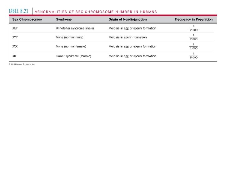
8. 21 Accidents during meiosis can alter chromosome number • Abnormal chromosome count is a result of nondisjunction – Either homologous pairs fail to separate during meiosis I Copyright © 2003 Pearson Education, Inc. publishing as Benjamin Cummings Nondisjunction in meiosis I Normal meiosis II Gametes n+1 n– 1 Number of chromosomes

– Or sister chromatids fail to separate during meiosis II Normal meiosis I Nondisjunction in meiosis II Gametes n+1 n– 1 n Number of chromosomes Copyright © 2003 Pearson Education, Inc. publishing as Benjamin Cummings n Figure 8. 21 B

• Fertilization after nondisjunction in the mother results in a zygote with an extra chromosome Egg cell n+1 Zygote 2 n + 1 Sperm cell n (normal) Figure 8. 21 C Copyright © 2003 Pearson Education, Inc. publishing as Benjamin Cummings

8. 23 Connection: Alterations of chromosome structure can cause birth defects and cancer • Chromosome breakage can lead to rearrangements that can produce genetic disorders or cancer – Four types of rearrangement are deletion, duplication, inversion, and translocation Copyright © 2003 Pearson Education, Inc. publishing as Benjamin Cummings

Deletion Inversion Duplication Reciprocal translocation Homologous chromosomes Nonhomologous chromosomes Copyright © 2003 Pearson Education, Inc. publishing as Benjamin Cummings
 2. Apoptosis (programmed cell death) Copyright © 2003 Pearson")
Cell death 1. Necrosis (괴사) 2. Apoptosis (programmed cell death) Copyright © 2003 Pearson Education, Inc. publishing as Benjamin Cummings
- Slides: 75