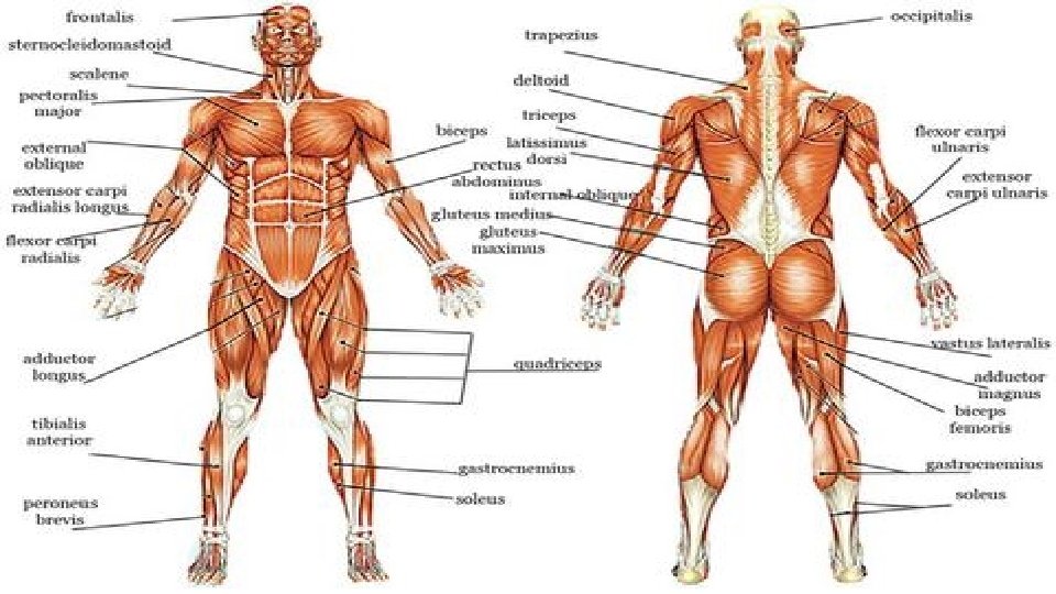
Chapter 8 Muscular System Section 1 Anatomy The


 have “heads” (-extensions, or")

 Muscle Fiber (cell) Myofibril Muscle Fiber (with many nuclei) Sarcomere Thin")

- Slides: 29
Chapter 8: Muscular System Section 1: Anatomy
The Muscular System • Muscles are responsible for all types of body movement Functions: • Producing movement • Maintaining posture • Stabilizing joints • Generating Heat
Characteristics of muscles • Muscle cells are elongated • muscle cell = muscle fiber • Contraction of muscles is due to the movement of microfilaments
Terminology of Muscles • All muscles share some terminology • Prefix myo refers to muscle • Prefix mys refers to muscle • Prefix sarco refers to flesh
3 types of muscles • Three basic muscle types are found in the body • Skeletal muscle • Cardiac muscle • Smooth muscle
1. • Most Skeletal Characteristics are. Muscle attached by tendons to bones • Cells are multinucleate • Striated – have visible banding • Voluntary – subject to conscious control • Cells are surrounded & bundled by connective tissue
2. Smooth Muscle Characteristics • Has no striations • Spindle-shaped cells • Single nucleus • Involuntary – no conscious control • Found mainly in the walls of hollow organs Figure 6. 2 a
3. Cardiac Muscle Characteristics • Has striations • Usually has a single nucleus • Joined to another muscle cell at an intercalated disc • Involuntary • Found only in the heart Figure 6. 2 b
Let’s look at skeletal muscles more closely….
Connective Tissue Wrappings of Skeletal Muscle • Endomysium – around single muscle fiber • Perimysium – around a fascicle (bundle) of fibers Figure 6. 1
CONNECTIVE TISSUE WRAPPINGS OF SKELETAL MUSCLE • Epimysium – covers the entire skeletal muscle • Fascia – on the outside of the epimysium Figure 6. 1
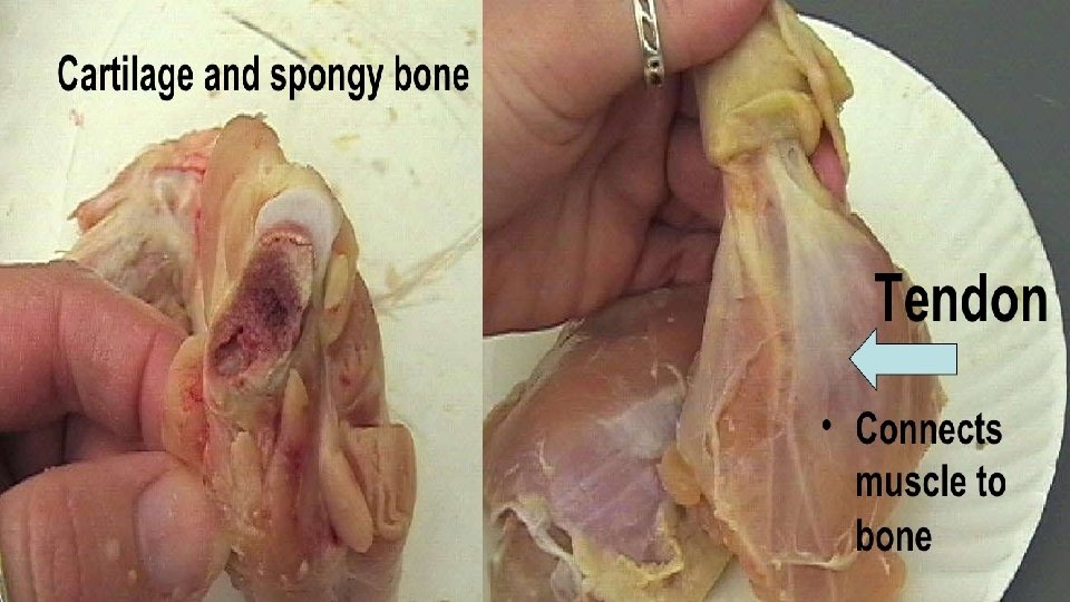
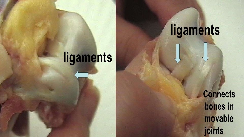
Skeletal Muscle Attachments • Epimysium blends into a connective tissue attachment • Tendon – cord-like structure • Aponeuroses – sheet-like structure • Sites of muscle attachment • Bones • Cartilages • Connective tissue coverings
Microscopic Anatomy of Skeletal Muscle • Cells are multinucleate Reminder* • Cell or fiber (same thing) Figure 6. 3 a
MICROSCOPIC ANATOMY OF SKELETAL MUSCLE Sarcolemma • specialized plasma membrane • Nuclei are just beneath the sarcolemma • Sarcoplasmic reticulum – specialized smooth endoplasmic reticulum for storage of calcium Figure 6. 3 a
Microscopic Anatomy of Skeletal Muscle • Myofibril • Bundles of myofilaments • Myofibrils are aligned to give distinct light and dark bands • I band = light band • A band = dark band Figure 6. 3 b
Microscopic Anatomy of Skeletal Muscle • Sarcomere • Contractile unit of a muscle fiber • Has thin and thick filaments Figure 6. 3 b
Anatomy • Microscopic Organization of the sarcomereof Skeletal Muscle • Thick filaments = myosin filaments **From Z line (disc) • Composed of the protein myosin to Z line (disc) • Has ATPase enzymes Figure 6. 3 c
MICROSCOPIC ANATOMY OF SKELETAL MUSCLE • Organization of the sarcomere • Thin filaments = actin filaments • Composed of the protein actin Figure 6. 3 c
Microscopic Anatomy of Skeletal Muscle • Myosin filaments (thick filaments) have “heads” (-extensions, or cross bridges) • Myosin and actin Thin & thick filaments overlap somewhat Figure 6. 3 d
MICROSCOPIC ANATOMY OF SKELETAL MUSCLE • At rest, there is a bare zone that lacks actin filaments Figure 6. 3 d
Fascicle Muscle (organ) Muscle Fiber (cell) Myofibril Muscle Fiber (with many nuclei) Sarcomere Thin Filament (actin) Thick Filament (myosin)
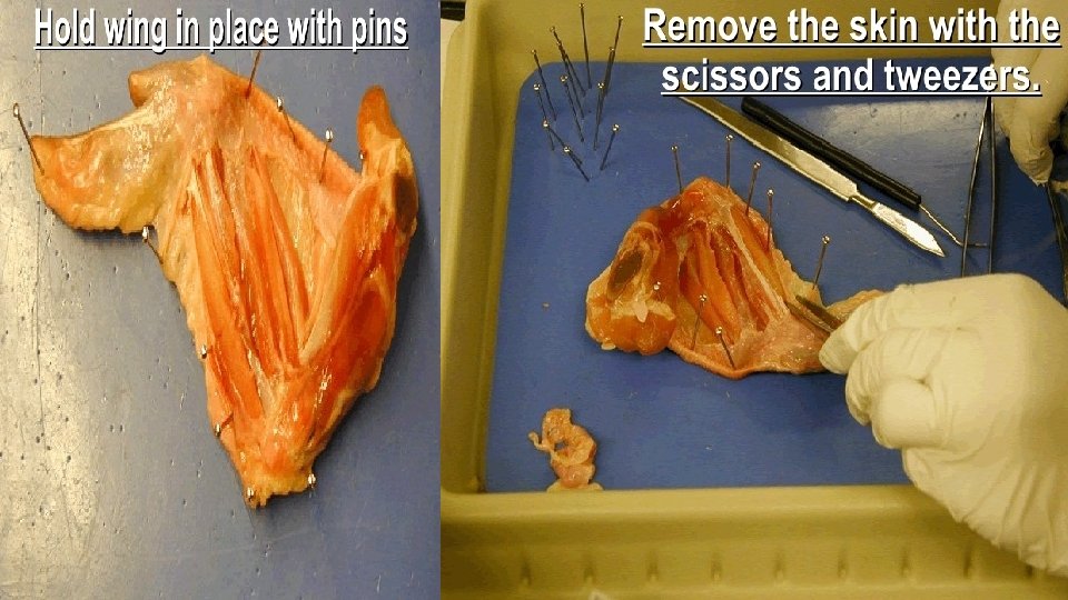
• Be prepared for some dissections!!!!