Chapter 8 Joints of the Skeletal System Articulations


 Ant.")


 • Most common chronic arthritis; often called “wear-and-tear” arthritis • Affects women")


 • Chronic, inflammatory, autoimmune disease of unknown cause, with an insidious")


- Slides: 54
Chapter 8 Joints of the Skeletal System • Articulations • Junctions between bones • Bind parts of skeletal system together • Make bone growth possible • Permit parts of the skeleton to change shape during childbirth • Enable body to move in response to skeletal muscle contraction
Joints = Articulations Articulation – site where two or more bones meet Two Fundamental Functions of Joints: Allow the skeleton to have mobility Hold the skeleton together
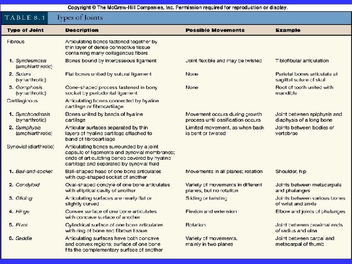
Joints – Structural and Functional Classes Three Structural Classifications: • Fibrous – suture, syndesomosis, gomphosis • Cartilaginous – synchondrosis, symphysis • Synovial Three Functional Classifications • Synarthrosis – immovable • Amphiarthrosis – slightly movable • Diarthrosis – freely movable
Classification of Joints • Fibrous Joints • dense connective tissues connect bones • between bones in close contact • Cartilaginous Joints • hyaline cartilage or fibrocartilage connect bones • Synovial Joints • most complex • allow free movement • synarthrotic • immovable • amphiarthrotic • slightly movable • diarthrotic • freely movable
08_02 a. jpg
08_02 b. jpg
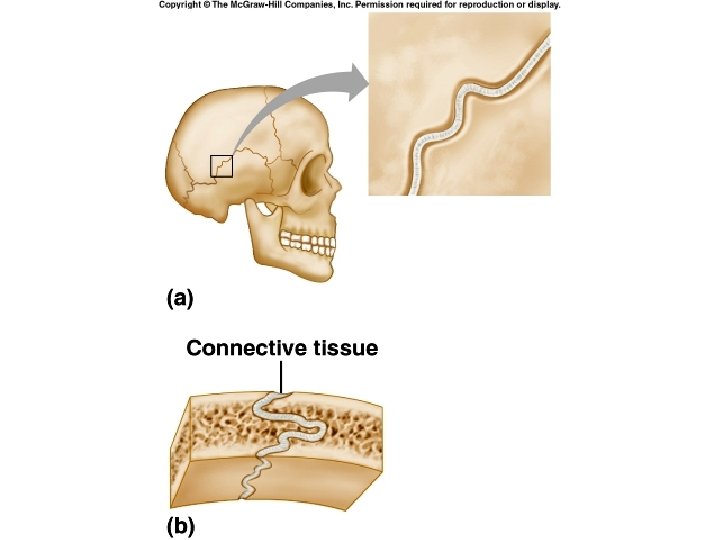
Fibrous Joints 3 Types • Syndesmosis • Suture • Gomphosis Syndesmosis • long fibers connect bones • amphiarthrotic • distal ends of tibia and fibula
Fibrous Joints Suture • between flat bones • synarthrotic • thin layer of connective tissue connects bones Gomphosis • cone-shaped bony process in a socket • tooth in jawbone • synarthrotic
Cartilaginous Joints 2 Types • Synchondrosis • Symphysis Synchondrosis • bands of hyaline cartilage unite bones • epiphyseal plate (temporary) • between manubrium and first rib • synarthrotic
Cartilaginous Joints Symphysis • pad of fibrocartilage between bones • pubis symphysis • joint between bodies of vertebrae • amphiarthrotic
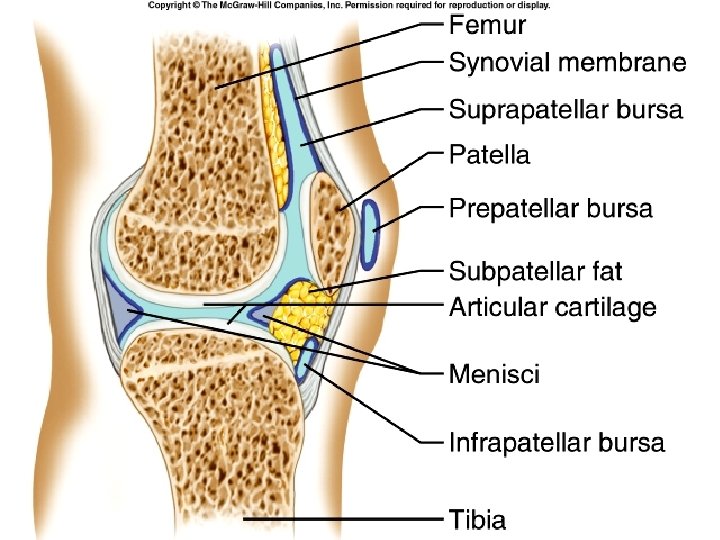
Synovial Joints • diarthrotic • joint cavity • synovial fluid • joint capsule • synovial membrane • bursae
Types of Synovial Joints Ball-and-Socket Joint • hip • shoulder Condyloid Joint • between metacarpals and phalanges
Types of Synovial Joints Gliding Joint • between carpals • between tarsals Hinge Joint • elbow • between phalanges
Types of Synovial Joints Pivot Joint • between proximal ends of radius and ulna Saddle Joint • between carpal and metacarpal of thumb
Angular Movement – Change of Angle Between Bones Flexion — bending movement that decreases the angle of the joint Extension — reverse of flexion; joint angle increases Dorsiflexion and Plantar flexion — up and down movement of the foot Abduction — movement of a limb away from the midline or median plane Adduction — movement of a limb toward the midline or median plane Circumduction — movement of a limb describing a cone in space
Types of Joint Movements • abduction/adduction • dorsiflexion/plantarflexion • flexion/extension/hyperextension
Rotation The turning of a bone around its own long axis Examples: Between first two vertebrae Hip and shoulder joints
Types of Joint Movements • rotation/circumduction • supination/pronation
Special Movements Supination and Pronation – refer to movements of radius around the ulna (also applied to foot movements)
Types of Joint Movements • eversion/inversion • protraction/retraction • elevation/depression
Special Movements Inversion and Eversion Protraction and Retraction
Special Movements Elevation and Depression Opposition
Shoulder Joint • ball-and-socket • head of humerus • glenoid cavity of scapula • loose joint capsule • bursae • ligaments prevent displacement • very wide range of movement
Shoulder Joint
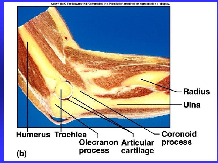
Elbow Joint • hinge joint • trochlea of humerus • trochlear notch of ulna • gliding joint • capitulum of humerus • head of radius • flexion and extension • many reinforcing ligaments • stable joint
Elbow Joint
Hip Joint • ball-and-socket joint • head of femur • acetabulum • heavy joint capsule • many reinforcing ligaments • less freedom of movement than shoulder joint
Hip Joint
Knee Joint • largest joint • most complex • medial and lateral condyles of distal end of femur • medial and lateral condyles of proximal end of tibia • femur articulates anteriorly with patella • modified hinge joint • flexion/extension/little rotation • strengthened by many ligaments and tendons • menisci separate femur and tibia • bursae
Knee Joint
Life-Span Changes • Joint stiffness is an early sign of aging • Regular exercise can prevent stiffness • Fibrous joints first to strengthen over a lifetime • Changes in symphysis joints of vertebral column diminish flexibility and decrease height • Synovial joints lose elasticity
Clinical Application Joint Disorders Sprains • damage to cartilage, ligaments, or tendons associated with joints • forceful twisting of joint Bursitis • inflammation of a bursa • overuse of a joint Arthritis • inflamed, swollen, painful joints • Rheumatoid Arthritis • Osteoarthritis • Gout
Joint Injuries – Sprains & Cartilage Injury Sprain - the ligaments in a joint are stretched or torn. Partially torn ligaments may repair themselves, but healing is slow due to lack of vascularization. Completely torn ligaments require surgical repair. Cartilage is mostly avascular and largely unable to repair itself when torn. Most cartilage injuries involve tearing of the menisci.
Dislocations - Luxation Occur when bones are forced out of alignment Usually accompanied by sprains, inflammation, and joint immobilization Subluxation – partial dislocation of a joint
Inflammatory Conditions Bursitis Inflammation of a bursa, usually caused by a blow or friction. Symptoms are pain and swelling. Treated with anti-inflammatory drugs; excessive fluid may be aspirated. Tendonitis Inflammation of tendon sheaths. Symptoms and treatment are similar to bursitis.
Ligament and Cartilage Tears: Example of the Knee Joint
Knee Ligaments and Tendons – Anterior View Tendon of the Quadriceps Femoris Lateral and Medial Patellar Retinacula Fibular and Tibial Collateral Ligaments Patellar Ligament
Knee Ligaments and other Supporting Structures Intracapsular Ligaments (but outside of synovial cavity) Ant. Cruciate Ligament Post. Cruciate Ligament Semilunar Cartilages Medial Meniscus Lateral meniscus
Knee Ligaments and other Supporting Structures Adductor Magnus Tendon Articular Capsule Oblique Popliteal Ligament Arcuate Popliteal Ligament Semimembranosus Tendon
Knee Injury
Arthritis • More than 100 different types of inflammatory or degenerative diseases that damage the joints • Most widespread crippling disease in the U. S. (1 out of every 7 people) • Symptoms – pain, stiffness, and swelling of a joint • Acute forms are caused by bacteria and are treated with antibiotics • Chronic forms include osteoarthritis, rheumatoid arthritis, and gouty arthritis
Arthritis: Causes and Symptoms
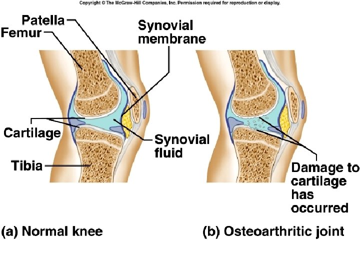
Osteoarthritis (OA) • Most common chronic arthritis; often called “wear-and-tear” arthritis • Affects women more than men • 85% of all Americans develop OA • More prevalent in the aged, and is probably related to the normal aging process
Osteoarthritis: Course • OA reflects the years of abrasion and compression causing increased production of metalloproteinase enzymes that break down cartilage • As one ages, cartilage is destroyed more quickly than it is replaced • The exposed bone ends thicken, enlarge, form bone spurs, and restrict movement • Crepitus – crunching noise as roughened articular surfaces rub together • Joints most affected are the cervical and lumbar spine, fingers, knuckles, knees, and hips
Osteoarthritis: Treatments • OA is usually slow and irreversible • Treatments include: – Mild pain relievers, along with moderate activity – Magnetic therapy? – Glucosamine sulfate? said to decrease pain and inflammation
Rheumatoid Arthritis (RA) • Chronic, inflammatory, autoimmune disease of unknown cause, with an insidious onset • Usually arises between the ages of 40 to 50, but may occur at any age • Signs and symptoms include joint tenderness, anemia, osteoporosis, muscle atrophy, and cardiovascular problems – The course of RA is marked with exacerbations and remissions
Rheumatoid Arthritis: Course • RA begins with synovitis of the affected joint • Inflammatory chemicals are inappropriately released • Inflammatory blood cells migrate to the joint, causing swelling • Inflamed synovial membrane thickens into a pannus • Pannus erodes cartilage, scar tissue forms, articulating bone ends fuse • The end result, ankylosis, produces bent, deformed fingers
Rheumatoid Arthritis: Treatment • Conservative therapy – aspirin, long-term use of antibiotics, and physical therapy • Progressive treatment – anti-inflammatory drugs or immunosuppressants • The drug Enbrel, a biological response modifier, neutralizes the harmful properties of inflammatory chemicals
Gouty Arthritis • Deposition of uric acid crystals in joints and soft tissues, followed by an inflammatory response • Typically, gouty arthritis affects the joint at the base of the great toe • In untreated gouty arthritis, the bone ends fuse and immobilize the joint • Treatment – colchicine, nonsteroidal antiinflammatory drugs, and glucocorticoids