
Chapter 8 Cell Reproduction AP Biology Exam Review
Chapter 8: Cell Reproduction AP Biology Exam Review
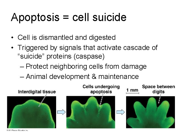
Apoptosis = cell suicide • Cell is dismantled and digested • Triggered by signals that activate cascade of “suicide” proteins (caspase) – Protect neighboring cells from damage – Animal development & maintenance
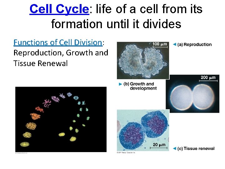
Cell Cycle: life of a cell from its formation until it divides Functions of Cell Division: Division Reproduction, Growth and Tissue Renewal
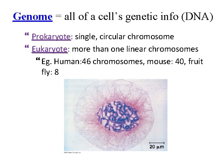
Genome = all of a cell’s genetic info (DNA) Prokaryote: Prokaryote single, circular chromosome Eukaryote: Eukaryote more than one linear chromosomes Eg. Human: 46 chromosomes, mouse: 40, fruit fly: 8
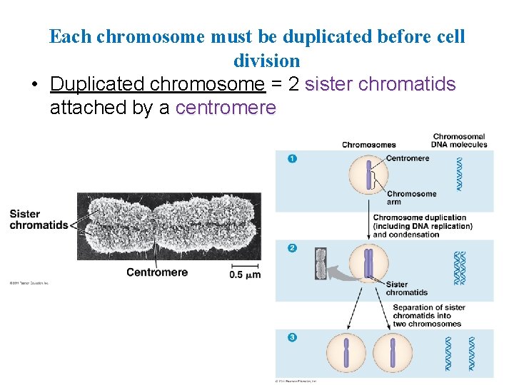
Each chromosome must be duplicated before cell division • Duplicated chromosome = 2 sister chromatids attached by a centromere
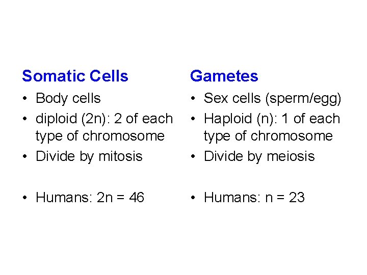
Somatic Cells Gametes • Body cells • diploid (2 n): 2 of each type of chromosome • Divide by mitosis • Sex cells (sperm/egg) • Haploid (n): 1 of each type of chromosome • Divide by meiosis • Humans: 2 n = 46 • Humans: n = 23
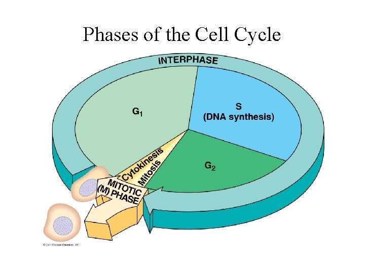
Phases of the Cell Cycle
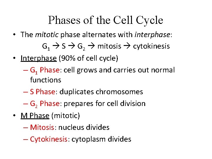
Phases of the Cell Cycle • The mitotic phase alternates with interphase: G 1 S G 2 mitosis cytokinesis • Interphase (90% of cell cycle) – G 1 Phase: cell grows and carries out normal functions – S Phase: duplicates chromosomes – G 2 Phase: prepares for cell division • M Phase (mitotic) – Mitosis: nucleus divides – Cytokinesis: cytoplasm divides
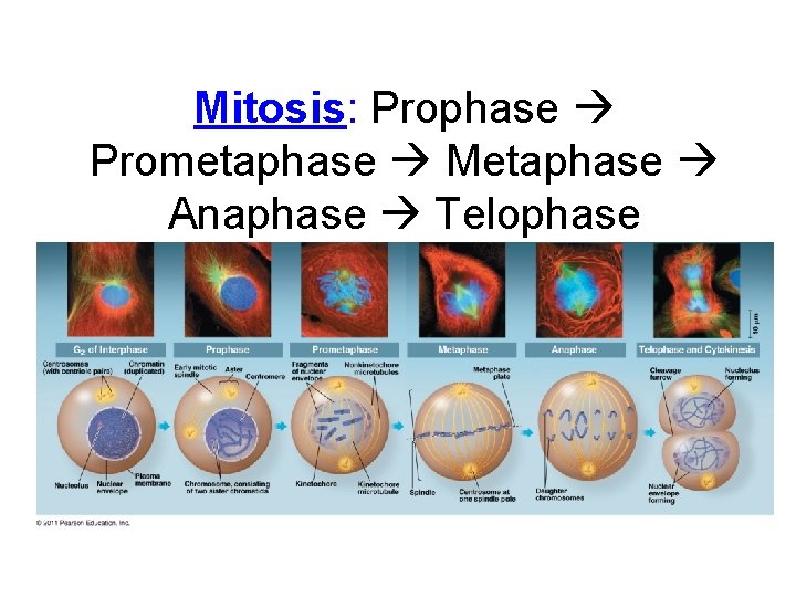
Mitosis: Prophase Prometaphase Metaphase Anaphase Telophase
Mitosis 1. Prophase – Chromatin fibers condense and coil – Nucleoli disappear – Spindle (microtubules) begins to form – Centrosomes begin to move to opposite ends 2. Prometaphase – Nuclear envelope fragments – Microtubules invade nucleus – Kinetochores attach to microtubules
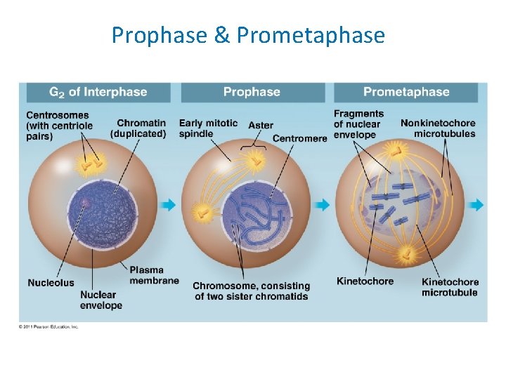
Prophase & Prometaphase
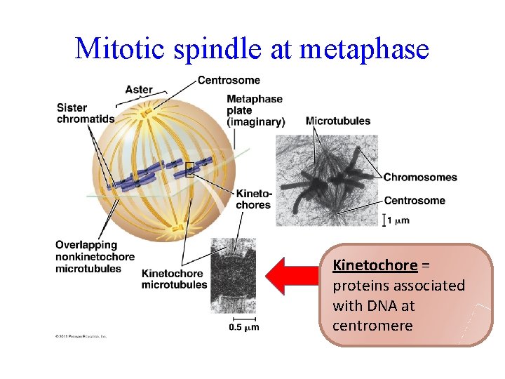
Mitotic spindle at metaphase Kinetochore = proteins associated with DNA at centromere
3. Metaphase – Chromosomes line up on metaphase plate at equator – Centrioles are at opposite poles (ends) 4. Anaphase (shortest phase) – Chromatids separate and pulled apart by motor proteins toward opposite ends of cell – Chromatids are called chromosomes now – Cell elongates
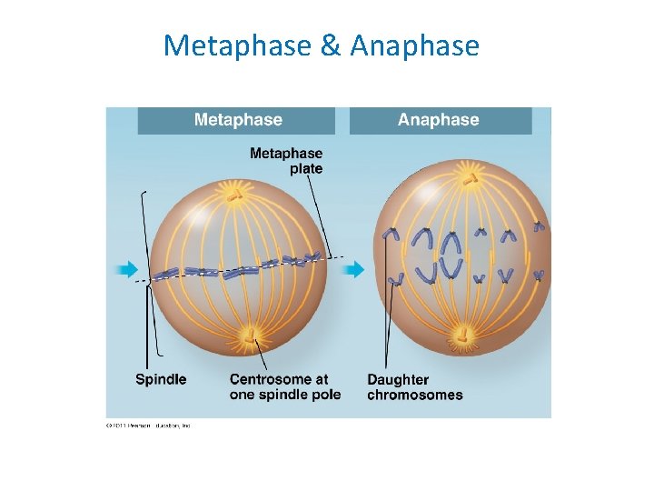
Metaphase & Anaphase
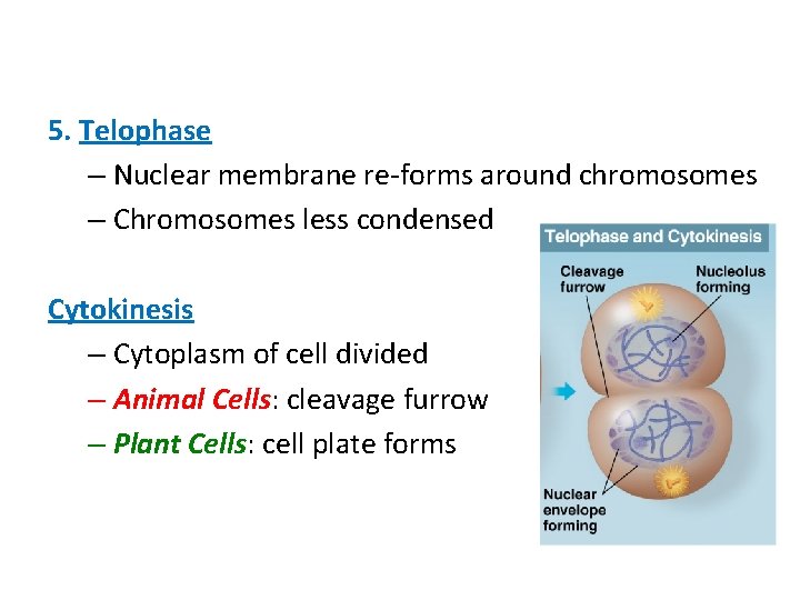
5. Telophase – Nuclear membrane re-forms around chromosomes – Chromosomes less condensed Cytokinesis – Cytoplasm of cell divided – Animal Cells: cleavage furrow – Plant Cells: cell plate forms
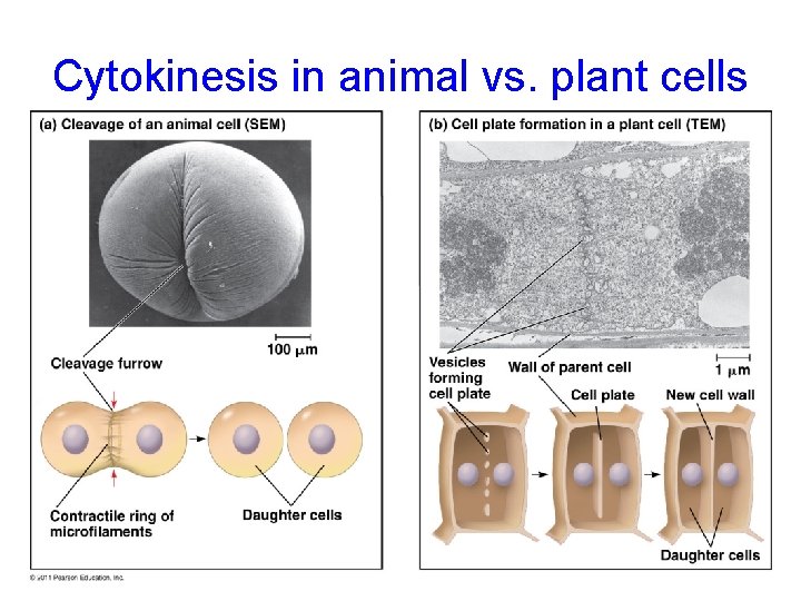
Cytokinesis in animal vs. plant cells
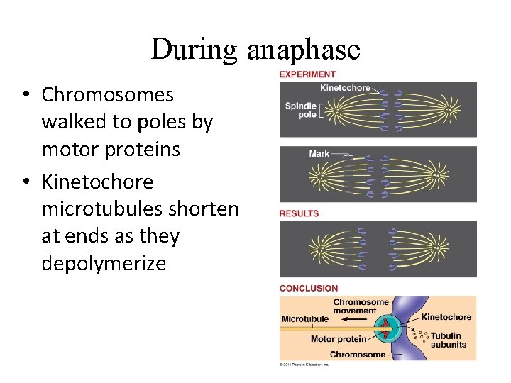
During anaphase • Chromosomes walked to poles by motor proteins • Kinetochore microtubules shorten at ends as they depolymerize
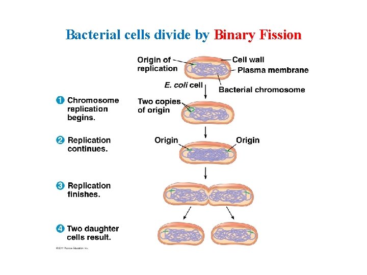
Bacterial cells divide by Binary Fission
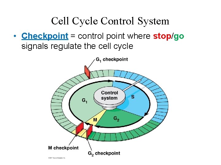
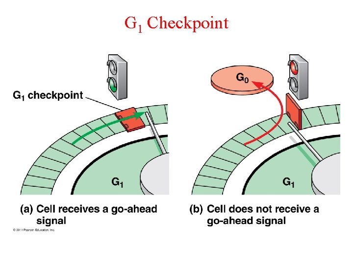
Cell Cycle Control System • Checkpoint = control point where stop/go signals regulate the cell cycle
Major Checkpoints 1. G 1 checkpoint (Most important!) – “Go” completes whole cell cycle – “Stop” cell enters nondividing state (G 0 Phase) • 2. 3. – Nerve, muscle cells stay at G 0; liver cells called back from G 0 G 2 checkpoint M Phase checkpoint Anaphase does not begin unless chromatids are properly attached to spindle at metaphase plate
G 1 Checkpoint
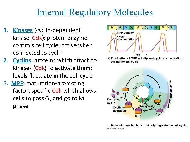
Internal Regulatory Molecules 1. Kinases (cyclin-dependent kinase, Cdk): protein enzyme controls cell cycle; active when connected to cyclin 2. Cyclins: proteins which attach to kinases (Cdk) to activate them; levels fluctuate in the cell cycle 3. MPF: maturation-promoting factor; specific Cdk which allows cells to pass G 2 and go to M phase
External Regulatory Factors
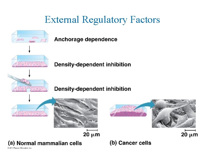
External Regulatory Factors Growth Factor: proteins released by other cells to stimulate cell division Density-Dependent Inhibition: crowded cells normally stop dividing; cell-surface protein binds to adjoining cell to inhibit growth Anchorage Dependence: cells must be attached to another cell or ECM to divide
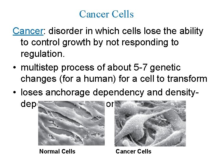
Cancer Cells Cancer: disorder in which cells lose the ability to control growth by not responding to regulation. • multistep process of about 5 -7 genetic changes (for a human) for a cell to transform • loses anchorage dependency and densitydependency regulation Normal Cells Cancer Cells
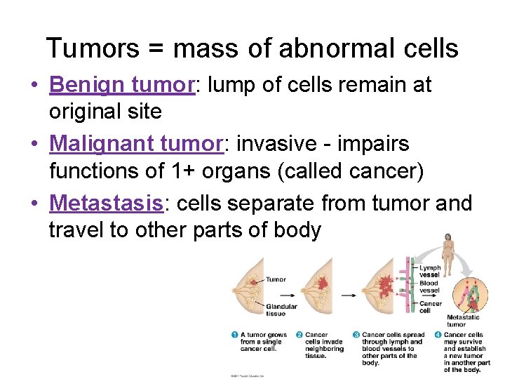
Tumors = mass of abnormal cells • Benign tumor: lump of cells remain at original site • Malignant tumor: invasive - impairs functions of 1+ organs (called cancer) • Metastasis: cells separate from tumor and travel to other parts of body
Types of Reproduction ASEXUAL • Produces clones (genetically identical) • Single parent • Little variation in population - only through mutations • Fast and energy efficient • Eg. budding, binary fission SEXUAL • Meiosis produces gametes (sex cells) • 2 parents: male/female • Lots of variation/diversity • Slower and energy consumptive • Eg. humans, trees
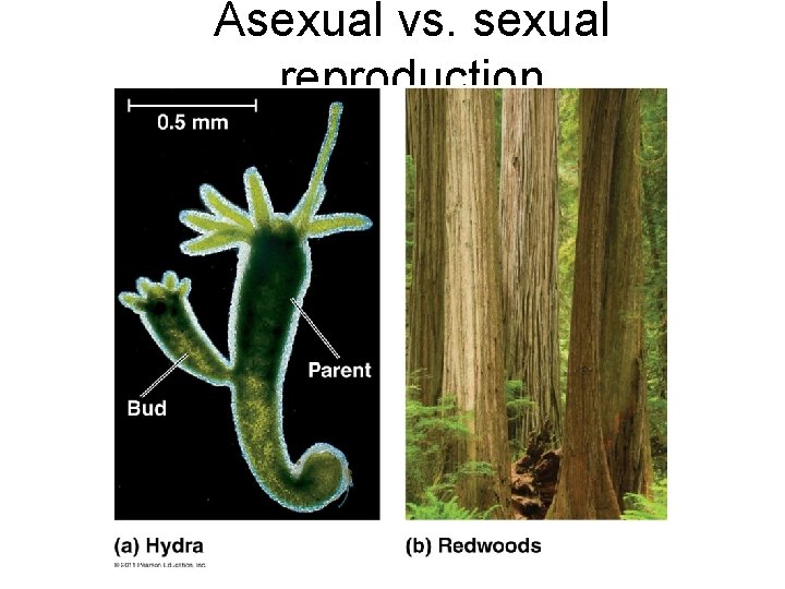
Asexual vs. sexual reproduction
Chromosomes • Somatic (body) cell: 2 n = 46 chromosomes • Each pair of homologous chromosomes includes 1 chromosome from each parent • Autosomes: 22 pairs of chromosomes that do not determine sex • Sex chromosomes: X and Y • Females: XX • Males: XY • Gametes (n=23): 22 autosomes + 1 sex chromosome • Egg: 22 + X • Sperm: 22 + X **or** 22 + Y
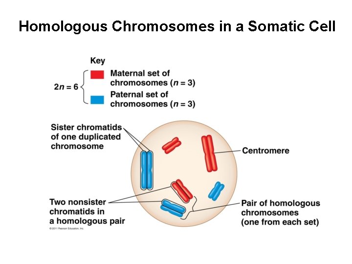
Homologous Chromosomes in a Somatic Cell
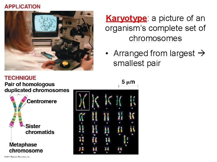
Karyotype: a picture of an organism’s complete set of chromosomes • Arranged from largest smallest pair
Life cycle: reproductive history of organism, from conception production of own offspring • Fertilization and meiosis alternate in sexual life cycles • Meiosis: cell division that reduces # of chromosomes (2 n n), creates gametes • Fertilization: combine gametes (sperm + egg) – Fertilized egg = zygote (2 n) • Zygote divides by mitosis to make multicellular diploid organism
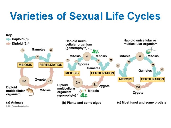
Varieties of Sexual Life Cycles
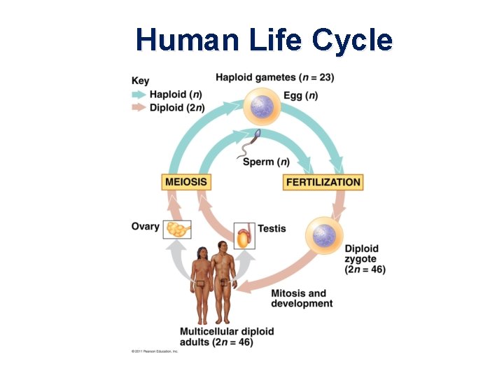
Human Life Cycle
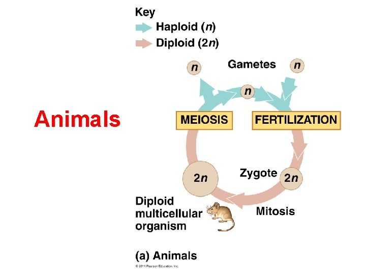
Animals
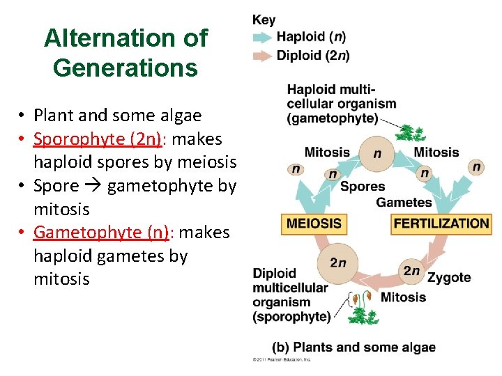
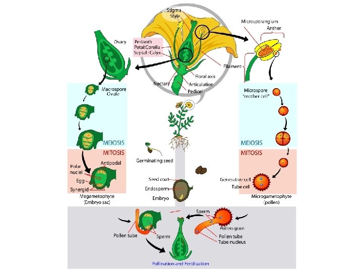
Alternation of Generations • Plant and some algae • Sporophyte (2 n): makes haploid spores by meiosis • Spore gametophyte by mitosis • Gametophyte (n): makes haploid gametes by mitosis
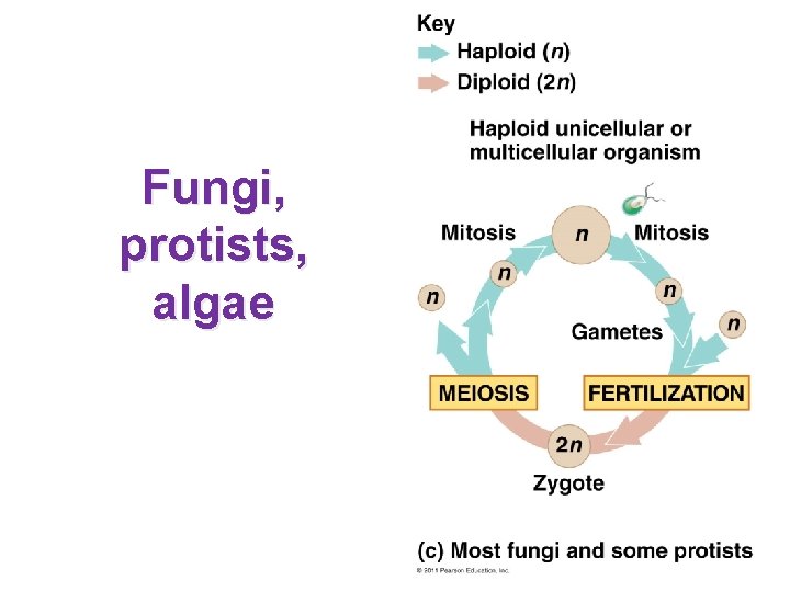
Fungi, protists, algae
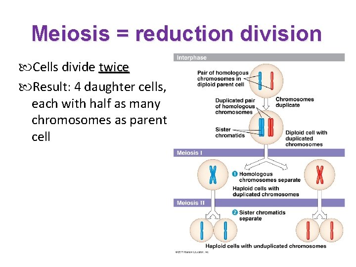
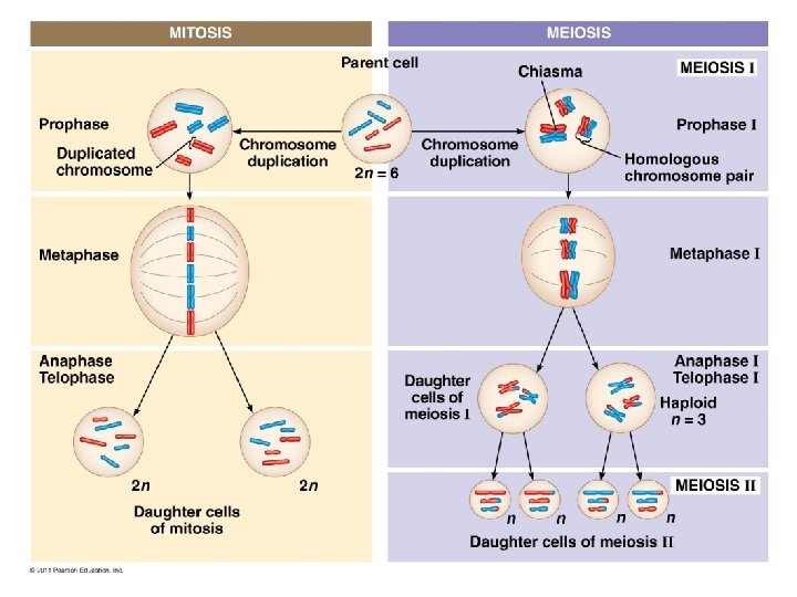
Meiosis = reduction division Cells divide twice Result: 4 daughter cells, each with half as many chromosomes as parent cell
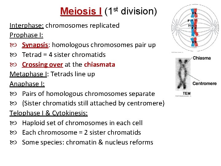
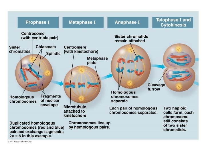
Meiosis I (1 st division) Interphase: chromosomes replicated Prophase I: Synapsis: homologous chromosomes pair up Tetrad = 4 sister chromatids Crossing over at the chiasmata Metaphase I: Tetrads line up Anaphase I: Pairs of homologous chromosomes separate (Sister chromatids still attached by centromere) Telophase I & Cytokinesis: Haploid set of chromosomes in each cell Each chromosome = 2 sister chromatids Some species: chromatin & nucleus reforms
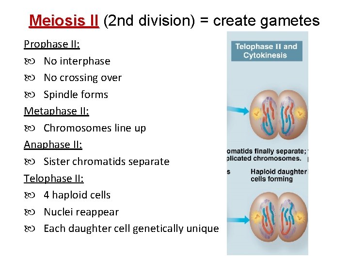
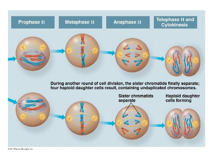
Meiosis II (2 nd division) = create gametes Prophase II: No interphase No crossing over Spindle forms Metaphase II: Chromosomes line up Anaphase II: Sister chromatids separate Telophase II: 4 haploid cells Nuclei reappear Each daughter cell genetically unique
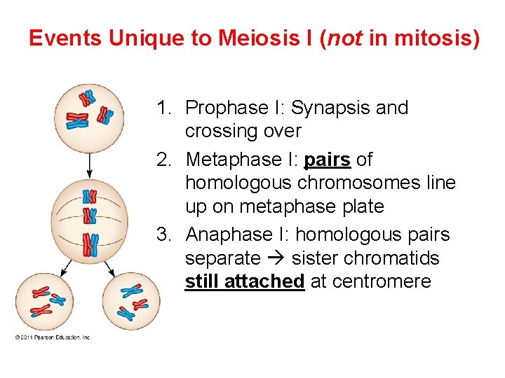
Events Unique to Meiosis I (not in mitosis) 1. Prophase I: Synapsis and crossing over 2. Metaphase I: pairs of homologous chromosomes line up on metaphase plate 3. Anaphase I: homologous pairs separate sister chromatids still attached at centromere
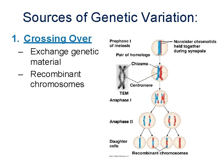
Sources of Genetic Variation: 1. Crossing Over – Exchange genetic material – Recombinant chromosomes
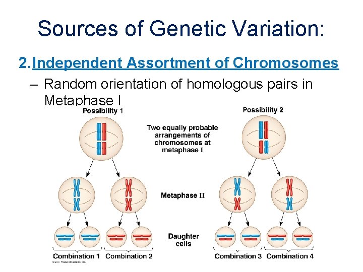
Sources of Genetic Variation: 2. Independent Assortment of Chromosomes – Random orientation of homologous pairs in Metaphase I
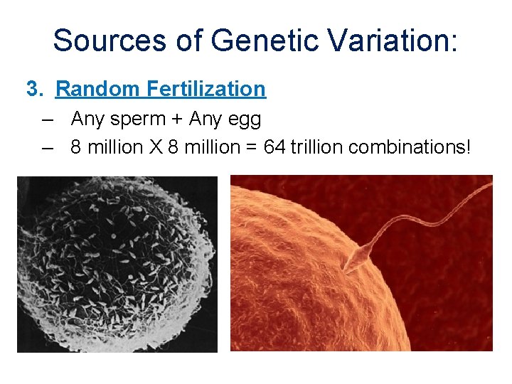
Sources of Genetic Variation: 3. Random Fertilization – Any sperm + Any egg – 8 million X 8 million = 64 trillion combinations!
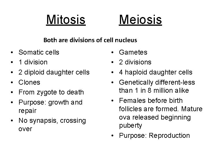
Mitosis Meiosis Both are divisions of cell nucleus • • • Somatic cells 1 division 2 diploid daughter cells Clones From zygote to death Purpose: growth and repair • No synapsis, crossing over • • Gametes 2 divisions 4 haploid daughter cells Genetically different-less than 1 in 8 million alike • Females before birth follicles are formed. Mature ova released beginning puberty • Purpose: Reproduction
- Slides: 49