Chapter 8 Cell Division Mitosis and Meiosis Asexual


- Slides: 54
Chapter 8 Cell Division : Mitosis and Meiosis
Asexual vs. Sexual reproduction �Asexual reproduction – new organisms/cells are genetically identical to parent cells/organisms �Sexual reproduction – offspring have a combination of genes from both parents.
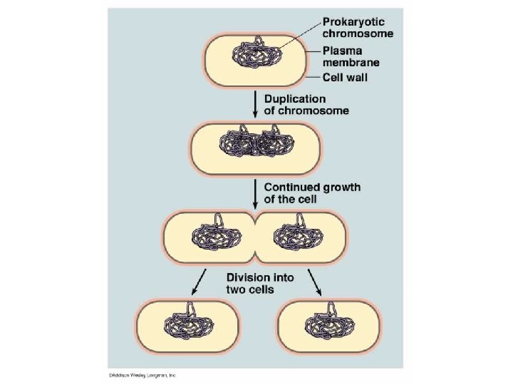
Asexual Reproduction �Budding - plants �Vegetative propagation -plants �Binary fission -bacteria �One cell dividing to become two –mitosis �Hermaphroditic organisms are NOT asexual!!!
Cells only come from other cells �To make more cells they must divide Mitosis – one cell divides to make two genetically identical cells for asexual reproduction and growth and repair. Meiosis – One cell divides twice to create 4 cells that are not genetically identical. These cells are eggs or sperm (gametes)
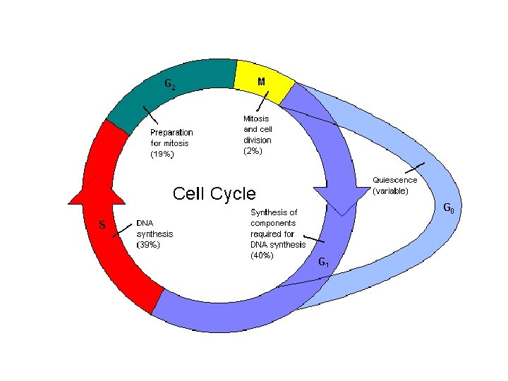
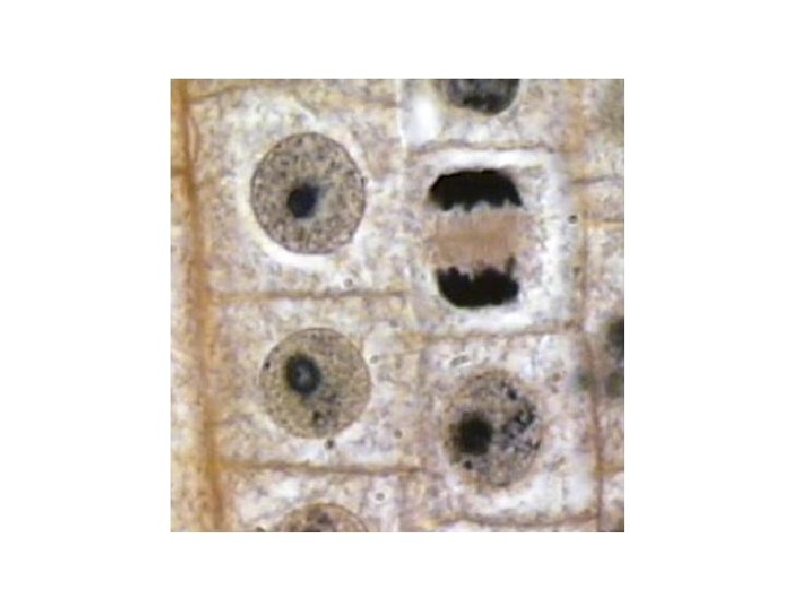
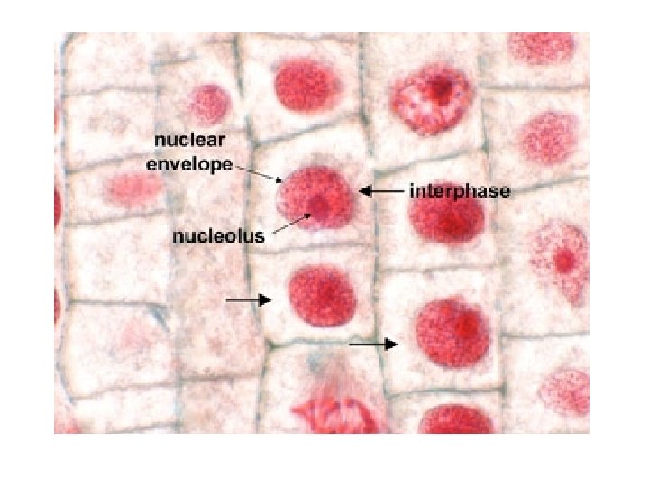
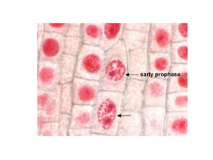
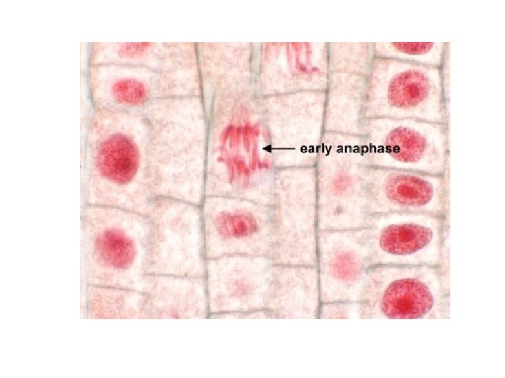
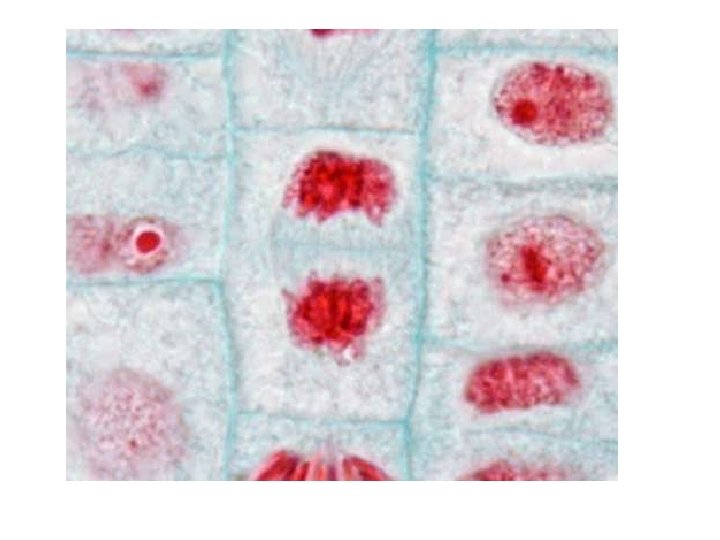
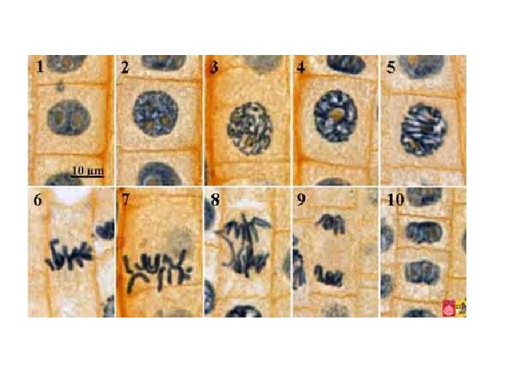
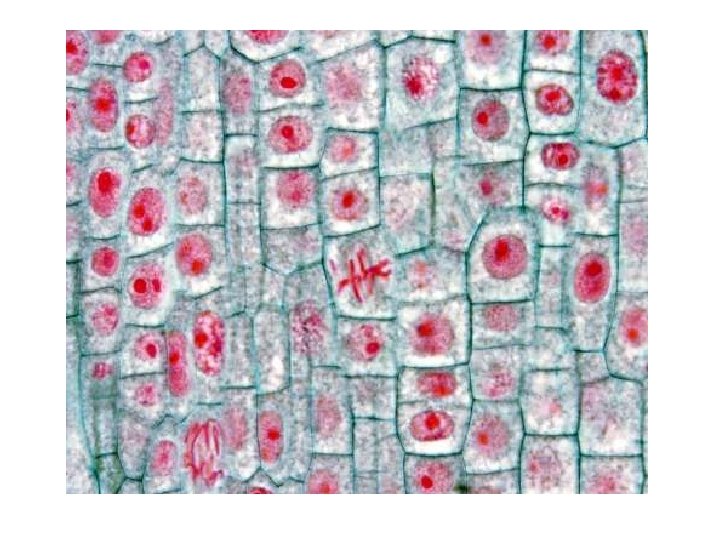
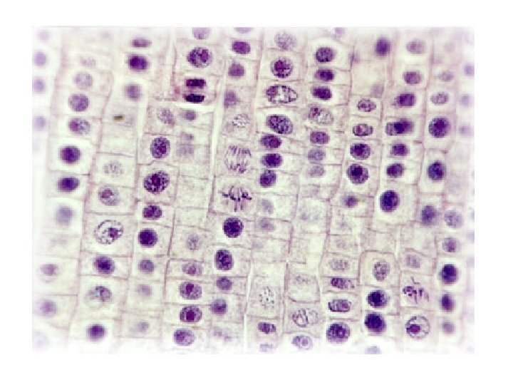
Cell Cycle �Interphase – cell does normal job, grows, and duplicates genetic material to prep for division, 90% of cell cycle G 1, S, G 2 �Mitosis – division of genetic material �Cytokinesis- division of cytoplasm, usually occurs after mitosis
Cell Cycle �Interphase G 1 - first gap, growth, normal function, makes proteins etc S phase- synthesis, cell copies DNA to prepare for division G 2 - second gap, growth and final preparation for division
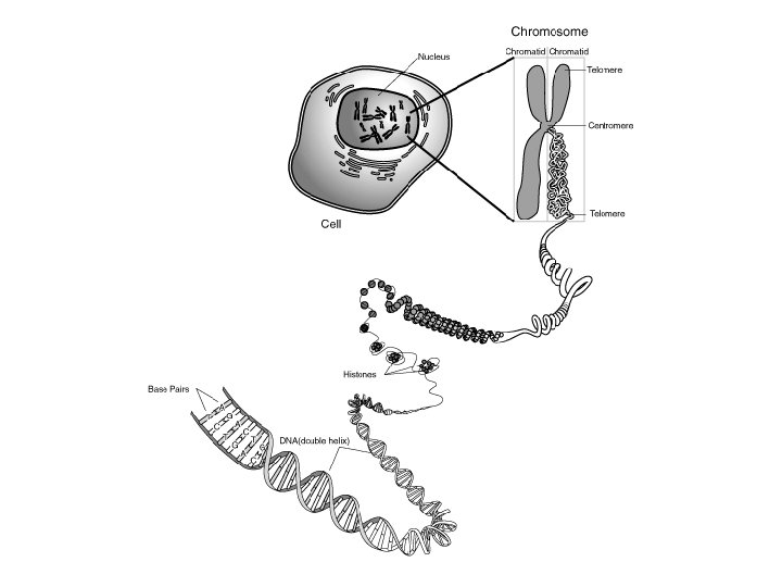
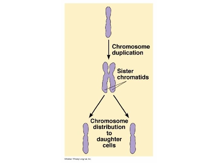
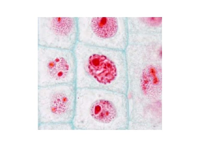
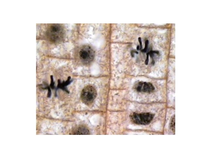
Eukaryotic cells have complex genomes �DNA in a non-dividing cell is disorganized �Called chromatin �When a cell prepares to divide the chromatin (DNA and proteins called histones) coil into chromosomes �Each chromosome is made of two identical halves called sister chromatids �Sister chromatids are connected by a centromere
Cytokinesis �In animal cells a cleavage furrow develops. The cell pinches from the outside �In plant cells a cell plate forms. A new cell wall develops from the inside and works out to the borders
Factors that affect cell division �Must be attached to a surface �Will stop dividing when they touch each other - density dependent inhibition �Secretion of proteins called growth factors �Three key checkpoints in the cell cycle G 1 G 2 M-
Cancer �Do not respond normally to checkpoints in the cell cycle �Excessive cell division, wasting of cellular resources, form masses called tumors Benign- stays in original location Malignant- spreads from original location- metastasis
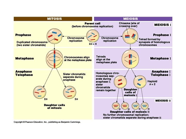
Mitosis Summary 8. 11 �Occurs in somatic or non-sex cells �Creates two genetically identical cells from one cell �The cells created are diploid (2 n)– having a full set of chromosomes. �Used for repair and growth
Human life cycle �Diploid cells in ovaries and testes divide by meiosis to create haploid gametes. �Gametes are egg and sperm. �Haploid cells have a half set of chromosomes �Haploid gametes combine to form a diploid zygote �Diploid zygotes divide by mitosis to form Multicellular organisms
Homologous chromosomes 8. 12 �Humans have 22 pairs of autosomes – non- sex chromosomes and one pair of sex chromosomes �Females have two X chromosomes �Males have an X and a Y chromosome
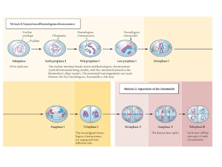
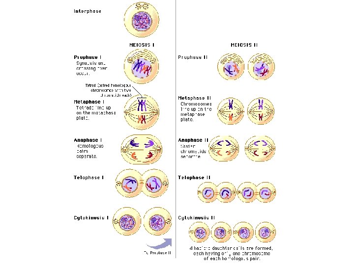
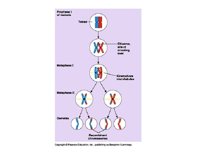
Meiosis- steps �Two divisions �Meiosis I; meiosis II �Major differences Prophase I- homologous chromosomes pair into a tetrad. Sometimes the homologous pairs exchange small pieces – crossing over Synapsis – the exchange of pieces
Meiosis increases genetic variation � 1. Crossing over – creates chromosomes that are mosaics of both maternal and paternal genes. �Is a random event and doesn’t happen for every meiotic cycle �Occurs in prophase I �Called genetic recombination
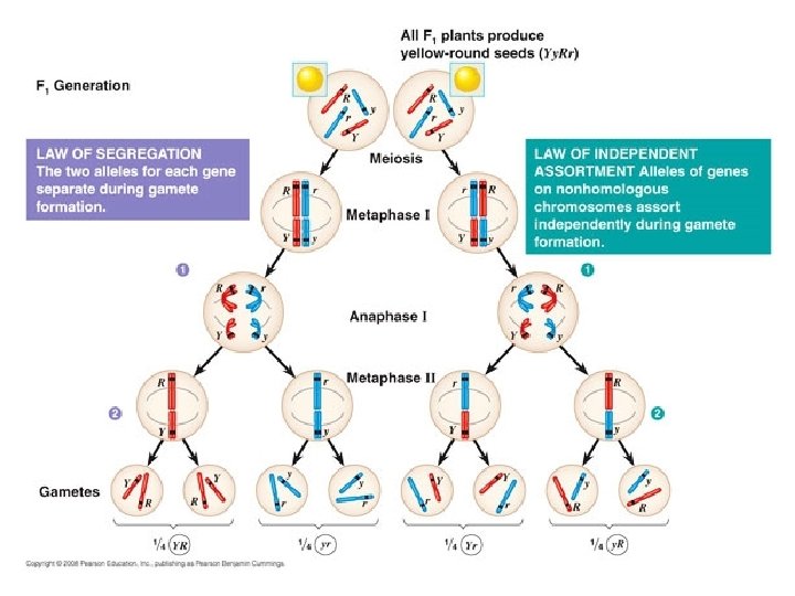
Meiosis- increases genetic variation 2. Law Of Segregation Each haploid cell inherits only one chromosome from each parent. Homologous chromosomes carry genes for the same trait but not necessarily the same gene – Law of Segregation. The physical process that underlies this law occurs in Anaphase I
Genetic variation � 3. Law of Independent assortment �Each homologous chromosome pair lines up side by side and separates randomly in metaphase I. �Creates many different random combinations of chromosomes in each egg or sperm �Different possibilities = 2 to the n power, where n= the haploid number
Genetic variation � 4. Random fertilization increases genetic variability in a species �Why is variation needed? �Organisms with very similar genomes have no raw material for natural selection should the environment abruptly change
Genes �Are carried on chromosomes �Each trait in your body is determined by at least two genes on two different homologous chromosomes – one from dad, and one from mom
Mistakes occur in meiosis �Crossing over �Separation in anaphase I and/or anaphase II �Nonreciprocal crossovers- exchange of pieces of DNA of different sizes �Inversion- pieces of chromosomes are reattached incorrectly �Non homologous crossovers �Failure to separate – nondisjunction
Crossing over mistakes �Chromosomes missing parts due to non reciprocal cross overs have deletions �Chromosomes with too much info have duplications. �Fragments reattached in the wrong sequence are inversions �Translocations occur when non-homologous chromosomes cross-over
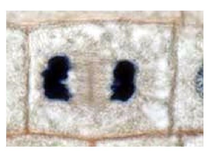
Disorders caused by CO mistakes �Cri du chat- deletion on # 5, “cry of the cat” in babies �Down Syndrome can be caused when #21 attaches to another chromosomes �Chronic myelogenous Leukemia (CML)- non homologous cross over activates a cancer gene. Call it the “Philadelphia chromosome”
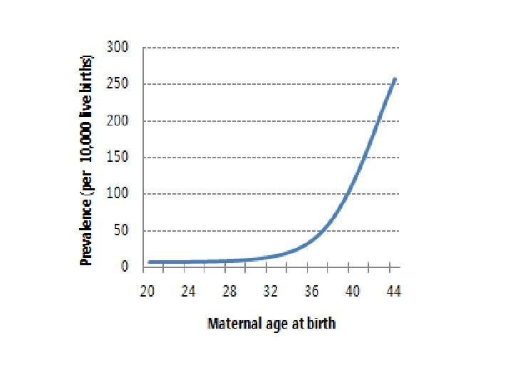
Nondisjunction �Can occur in anaphase I or anaphase II �Results in organisms with the wrong number of chromosomes for their species – aneuploid individuals �Most situations with missing or extra chromosomes lead to spontaneous miscarriage
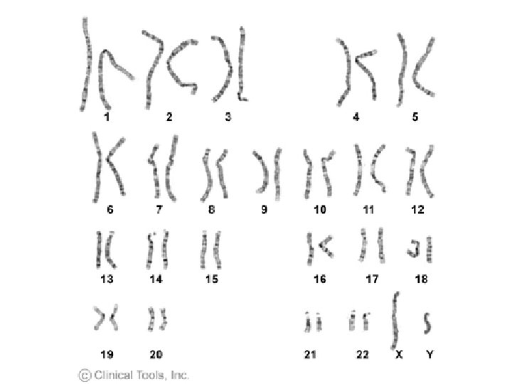
Extra Autosomes �Trisomy #21 – Down’s Syndrome Trisomy- # 18 - Edward’s Syndrome Trisomy #13 - Patau’s Syndrome
Extra or missing sex chromosomes �Individual with only one X- Turner Syndrome, female, sterile, can have other physical traits XO �Individual with two X chromosomes and one Y, - Klinefelter’s Syndrome male, sterile, some female characteristics, taller than normal XXY
Prenatal diagnosis of defects �Amniocentesis – performed at 14 -20 weeks, a needle is inserts into the uterus to extract amniotic fluid which contains fetal cells. Cells are cultured for a few weeks �Chorionic villi sampling- placental tissue is removed and cultured within 24 hours, can be performed at the 8 th week �Both carry a small risk of miscarriage
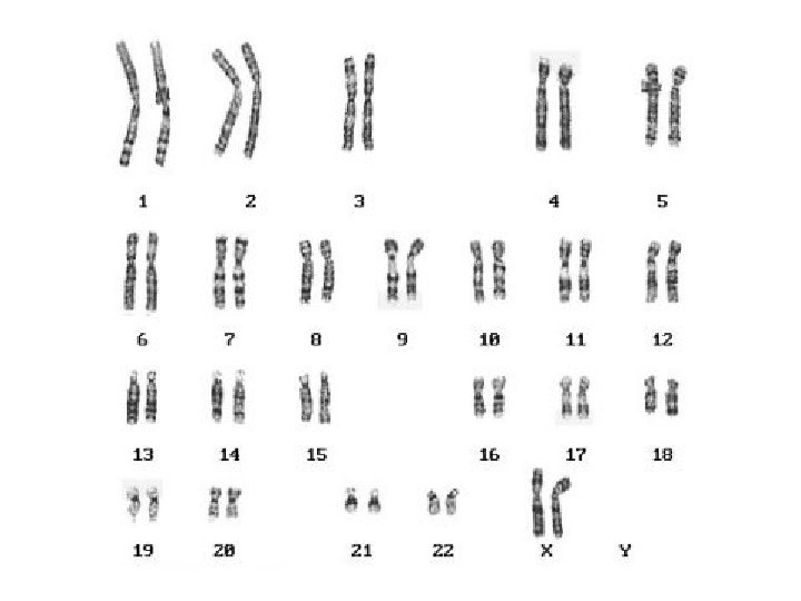
Karyotyping �Fetal cells or blood and tissue samples are cultured and are used to make pictures of chromosomes called karyotypes �White blood cells are useful for karyotyping