Chapter 7 The Respiratory System Chapter 7 The


 facilitates diffusion into")


 end at a group of")


 where one breath is")

 of the average male is 5.")


 External respiration - occurs between alveoli and the capillaries")


 These normal lungs have healthy red tissue. (The")


- Slides: 44
Chapter 7 The Respiratory System
Chapter 7 The Respiratory System 7. 1 Structures of the Respiratory System 7. 2 Breathing and Respiration 7. 3 Respiratory Health
Chapter 7 The Respiratory System In this chapter, you will learn: The upper respiratory tract filters, warms, and moistens oxygen-containing air, and channels it into the lungs. The lower respiratory tract is made up of specialized structures that exchange oxygen for carbon dioxide in the bloodstream. Humans ventilate their lungs by the mechanism of breathing, which involves inspiration and expiration. The volume of air that is taken into the lungs can increase if the need for oxygen increases, such as during exercise. External respiration takes place in the lungs, between the air in the alveoli and the blood in the capillaries. Internal respiration takes place between the blood in the capillaries and tissue cells. Gas exchange occurs through the processes of simple diffusion and facilitated diffusion. Some disorders are specific to the respiratory system. Technologies are available to treat respiratory disorders, but they may not be able to restore the respiratory system to optimal health. Smoking causes respiratory diseases. Technologies can help some symptoms of smoking, but many symptoms are untreatable.
7. 1 Structures of the Respiratory System In this section, you will: identify the principal structures of the respiratory system identify the principal functions of the respiratory system observe and identify the major respiratory structures
Respiratory System Respiration cellular respiration – at the cellular level involves the oxidation of glucose respiration at the multicellular level (breathing) – involves taking oxygen from the environment and returning carbon dioxide to it (breathing) – breathing is necessary to bring enough nutrients and oxygen to all cells in a multicellular organism (where diffusion just isn’t enough)
Respiration: Stages & Structures • Breathing involves two basic processes: inspiration (breathing in, or inhaling) and expiration (breathing out, or exhaling). Inspiration moves air from the external environment to the lungs inside the body. Expiration moves air from the lungs back to the external environment. • External respiration is the exchange of oxygen and carbon dioxide between the air and the blood. • Internal respiration is the exchange of oxygen and carbon dioxide between the body’s tissue cells and the blood. • Cellular respiration is the series of energy-releasing chemical reactions that take place inside the cells. Cellular respiration is the final stage in respiration. It is the sole means of providing energy for all cellular activities, and it helps the body maintain homeostasis.
Breathing brings oxygen to where diffusion can take place (bulk flow) facilitates diffusion into the transport system the circulatory system brings the oxygen to the cells (bulk flow) oxygen is brought into cells by diffusion across the membrane
The Respiratory System as air passes through the nasal cavity, it is warmed and moistened the passages are lined with hair and later cilia and mucous to help trap foreign invaders and sweep them into the pharynx where they are swallowed, sneezed or coughed out
the warmed air passes by the epiglottis and down the trachea which is lined with cartilage to prevent the trachea from collapsing or being damaged – the opening (slit) to the trachea is called the glottis – the air passes by the enlargement of the trachea, the larynx, where the vocal cords are located
the trachea is also lined with cilia and mucous-secreting cells – which beat 20 x per minute to move the trapped particles up to the pharynx Smokers cilia just above the heart, the trachea branches into two bronchi Bronchi branch again into bronchioles which branch smaller and smaller – only the smallest of the bronchioles lack cartilage
at the end, the terminal bronchioles (the last ones) end at a group of alveolar ducts and sacs called alveoli – the specialized structures for gas exchange the lungs are each encased in a doublemembraned sac, the pleura which allow the lungs to expand contract freely the diaphragm separates the lungs (in the thoracic cavity) from the peritoneal cavity, where the digestive organs are located
Alveoli are specialized for optimal diffusion – moist membrane – large surface area – thin walls for diffusion (1 m across) – immediately adjacent to pulmonary capillaries, which are just large enough for a RBC to get through – the inner surface of the alveoli are covered with a single layer of lipid called surfactant which reduces the surface tension in the alveoli allowing them to easily expand to twice their size with each breath
when a baby is born, the first cry and breath must expand the alveoli without the aid of surfactant – the lipid layer is immediately formed making breathing easier, and oxygen diffusion more efficient (by surface area since surfactant aids in the expansion of the alveoli) – for premature babies, the lungs may not be completely mature. Doctors must support breathing until the baby’s lungs develop by giving them air with a higher oxygen concentration
-Each bronchiole ends in several clusters of alveoli. -Surrounding each alveolus is a fine network of capillaries from the circulatory system. -Gas exchange occurs between the blood in the capillaries and the air in the alveolus, so that blood leaving the lungs has a high oxygen content.
7. 2 Breathing and Respiration In this section, you will: explain the mechanics of breathing explain how gases are exchanged between the human respiratory system and the external environment perform an experiment to determine your respiratory volume perform an experiment to examine factors that affect the rate of respiration
Mechanics of Breathing breathing involves two processes, inhalation and exhalation – Inhalation: an active process (using energy) where one breath is drawn into the lungs with the aid of muscle contractions air is drawn into the lungs when the thoracic cavity expands in size, and since the pleural cavity is sealed, decreasing the pressure of the cavity, which pulls air in from the environment
depending on the depth of breathing, a number of muscles will be involved – Diaphragm: contracts, moving downward – intercostal muscles (between the ribs): contract, expanding the chest cavity – neck muscles: contract, raising the top two ribs
– Exhalation: generally a passive process (unless breathing very deeply) where one breath is expelled as the muscles above relax as the muscles relax, the size of the thoracic cavity is decreased, increasing the pressure, forcing the gas out of the lungs when breathing deeply, more air may be exhaled with the contraction of the abdominal muscles
The Mechanics of Breathing
Lung Volumes the lung volume (total lung capacity) of the average male is 5. 7 L, the average female, 4. 2 L, including the residual volume is the amount of air that remains in the lungs after maximum expiration, keeping the lungs partially inflated (about 1 L) during quiet breathing, lungs generally inflate from 2. 2 L to 2. 7 L (the amount of air inhaled during quiet breathing, about 500 m. L is the tidal volume)
The largest breath you can take is called vital capacity – involves expanding the lungs to a greater extent than normal (recruiting the diaphragm, intercostals and neck muscles), and exhaling actively by recruiting the abdominal muscles Vital capacity includes tidal volume, the extra you can inhale (inspiratory reserve volume) and the extra you can exhale (expiratory reserve volume)
Of the 500 m. L inhaled with every normal breath, only about 0. 35 L reaches the alveoli (the rest fills the trachea, bronchi and bronchioles) – with an average respiratory rate (# breaths/minute) of 10, the amount of air exchanged per minute is 3. 5 L Lung volumes will be affected by height and gender in that thoracic cavity size will be affected which affects the lung size
Exercise performed over an extended period of time will strengthen the muscles involved in breathing which will increase tidal volume and vital capacity Illness can reduce vital capacity and tidal volume by affecting the number of alveoli that can successfully exchange oxygen, or by weakening the muscles in breathing
A Typical Spirograph
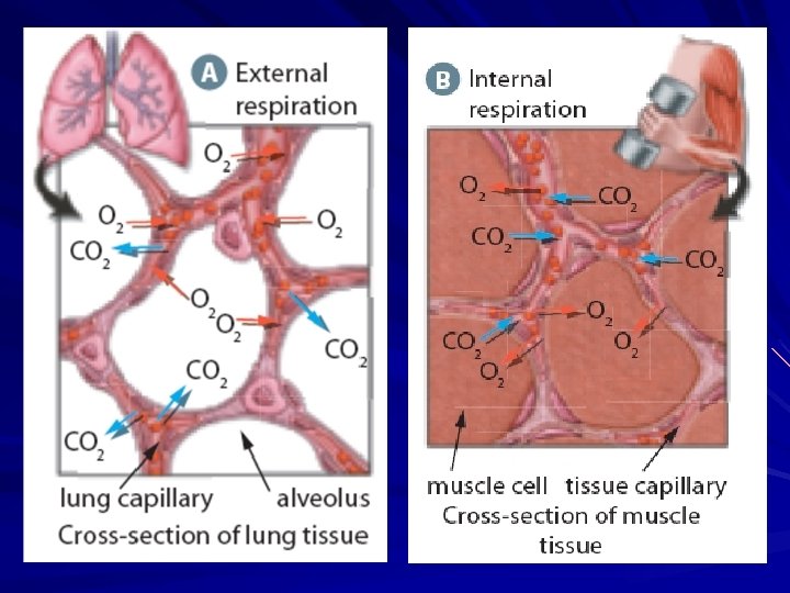
External vs. Internal Respiration A) External respiration - occurs between alveoli and the capillaries next to them. - As blood moves away from the body tissues, it is oxygen-poor and carbon dioxide-rich. - As it moves through the lung capillaries, oxygen from the air in the alveoli diffuses into the capillaries and carbon dioxide diffuses out of the blood. B) Internal respiration - occurs between the capillaries and the body tissues. - Oxygen diffuses from the blood into the oxygen-poor tissues while carbon dioxide diffuses from the tissues into the blood.
Gas Exchange and Transport oxygen and carbon dioxide must diffuse through the layer of cells lining the alveolus and the layer of cells lining the pulmonary capillary to enter/exit the blood. The gas must also diffuse across the membrane of the RBC to attach to a hemoglobin molecule
Oxygen 98. 5% of oxygen is carried on the heme group of the hemoglobin on the RBC oxygen binds reversibly to hemoglobin to form oxyhemoglobin (Hb. O 2) in a reversible reaction
the attaching of oxygen to hemoglobin is affected by the following – concentration of oxygen – oxygen, binding, oxygen, binding (allows oxygen to be released and diffuse into tissues over the entire capillary length – gradually) – p. H - p. H, binding ( p. H means CO 2) – temperature - temp, binding – these conditions favour the release of oxygen in tissues that are metabolically active (undergoing lots of cellular respiration)
Carbon Dioxide is transported three ways in the blood – 7% dissolved in the blood plasma – 23% carried on the hemoglobin molecule as carbaminohemoglobin (Hb. CO 2) – 70% is carried as carbonic acid/carbonate ion equilibrium – Equation CO 2(g) + H 2 O(l) → H 2 CO 3(aq) → HCO 3 -(aq) + H+(aq)
with the aid of the enzyme carbonic anydrase, the reaction between carbon dioxide and water is enhanced, which removes the carbon dioxide from the plasma, maintaining the concentration gradient from the tissues to the plasma, ensuring diffusion the HCO 3 -(aq)/ H 2 CO 3(aq) behaves as a blood buffer to help maintain the appropriate p. H for enzyme activity
Controlling Breathing Rate to deliver the optimal quantity of oxygen to the tissues, the body will HR and alter BP (via baroreceptors and chemosensors) but unless the breathing rate is matched, oxygen transport will not be effective the nervous system will match rate and magnitude of breathing to the heart rate and blood pressure – the medulla oblongata will control breathing rate – the pons smoothes out the rhythm of the respiration – chemoreceptors monitor p. H ( CO 2(g) p. H) – receptors in the aorta and carotid arteries monitor O 2(g) in arterial blood
7. 3 Respiratory Health In this section, you will: identify specific diseases that are associated with the respiratory system identify technologies that may be used to treat these respiratory diseases summarize the physiological effects of smoking and the limitation of technologies to address these effects
Upper Respiratory Tract Infections Tonsillitis is an infection of the tonsils, which are located in the pharynx. A viral infection, rather than a bacterial infection, is the more common cause of tonsillitis. The tonsils can be removed surgically if the infections are frequent and breathing is impaired. In the past, many children had their tonsils removed as a precaution, but this surgery is no longer as common. The tonsils help to prevent bacteria and other foreign pathogens from entering the body, so removing them can increase the number of infections later in life. Laryngitis is an inflammation of the larynx. Recall that the larynx contains the vocal cords. The most common cause of laryngitis is a viral infection; allergies and overstraining of the voice can also lead to laryngitis. When the larynx is inflamed, the vocal cords are not able to vibrate as they normally do. This reduces the ability to speak in a normal voice or even to speak at all. Symptoms of laryngitis include a sore throat and hoarseness.
Lower Respiratory Tract Infections Bronchitis is a disorder that causes the bronchi to become inflamed and filled with mucus, which is expelled by coughing. Pneumonia is a disease that occurs when the alveoli in the lungs become inflamed and fill with liquids. This interferes with gas exchange, and the body becomes starved for oxygen. Pleurisy is a lung disorder that is caused by the swelling and irritation of the pleura, the membranes that surround the lungs. Emphysema is an obstructive respiratory disorder in which the walls of the alveoli break down and lose their elasticity. This reduces the surface area for gas exchange and causes oxygen shortages in the tissues. Cystic fibrosis is a serious genetic condition that affects the lungs. Cystic fibrosis is caused by an abnormal gene that disrupts the function of the cells lining the passageways of the lungs. Asthma is a chronic obstructive lung disease that affects the bronchi and bronchioles, making breathing difficult or impossible because of reduced air flow. Lung cancer is the uncontrolled and invasive growth of abnormal cells in the lungs. It is the leading cause of cancer deaths for men and women in Canada.
Lower Respiratory Tract Disorders
Normal Lungs vs. Diseased Lungs (A) These normal lungs have healthy red tissue. (The heart is visible near the lower centre. ) (B) These diseased lungs have black tissue caused by heavy smoking. The white areas are tumours, or carcinomas.
Carcinoma of the Lung The large ball of cells in the centre of the image is a carcinoma that has developed from the interior surface cells of the human lung. The carcinoma continues to grow and invade surrounding tissues, including the lymphatic and blood vessels in the lung. The lymphatic and blood vessels circulate through the body and carry the cancerous cells, or metastatic cells, to new locations where they can grow and invade new tissues.
Chapter 7 Review Create a flowchart or diagram showing the path of oxygen through the respiratory system. Explain how each of the major respiratory structures function. What is cellular respiration? Compare and contrast a normal lung with smoker’s lung. Identify three respiratory diseases. Briefly describe their symptoms and how they are diagnosed.
Concept Organizer
Chapter 7 Summary Respiration enables the body to take oxygen from the external environment and process it for delivery to the cells and, at the same time, rid itself of carbon dioxide.
Chapter 7 Summary Oxygen is delivered to the cells and carbon dioxide is removed from the cells and the body in a number of exchanges. Inspiration (breathing in, inhaling) and expiration (breathing out, exhaling) exchange air between the environment and the lungs. External respiration exchanges oxygen and carbon dioxide between the air in the lungs and the blood. Internal respiration exchanges oxygen and carbon dioxide between the blood and the body’s tissue cells. Cellular respiration is the final step, when the oxygen delivered to the cells is used to provide the energy for all cellular activities; carbon dioxide is the waste product of cellular respiration.
Chapter 7 Summary The respiratory tract is the passageway for air to move from the external environment into the lungs. The upper respiratory tract begins at the nostrils and includes the nasal passages, pharynx, larynx, and trachea. These passageways all clean and warm the air as it passes through. The lower respiratory tract consists of two bronchi that each lead to a lung. Within the lungs are small, fine tubes called bronchioles, where the air continues to be cleaned and warmed. The exchange of gases takes place in a cluster of tiny sacs at the end of each bronchiole, called alveoli, where the oxygen diffuses through the membranes of the alveoli into the capillaries of the circulatory system.
Chapter 7 Summary A number of disorders of the respiratory tract can impair the delivery of oxygen to the cells, including bronchitis, pneumonia, pleurisy, emphysema, cystic fibrosis, asthma, and lung cancer. These are all disorders of the lower respiratory tract. Infections of the upper respiratory tract, such as tonsillitis and laryngitis are short term infections that do not obstruct breathing.