Chapter 7 The Nervous System Neurons and Synapses

 2. Electroactivity (7. 2) 3. The")
")
 Brain Spinal cord 2. Peripheral")


 of the CNS • Four types are found in the CNS:")
 of the PNS • Two types are found in the PNS:")
 of the PNS Page 167 Posterior root ganglion Neurofibril nodes (nodes")


 are")


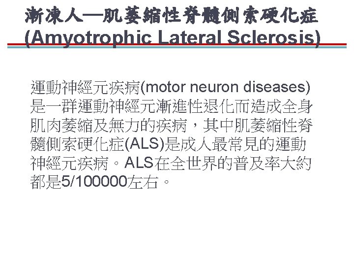
 vs 漸凍人─肌萎縮性脊髓側索硬化症 (Amyotrophic Lateral Sclerosis)")


- Slides: 54
Chapter 7 The Nervous System: Neurons and Synapses
Outline 1. Neuron and Glial cells (7. 1) 2. Electroactivity (7. 2) 3. The synapse (7. 3) 4. Neurotransmitter (7. 4 -7. 6) 5. Synaptic integration (7. 7)
I. Neurons and Supporting Cells (Glial cells or neuroglia)
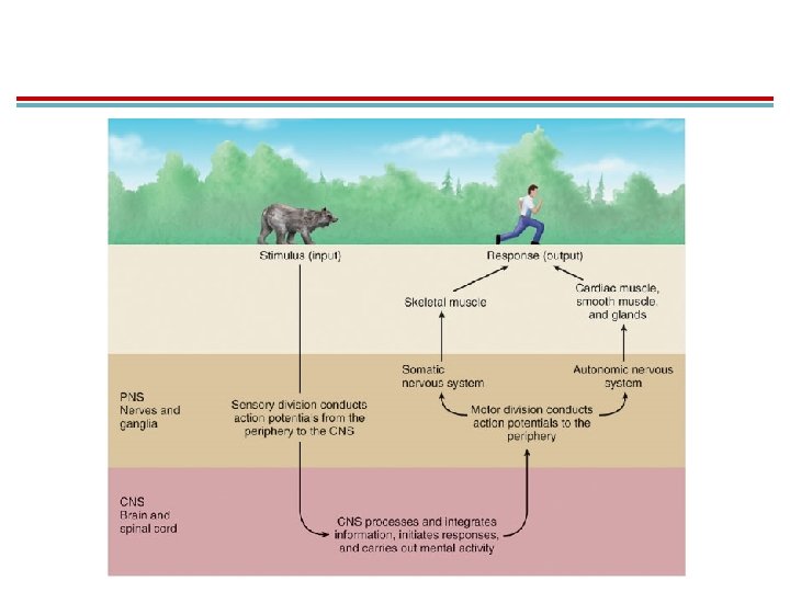
Nervous System Page 163 1. Central nervous system (CNS) Brain Spinal cord 2. Peripheral nervous system (PNS) Nerve outside the brain and spinal cord
Page 163 • Nerve tissue is composed of two types of cells: a. Neurons that conduct impulses but generally can not divide. b. Glial cells (neuroglia) that support the neurons and can not conduct impulses, but can divide
Neurons Page 163 • Neurons vary in size and shape, but they all have: – A cell body: cluster in groups called nuclei in the CNS and ganglia in the PNS – Dendrites: receive signals – Axon: conducts impulses
Neurons Page 163 Motor neuron Sensory neuron
Axons Page 164
Axons Page 163 • Vary in length from a few millimeters to a meter • The region closest to the cell body is the axon hillock where action potentials are generated. • Covered in myelin with a node of Ranvier
Neurons • Structural and functional units of the nervous system • General functions a. Respond to chemical and physical stimuli b. Conduct electrochemical impulses c. Release chemical regulators d. Enable perception of sensory stimuli, learning, memory, and control of muscles and glands • Most can not divide, but can repair
Structural Classification of Neurons Page 165 &166
Page 164 -166
Functional Categories of Neurons Page 165
Page 164
Functional Categories of Neurons Page 165 • Functional classification: – Sensory neurons: conduct impulses from sensory receptors to the CNS – Motor neurons: conduct impulses from the CNS to target organs (muscles or glands) – Association/interneurons: located completely within the CNS and integrate functions of the nervous system
Motor Neurons • Two types: Page 165 – Somatic motor neurons: responsible for reflexes and voluntary control of skeletal muscles – Autonomic motor neurons: innervate involuntary targets such as smooth muscle, cardiac muscle, and glands • Sympathetic • Parasympathetic
Functional Categories of Neurons Page 165
Nerves Page 166 SEM tracts nerves
Nerves Page 166 • Bundles of axons located outside the CNS – Most are composed of both sensory and motor neurons and are called mixed nerves. – Some of the cranial nerves have sensory fibers only. – A bundle of axons in the CNS is called a tract.
Glial Cells of the CNS Page 166
Page 167
Glial Cells (Neuroglia) of the CNS • Four types are found in the CNS: Page 166 – Oligodendrocytes: form myelin sheaths around the axons of CNS neurons – Microglia: migrate around tissue and phagocytize foreign and degenerated material – Astrocytes: regulate the external environment of the neurons – Ependymal cells: line the ventricles and secrete cerebrospinal fluid
Glial Cells (Neuroglia) of the PNS • Two types are found in the PNS: Page 167 – Schwann cells (neurolemmocytes): form myelin sheaths around peripheral axons – Satellite cells (ganglionic gliocytes): protect cell bodies within the ganglia of the PNS
Glial Cells (Neuroglia) of the PNS Page 167 Posterior root ganglion Neurofibril nodes (nodes of Ranvier) Satellite cells Axon Nucleus Axon Cell body Dorsal root (sensory neuron) (e) Satellite cells Neurolemmocyte (Schwann cell) (f) Neurolemmocytes
Myelin sheaths in the PNS and CNS Page 166 & 168
Myelin Sheath in CNS Page 168 Oligodendrocyte Node of Ranvier Myelin sheath Axon
Comparison of Myelinated and Unmyelinated Axons Page 168
Neurilemma and Myelin Page 167 • All axons in the PNS are surrounded by a sheath of Schwann cells called the neurilemma, or sheath of Schwann. • These cells wrap around to form the myelin sheath in the PNS. • Node of Ranvier is left open.
Myelin Sheath in the PNS Small Page 167 axons (2 micrometers in diameter) are usually unmyelinated. Even unmyelinated axons in the PNS have a neurilemma but lack the multiple wrappings of the Schwann cell plasma membrane Myelinated axons conduct impulses more rapidly.
Myelin Sheath in CNS Page 168 • In the CNS, the myelin sheath is produced by oligodendrocytes. • One oligodendrocyte sends extensions to several axons. • Myelin gives these tissues a white color = white matter. • Gray matter is cell bodies and axons.
Myelin Sheath Page 168
Neurilemma and Myelin Sheath Page 165
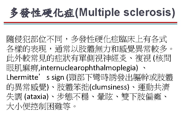
Page 168 多發性硬化症 Multiple sclerosis
多發性硬化症(Multiple sclerosis) vs 漸凍人─肌萎縮性脊髓側索硬化症 (Amyotrophic Lateral Sclerosis)
神經再生帶來治療癱瘓契機 Page 169 Regeneration of a Cut Neuron in PNS
A Cut Neuron in CNS Page 169 CNS axons are not as able to regenerate. 1)Death receptors form that promote apoptosis of oligodendrocytes 2)Inhibitory proteins in the myelin sheath prevents regeneration 3)Glial scars from astrocytes form that also prevent regeneration
Regeneration of a Cut Neuron in PNS Page 169
Regeneration of a Cut Neuron in PNS Page 169 When an axon in the PNS is cut, the severed part degenerates, and a regeneration tube is formed by Schwann cells. 1)Growth factors are leased that stimulate growth of axon sprouts within the tube 2)New axon eventually connects to the undamaged axon or the effector
Neurotrophins Page 170 • Promote neuronal growth in the fetal brain – Nerve growth factor (NGF) – Brain-derived neurotrophic factor (BDNF) – Glial-derived neurotrophic factor (GDNF) – Neurotrophin-3, neurotrophin-4/5 • In adults, neurotrophins aid in the maintenance of sympathetic ganglia and the regeneration of sensory neurons.
Astrocytes Page 170
Astrocytes Page 170 • Most abundant glial cell • Processes with end-feet associate with blood capillaries and axon terminals • Influences interactions between neurons and blood
Astrocyte Functions Page 170 & 171 Take up K+ from the extracellular environment to maintain ionic environment for neurons Take up extra neurotransmitter released from axon terminals, particularly glutamate. Chemicals are recycled. End-feet around capillaries take up glucose from blood for use by neurons to make ATP; converted first to lactic acid Can store glycogen and produce lactate for neurons to use
Astrocyte Functions Page 170 & 171 Needed for the formation of synapses in the CNS Regulate neurogenesis in regions of the adult brain Form the blood-brain barrier Release transmitter molecules (gliotransmitters) that can stimulate or inhibit neurons; includes glutamate, ATP, adenosine, D-serine
Astrocytes and neural activity Page 171 Although astrocytes do not produce action potentials, they are excited by changes in intracellular Ca 2+ concentration. When some neurons are active, they release ATP, which increases the Ca 2+ of adjacent astrocytes; creates a Ca 2+ wave A rise in Ca 2+ can also cause the astrocyte release prostaglandin E 2 from the end-feet on a blood capillary, increasing blood flow.
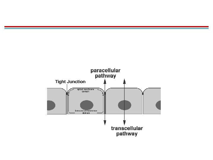
Blood-Brain Barrier Page 171 Capillaries in the brain do not have pores between adjacent cells but are joined by tight junctions. Substances can only be moved by very selective processes of diffusion through endothelial cells, active transport, and bulk transport Movement is transcellular not paracellular
Blood-Brain Barrier Page 171 Astrocytes influence the production of ion channels and enzymes that can destroy toxic substances by secreting glial-derived neurotrophic factor. Creates problems with chemotherapy of brain diseases because many drugs can not penetrate the blood-brain barrier.
Page 164