CHAPTER 7 THE MICROSCOPE The microscope An optical


 2) 3) 4) 5) 6) 7) 8) 9) 10) 11) 12) 13) What")


 : mm")

")


 n n SEM forms an image by aiming a beam")
")
")
")

")
")
- Slides: 28
CHAPTER 7 THE MICROSCOPE
The microscope: An optical instrument that uses a lens or a combination of lenses to magnify and resolve the fine details of an object. § §An indispensable tool for the forensic scientist. -One of the first tools used, yet still used today. Earliest & Simplest is a handheld magnifying glass. (5 X – 10 X) §
1) 2) 3) 4) 5) 6) 7) 8) 9) 10) 11) 12) 13) What do you remember about a compound microscope? 1 13 12 10 11 4 8 7 5 9 2 3 6
What do you remember about a compound microscope? n n n n 1. ocular 2. Coarse adjustment knob 13 3. Fine adjustment knob 4. Stage clips 5. Arm 12 6. Base 10 7. Light source 11 8. Diaphragm 4 9. Stage 8 10. Low power objective 7 11. High power objective 12. Nosepiece 13. Body tube 1 5 9 2 3 6
Virtual image is not viewed directly but can only be observed when you look through the lens. A magnified or enlarged image. Real image is seen directly, like the image that is projected onto a motion picture screen
The Compound Microscope Utilizes transmitted light through a specimen – therefore specimens must be very thin. § n. Ocular lens is closest to the eye; Inscribed with its magnifying power (usually 10 X) Monocular: One eyepiece/ocular lens Binocular: Two eyepieces/ocular lens
Total Magnification Total magnification = Mag. of objective lens X Mag. of ocular lens What is the total magnification of the following microscopes? n 10 X ocular and 40 X objective = ______ n 10 X ocular and 10 X objective = ______ n
Field Of View The area that ocular of is seen through the microscope. You can measure the field of view with a ruler if the area is big enough. If not, go by… Low power = 2 mm 2000 um High Power = 0. 5 mm 500 um (If different, values given) n
Field of View Micrometers: 1/1000 of a millimeter mm um (micrometers/ microns) : mm x 1000 2 mm How big is the letter “e”? The field of view is 2 mm or 2000 Um There are 4 letter “e” in the field Field of View/# of objects Diameter “e” = 2000/4 500 Um
Field of View decreases as Magnification increases Depth of Focus decreases As Magnification increases
Compound Microscope, cont. n Images appear enlarged, upside down and backwards (180 degree turn) View under microscope (not through lens) n view through lens Images move in opposite direction of the stage A. B. To center the “e” you have to move the stage down in figure A. and to the left in figure B.
Comparison Microscope: Two compound microscopes combined into one unit. Two independent objective lenses joined by an optical bridge to a common eyepiece lens
Stereo/Dissecting Microscope § Utilizes reflected light Can have magnification ranges from 10 X – 125 X § § Produces a right-side-up 3 -D image Most frequently used and versatile microscope found in the crime laboratory. -Wide field of view -Great depth of focus §
Stereo/Dissecting Microscope, cont. Advantages… *3 -D image of an object that is rightside-up. (Unlike the compound microscope) *Has a large working distance (the distance between the objective lens and the specimen) *Useful for opaque objects, locating trace evidence.
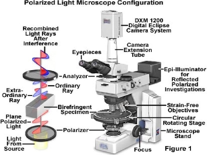
Polarizing Microscope n n Passes light through a polarizer which puts light waves in a single direction. Polarized light passes through the specimen through the analyzer, which sends it to the eyepiece. This results in very vivid colors and contrasts that make a specimen very unique. Is used on specimens that exhibit birefrigence as a physical property. Substances include… *crystalline substances (minerals) *synthetic fibers *many plastics (cellophane)
Polarizer – a device that permits the passage of light wave vibration in only one direction (plane). Analyzer – a second polarizing crystal placed between a polarized beam Analyzer is parallel to polarized beam = light passes through (when a specimen is observed) Analyzer is perpendicular to polarized beam = no light passes through (this is when there’s no specimen)
Microspectrophotometer n n Links a microscope to a computerized spectrophotometer Allows the user to view a particle under the microscope while a beam of light is directed at the particle in order to obtain its absorption spectrum. take absorption and/or fluorescence measurements directly.
Uses: Paint, fiber and ink evidence §Comparison completed with the microscope, then the absorption spectra is determined at the same time. §Different inks/pigments will have a different absorption pattern. §IR spectrophotometry will produce a unique ‘fingerprint’ for a substance (confirmation test).
Infrared absorption spectra
Scanning Electron Microscope (SEM) n n SEM forms an image by aiming a beam of electrons, which is focused by means of magnets, onto the surface of a specimen. This results in backscatter and the emission of secondary electrons from the surface elements of the specimen. Has a magnifying range From 10 X to 100, 000 X. n n Great depth of focus (3 -D) Scanning Electron Microscope
Human hair (S. E. M. )
Head of a Termite (S. E. M. )
Spider (S. E. M. )
146 X S. E. M. 392 X Sponge x 400
Bacteria (T. E. M. )
Golgi Apparatus (T. E. M. )