
CHAPTER 7 MEMBRANES THEIR STRUCTURE FUNCTION AND CHEMISTRY
CHAPTER 7 MEMBRANES: THEIR STRUCTURE, FUNCTION, AND CHEMISTRY 7. 1 The Functions of Membranes 7. 2 The Models of membrane structure 7. 3 The Membrane lipids 7. 4 The Membrane proteins
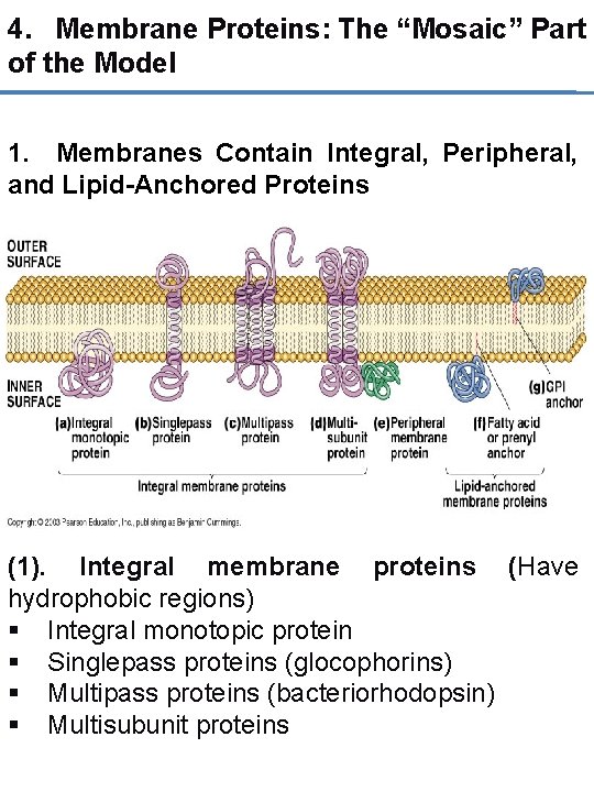
4. Membrane Proteins: The “Mosaic” Part of the Model 1. Membranes Contain Integral, Peripheral, and Lipid-Anchored Proteins (1). Integral membrane proteins (Have hydrophobic regions) § Integral monotopic protein § Singlepass proteins (glocophorins) § Multipass proteins (bacteriorhodopsin) § Multisubunit proteins
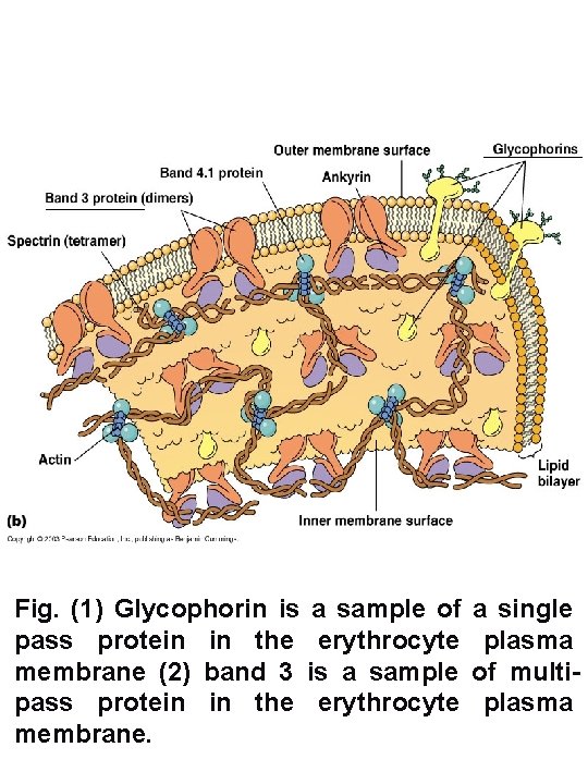
Fig. (1) Glycophorin is a sample of pass protein in the erythrocyte membrane (2) band 3 is a sample pass protein in the erythrocyte membrane. a single plasma of multiplasma
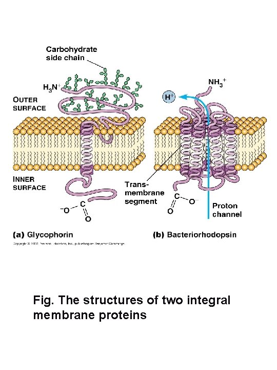
Fig. The structures of two integral membrane proteins
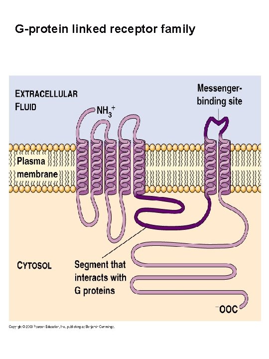
G-protein linked receptor family
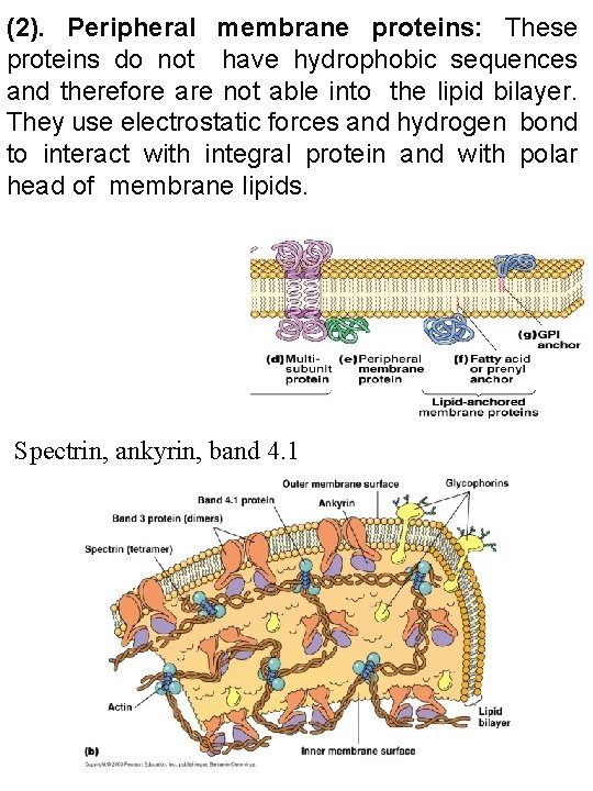
(2). Peripheral membrane proteins: These proteins do not have hydrophobic sequences and therefore are not able into the lipid bilayer. They use electrostatic forces and hydrogen bond to interact with integral protein and with polar head of membrane lipids. Spectrin, ankyrin, band 4. 1
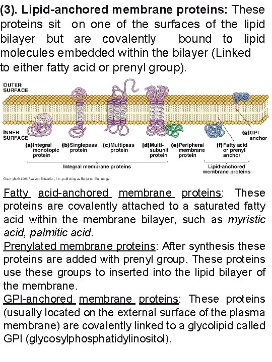
(3). Lipid-anchored membrane proteins: These proteins sit on one of the surfaces of the lipid bilayer but are covalently bound to lipid molecules embedded within the bilayer (Linked to either fatty acid or prenyl group). Fatty acid-anchored membrane proteins: These proteins are covalently attached to a saturated fatty acid within the membrane bilayer, such as myristic acid, palmitic acid. Prenylated membrane proteins: After synthesis these proteins are added with prenyl group. These proteins use these groups to inserted into the lipid bilayer of the membrane. GPI-anchored membrane proteins: These proteins (usually located on the external surface of the plasma membrane) are covalently linked to a glycolipid called GPI (glycosylphosphatidylinositol).
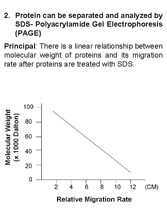
2. Protein can be separated analyzed by SDS- Polyacrylamide Gel Electrophoresis (PAGE) Principal: There is a linear relationship between molecular weight of proteins and its migration rate after proteins are treated with SDS.
Function of Sodium Dodecyl Sulfate (SDS) Sodium dodecylsulfate (SDS) is a detergent with a highly negatively charged sulfate group. SDS binds strongly to most proteins and causes them to unfold to a random, rod-like chain. No covalent bonds are broken in this process. Therefore, the amino acid composition and sequence remains the same. (1) Denature proteins: SDS unfold protein into stiff polypeptide rods. However, SDS does not broke covalent bonds including disulfide bonds. Other reducing agents (beta-ME) can disrupt the disulfide bonds. (2) SDS carries lager amount of negative charges compared with proteins (therefore, the small differences of electric charges between proteins have no effect on migration rate).
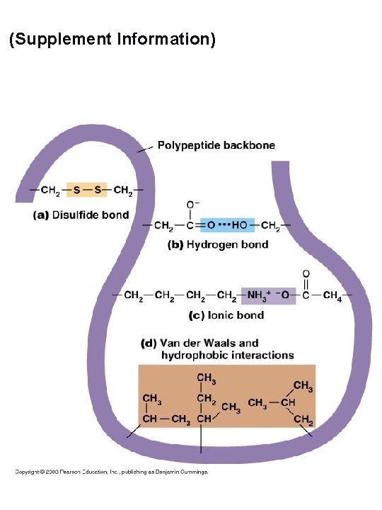
(Supplement Information)
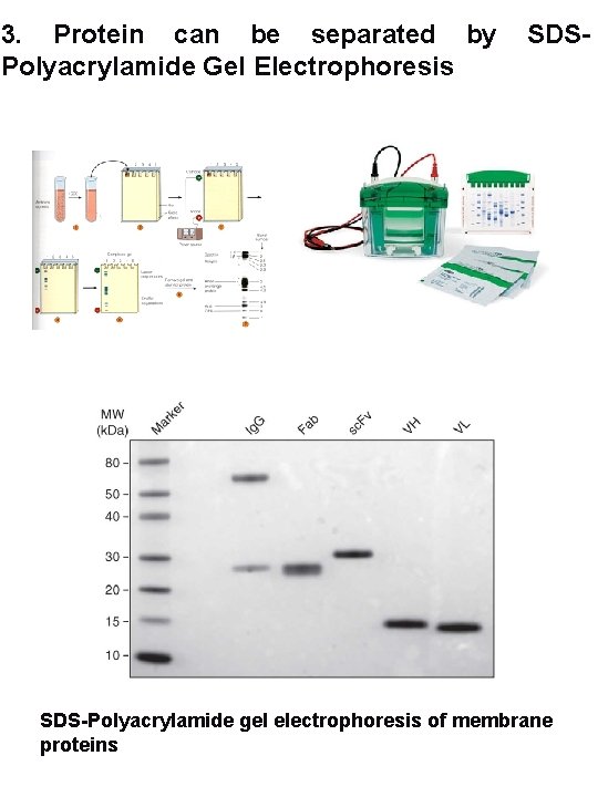
3. Protein can be separated by Polyacrylamide Gel Electrophoresis SDS-Polyacrylamide gel electrophoresis of membrane proteins
4. Determination of the three-dimensional structure of integral membrane protein 1. X-ray crystallography 2. Cryo-electron microscopy electron cryomicroscopy 3. Hydropathic analysis (cryo-EM), or
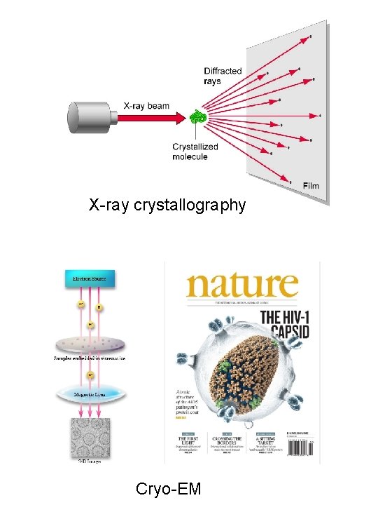
X-ray crystallography Cryo-EM
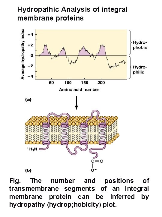
Hydropathic Analysis of integral membrane proteins Fig. The number and positions of transmembrane segments of an integral membrane protein can be inferred by hydropathy (hydrop; hobicity) plot.
Nucleic Acid Sequence of GM-CSF receptor (alpha subunit. 1) agcaggtggaaggagaggaagcggatgcc 29 gtggggtttacagcaggaaaatccgtggagacagcagatccgagaagcggcgatgtttgc 89 gtagaaccctgtacgtgcttcggcctgtcgctcttcccttctctctgaccagcacc 149 atgcttctcctggtgacaagccttctgctctgtgagttaccacacccagcattcctcctg 209 atcccagagaaatcggatctgcgaacagtggcaccagcctctagtctcaatgtgaggttt gactccaggacgatgaatttaagctgggactgccaagaaaacacaaccttcagcaagtgt 329 ttcttaactgacaagaagaacagagtcgtggaacccaggctcagtaacaacgaatgttcg 389 tgcacatttcgtgaaatttgtctgcatgaaggagtcacatttgaggttcacgtgaatact 449 agtcaaagaggagttcaacagaaactgctttatccaaattcaggaagggtaccgct 509 gctcagaatttctcctgtttcatctacaatgcggatttaatgaactgtacctgggcgagg 569 ggtccgacggccccccgtgacgtccagtattttttgtacatacgaaactcaaagagaagg 629 agggagatccggtgtccttattacaagactcaggaacccatgtgggatgtcacctg 689 gataacctgtcaggattaacgtctcgcaattactttctggttaacggaaccagccgagaa 749 attggcatccaattctttgattcacttttggacacaaagaaaatagaacgattcaaccct 809 cccagcaatgtcaccgtacgttgcaacacgacgcactgcctcgtacggtggaaacagccc 869 aggacctatcagaagctgtcgtacctggactttcagtaccagctggacgtccacagaaag 929 aatacccagcctggcacggaaaacctactcattaatgtttctggtgatttggaaaataga 989 tacaactttccaagctctgagcccagagcaaaacacagtgtgaagatcagagctgcagac 1049 gtccgcatcttgaattggagctcctggagtgaagccattgaatttggttctgacgacggg 1109 aacctcggctctgtgtacatttatgtgctcctaatcgtgggaacccttgtctgtggcatc 1169 gtcctcggcttcctctttaaaaggttccttaggatacagcggctgttcccgccagttcca 1229 cagatcaaagacaaactgaatgataaccatgaggtggaagacgagatcatctgggaggaa 1289 ttcaccccagaggaagggaaaggctaccgcgaagaggtcttgaccgtgaaggaaattacc 1349 tgagacccagagggtgtaggaatggcatggacatctccgcgacacgggggaact 1409 ttttcttgatgatgctgtgaacctttatatcattttctatgtttttaaaaacatg 1469
5. Membrane proteins have a variety of functions 1. Some membrane proteins are enzymes 2. Membrane proteins regulate the transport of solutes. (1) Small and lager molecules: simple diffusion, facilitated diffusion, active transport/pumps, (2) Very large molecules: Endocytosis: Exocytosis: 3. Membrane proteins detect and transmit electrical and chemical signals Ligands and receptors/channels Chemical signal transduction 4. Membrane proteins mediate cell adhesion and cell-to-cell communication gap junctions in animal cells), Plasmodesma (in plant cells) 5. Membrane proteins are strurcture components of membrane
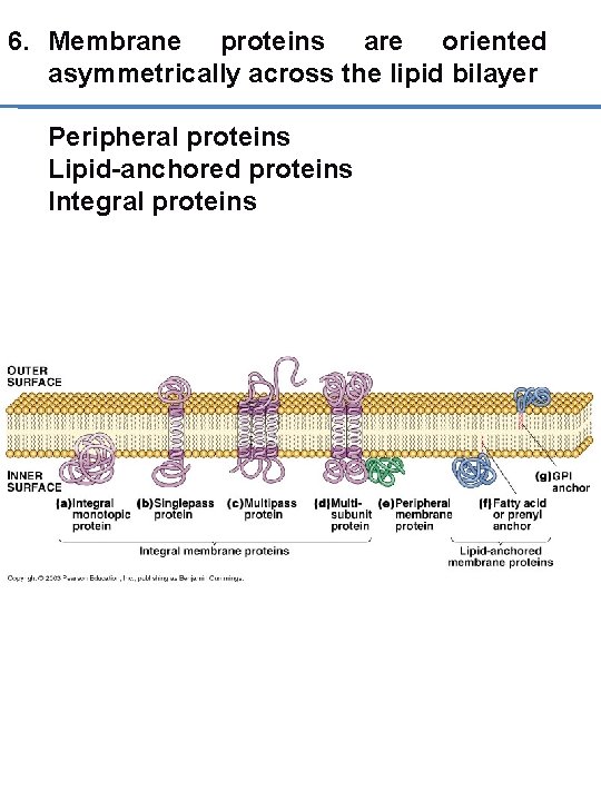
6. Membrane proteins are oriented asymmetrically across the lipid bilayer Peripheral proteins Lipid-anchored proteins Integral proteins
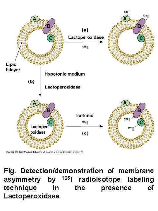
Fig. Detection/demonstration of membrane asymmetry by 125 I radioisotope labeling technique in the presence of Lactoperoxidase
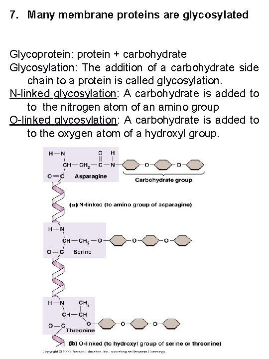
7. Many membrane proteins are glycosylated Glycoprotein: protein + carbohydrate Glycosylation: The addition of a carbohydrate side chain to a protein is called glycosylation. N-linked glycosylation: A carbohydrate is added to to the nitrogen atom of an amino group O-linked glycosylation: A carbohydrate is added to to the oxygen atom of a hydroxyl group.
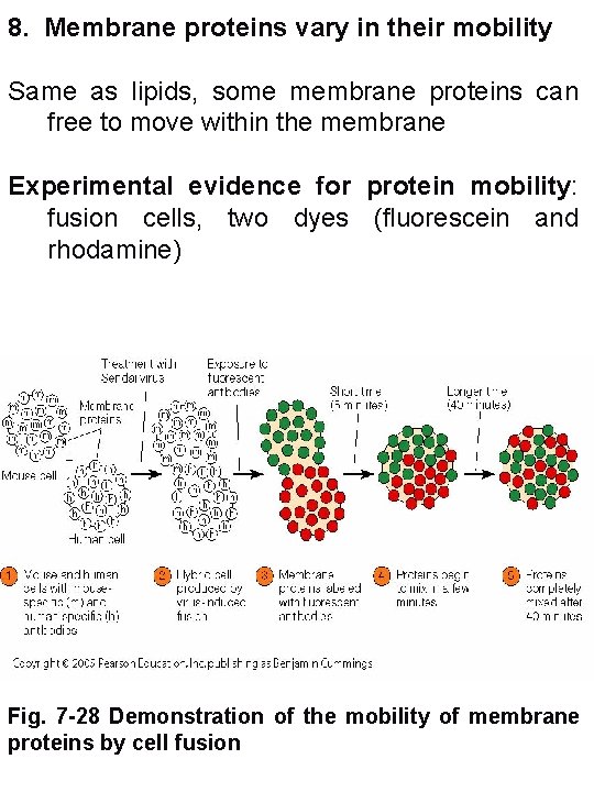
8. Membrane proteins vary in their mobility Same as lipids, some membrane proteins can free to move within the membrane Experimental evidence for protein mobility: fusion cells, two dyes (fluorescein and rhodamine) Fig. 7 -28 Demonstration of the mobility of membrane proteins by cell fusion
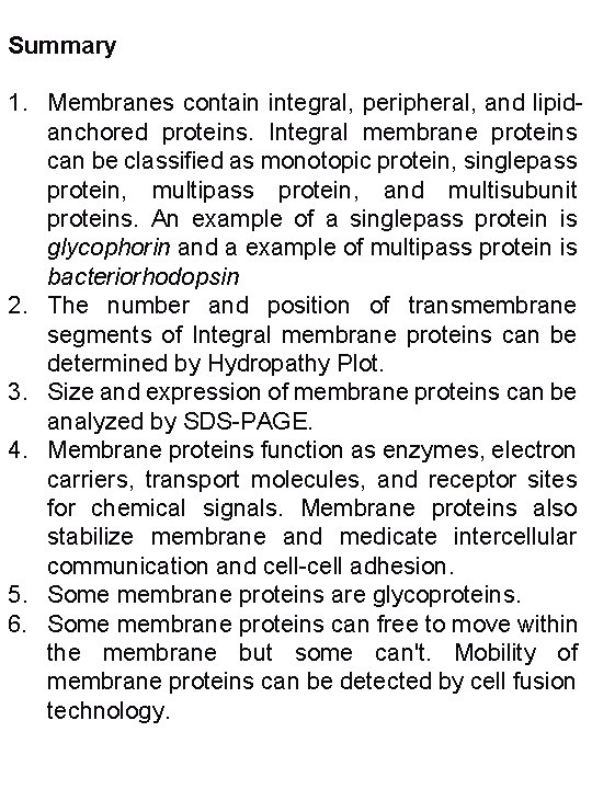
Summary 1. Membranes contain integral, peripheral, and lipidanchored proteins. Integral membrane proteins can be classified as monotopic protein, singlepass protein, multipass protein, and multisubunit proteins. An example of a singlepass protein is glycophorin and a example of multipass protein is bacteriorhodopsin 2. The number and position of transmembrane segments of Integral membrane proteins can be determined by Hydropathy Plot. 3. Size and expression of membrane proteins can be analyzed by SDS-PAGE. 4. Membrane proteins function as enzymes, electron carriers, transport molecules, and receptor sites for chemical signals. Membrane proteins also stabilize membrane and medicate intercellular communication and cell-cell adhesion. 5. Some membrane proteins are glycoproteins. 6. Some membrane proteins can free to move within the membrane but some can't. Mobility of membrane proteins can be detected by cell fusion technology.
- Slides: 21