Chapter 7 Membrane Structure and Function Plasma Membrane

Chapter 7 Membrane Structure and Function Plasma Membrane Rap

What you will be responsible to know… �The plasma membrane is the boundary that separates the living cell from its surroundings �The plasma membrane exhibits selective permeability, allowing some substances to cross it more easily than others

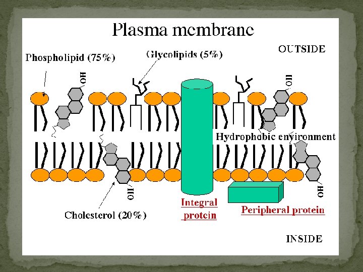
Makeup of the PM ① Phospholipids ② Cholesterol ③ Embedded Proteins ④ Glycoproteins ⑤ Glycolipids CARBOHYDRATES
 Phospholipids �most abundant lipid in the plasma membrane � amphipathic molecules, containing hydrophobic")
1) Phospholipids �most abundant lipid in the plasma membrane � amphipathic molecules, containing hydrophobic and hydrophilic regions � J. Singer & G. Nicolson (1972) proposed that the membrane is a mosaic of proteins dispersed within the bilayer, with only the hydrophilic regions exposed to water (fluid mosaic model) Plasma Membrane Video

Phospholipids Cont’d � Freeze-fracture studies of the plasma membrane supported the fluid mosaic model � Freeze-fracture is a specialized preparation technique that splits a membrane along the middle of the phospholipid bilayer TECHNIQUE RESULTS Extracellular layer Knife Plasma membrane Proteins Inside of extracellular layer Cytoplasmic layer Inside of cytoplasmic layer

Phospholipids Cont’d �Phospholipids can move within the bilayer � Most of the lipids, and some proteins, drift laterally � Rarely does a molecule flip-flop transversely across the membrane �As temperatures cool, membranes become more solid � The temperature at which a membrane solidifies depends on the types of lipids �Membranes rich in unsaturated fatty acids are more fluid that those rich in saturated fatty acids �Membranes must be fluid to work properly; they are usually about as fluid as salad oil
 Cholesterol �Ring structure reduces membrane fluidity at moderate temperatures by reducing phospholipid movement")
2) Cholesterol �Ring structure reduces membrane fluidity at moderate temperatures by reducing phospholipid movement � low temperatures hinders solidification by disrupting the regular packing of phospholipids. � warm temperatures (such as 37°C), cholesterol restrains movement of phospholipids �Cholesterol is known as “temperature buffer”. Cholesterol (c) Cholesterol within the animal cell membrane
 Proteins �collage of different proteins embedded in the fluid matrix of the lipid")
3) Proteins �collage of different proteins embedded in the fluid matrix of the lipid bilayer � Proteins determine most of the membrane’s specific functions �The two sides of a membrane have different protein and lipid compositions. • Peripheral proteins are bound to the surface of the membrane Not embedded • Loosely bound to surface of membrane • Integral proteins penetrate the hydrophobic core • transmembrane proteins span the membrane • hydrophobic regions consist of one or more stretches of nonpolar amino acids, often coiled into alpha helices • Hydrophilic regions exposed to inside/outside of cell

Proteins Cont’d
 substances and ions across")
Proteins Cont’d �Transport proteins allow passage of large hydrophilic (polar) substances and ions across the membrane (FACILITATED DIFFUSION) 1) channel proteins have a hydrophilic channel that certain molecules or ions can use as a tunnel �Channel proteins called aquaporins facilitate the passage of water 2) carrier proteins bind to molecules and change shape to shuttle them across the membrane �A transport protein is specific for the substance it moves

The Permeability of the Lipid Bilayer �Small, uncharged polar and nonpolar molecules can freely pass through a cell membrane � Hydrophobic (nonpolar) molecules, such as hydrocarbons, can dissolve in the lipid bilayer and pass through the membrane rapidly �Hydrocarbons, CO 2 and O 2 � Polar molecules, such as sugars, do not cross the membrane easily �C 6 H 12 O 6, or charged molecules

Proteins Cont’d �Six major functions � Transport � Enzymatic activity � Signal transduction � Cell-cell recognition � Intercellular joining � Attachment to the cytoskeleton and extracellular matrix (ECM)

The Role of Membrane Carbohydrates in Cell-Cell Recognition �Cells recognize each other by binding to surface molecules, usually carbohydrates �Membrane carbohydrates may be covalently bonded to lipids (forming glycolipids) or more commonly to proteins (forming glycoproteins) �Carbohydrates on the external side of the plasma membrane vary among species, individuals, and even cell types in an individual Copyright © 2008 Pearson Education, Inc. , publishing as Pearson Benjamin Cummings
 Glycoproteins & 5) Glycolipids � Carbohydrates are third major component of plasma membranes")
4) Glycoproteins & 5) Glycolipids � Carbohydrates are third major component of plasma membranes � found on the exterior surface of cells � bound either to proteins (forming glycoproteins) � or bound to lipids (forming glycolipids) � Chains can be either straight or branched � Form specialized sites on the cell surface that allow cells to recognize each other. � unique patterns that allow the cell to be recognized � allows the immune system to differentiate between body cells (called “self”) and foreign cells or tissues (called “non-self”). � Similar types of glycoproteins and glycolipids are found on the surfaces of viruses and may change frequently, preventing immune cells from recognizing and attacking them.

Glycoproteins & Glycolipids Cont’d

Fun Facts about the PM �Membranes have distinct inside and outside faces � The two sides of a membrane have different protein and lipid compositions. �The asymmetrical distribution of proteins, lipids, and associated carbohydrates in the plasma membrane is determined when the membrane is built by the ER and Golgi apparatus � Molecules that start out on the inside face of the ER end up on the outside face of the plasma membrane

Passive & Active Transport Import of resources and export of wastes

Passive Transport: � Substances diffuse down their concentration gradient (the difference in concentration of a substance from one area to another) � From an area of high concentration to low concentration � No work must be done to move substances down the concentration gradient � O 2 gets into cells this way for cellular respiration � Diffusion is the tendency for molecules to spread out evenly into the available space � At dynamic equilibrium, as many molecules cross one way as cross in the other direction � This is not static – even at equilibrium molecules are still moving.
 WATER Net diffusion (a) Diffusion of one solute")
Molecules of dye Membrane (cross section) WATER Net diffusion (a) Diffusion of one solute Net diffusion Equilibrium
 Diffusion of two solutes The diffusion")
Fig. 7 -11 b Net diffusion Equilibrium (b) Diffusion of two solutes The diffusion of one solute is unaffected by the diffusion of another solute.

Passive Transport Cont’d �Osmosis is the diffusion of water across a selectively permeable membrane � The direction of osmosis is determined only by a difference in total solute concentration. �Water diffuses across a membrane from the region of lower solute concentration to the region of higher solute concentration � or you can think [high water] to [low water]
 Higher concentration of sugar H 2")
Fig. 7 -12 Lower concentration of solute (sugar) Higher concentration of sugar H 2 O Selectively permeable membrane Osmosis Same concentration of sugar

Passive Transport Cont’d �Tonicity is the ability of a solution to cause a cell to gain or lose water � Isotonic solution: Solute concentration is the same as that inside the cell; no net water movement across the plasma membrane � Hypertonic solution: Solute concentration is greater than that inside the cell; cell loses water � Hypotonic solution: Solute concentration is less than that inside the cell; cell gains water
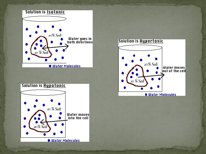

Fig. 7 -13 Hypotonic solution H 2 O Isotonic solution Hypertonic solution H 2 O (a) Animal cell Lysed H 2 O Normal H 2 O Shriveled H 2 O (b) Plant cell Turgid (normal) Flaccid Plasmolyzed

Passive Transport Cont’d �Hypertonic or hypotonic environments create osmotic problems for organisms �Osmoregulation, the control of water balance, is a necessary adaptation for life in such environments � Ex) The protist Paramecium, which is hypertonic to its pond water environment, has a contractile vacuole that acts as a pump

Passive Transport Cont’d �Cell walls of plants, bacteria, fungi, algae, and some archaea help maintain water balance � plant cell in a hypotonic solution cell swells � turgid (firm) � plant cell and its surroundings are isotonic �there is no net movement of water into the cell; the cell becomes flaccid (limp), and the plant may wilt � Plant cell in hypertonic environment � cells lose water; eventually, the membrane pulls away from the wall, a usually lethal effect called plasmolysis

Passive Transport Cont’d: Facilitated Diffusion � In facilitated diffusion, transport proteins speed the passive movement of molecules across the plasma membrane � Most transport proteins are very specific � 2 types of transport proteins �Channel proteins and carrier proteins

Passive Transport Cont’d: Facilitated Diffusion via Channel Proteins � Channel proteins provide corridors that allow a specific molecule or ion to cross the membrane � Aquaporins, for facilitated diffusion of water – speed up osmosis � Ion channels that open or close in response to a stimulus (gated channels)

Passive Transport Cont’d: Facilitated Diffusion via Carrier Proteins � Carrier proteins transport substances out of or into the cell by facilitated diffusion and active transport. � Each designed to recognize only ONE substance or ONE group of very similar substances. � Diffusion of sugars, amino acids, nucleoside. � Uptake of glucose. � Transportation of salts, glucose, and amino acids undergo a subtle change in shape that translocates the solute-binding site across the membrane

Active Transport �Substances diffuse against their concentration gradient � From an area of low concentration to high concentration �Work must be done to move substances from low to high � performed by specific proteins embedded in the membranes � Allows cells to maintain concentration gradients that differ from their surroundings � Ex) sodium-potassium pump in nerve conduction

Active Transport Cont’d: Ion Pumps � Membrane potential is the voltage difference across a membrane � Voltage is created by differences in the distribution of positive and negative ions � Cells have a net negative internal charge � Favors passive transport of cations into the cell and anions out � Two combined forces, collectively called the electrochemical gradient, drive the diffusion (PASSIVELY) of ions across a membrane: � A chemical force (the ion’s concentration gradient) � An electrical force (the effect of the membrane potential on the ion’s movement) VIDEO
")
Figure 5 -31 c Potassium equilibrium potential Concentration gradient Electrical gradient (c)

�An electrogenic pump is a transport protein that generates voltage across a membrane (increasing membrane potential) � EX: Sodium-Potassium pump – major electrogenic pump of animals � 3 Na+out 2 K+ in = overall positive charge to the extracellular fluid �The main electrogenic pump of plants, fungi, and bacteria is a proton pump � These pumps generate voltage across membranes which stores energy for use for the cell (mitochondria and chloroplasts)

Figure 5 -14 Mechanism of the Na+-K+-ATPase 1 ECF ATP ADP 5 2 3 Na+ from ICF bind ICF K+ concentration less outside, more inside Na+ concentration is more outside, less inside Na+/K+ maintains these concentration gradients using ATP to pump 3 Na+ out and 2 K+ Nerve cells depend on this pump for responding to stimuli and transmitting impulses 2 K+ released into ICF Protein changes conformation. 4 2 K+ from ECF bind ATPase is phosphorylated with Pi from ATP. Protein changes conformation. 3 3 Na+ released into ECF

Cotransport � A single ATP-powered pump that transports one solute can indirectly drive the active transport of other solutes � Actively transported substance diffuses back passively through protein, its movement can be actively paired with another substance to move it against the concentration gradient

Active Transport Cont’d: Bulk transport: exocytosis and endocytosis �Large molecules cross the membrane in bulk via vesicles � polysaccharides and proteins �Bulk transport requires energy

Exocytosis �Transport vesicles migrate to the membrane, fuse with it, and release their contents �Many secretory cells use exocytosis to export their products � Pancreatic beta cells releasing insulin � Neurons releasing neurotransmiters � Plants making cell walls

Endocytosis �Cell takes in macromolecules by forming vesicles from the plasma membrane � reversal of exocytosis, involving different proteins �There are three types of endocytosis: � Phagocytosis (“cellular eating”) � Pinocytosis (“cellular drinking”) � Receptor-mediated endocytosis phagocytosis pinocytosis receptor-mediated endocytosis fuse with lysosome for digestion non-specific process triggered by ligand signal
 a cell engulfs a particle in a vacuole �")
� In phagocytosis (cellular eating) a cell engulfs a particle in a vacuole � The vacuole fuses with a lysosome to digest the particle � In pinocytosis, ( cellular drinking) phagocytosis fuse with lysosome for digestion molecules are taken up when extracellular fluid is “gulped” into tiny vesicles � In receptor-mediated endocytosis, (picky eater) binding of ligands to receptor-mediated endocytosis receptors triggers vesicle formation � A ligand is any molecule that binds specifically to a receptor site of another molecule pinocytosis non-specific process triggered by ligand signal

Figure 5 -20 Phagocytosis Bacterium Phagocyte Lysosome 1 The phagocytic white blood cell encounters a bacterium that binds to the cell membrane. 2 The phagocyte uses its cytoskeleton to push its cell membrane around the bacterium, creating a large vesicle, the phagosome. 3 The phagosome containing the bacterium separates from the cell membrane and moves into the cytoplasm. 4 The phagosome fuses with lysosomes containing digestive enzymes. 5 The bacterium is killed and digested within the vesicle.

Figure 5 -21 Receptor-mediated endocytosis and exocytosis 1 Ligand binds to membrane receptor. 9 Exocytosis Extracellular fluid 2 Receptor-ligand migrates to clathrin-coated pit. 8 Transport vesicle and cell membrane fuse (membrane recycling). Clathrin-coated pit 3 Endocytosis Receptor Clathrin 4 Vesicle loses clathrin coat. 7 Transport vesicle with receptors moves to the cell membrane. 5 Receptors and ligands separate. To lysosome or Golgi complex 6 Ligands go to lysosomes or Golgi for processing. Endosome Intracellular fluid

SUMMARY OF TRANSPORT Molecules move from a region of high concentration, spontaneously. In other words, this happens because it results in a release of energy. Molecules in this situation are described as going down their concentration gradient. The direction of the electrical driving force results in the ion moving toward a region where the opposite charge exists. The magnitude of the electrical force depends upon the size of the membrane potential and the charge of the ion. The greater the membrane potential or the charge of the ion the greater the electrical driving force. The total forces acting upon ions across a membrane is a combination of both chemical and electrical forces and is referred to as the electrochemical driving force. The net direction of this force is equal to the sum of both forces. If the chemical and electrical driving forces are in opposite directions, the point at which each equals the other is the equilibrium potential.

You should now be able to: Define the following terms: amphipathic molecules, aquaporins, diffusion 2. Explain how membrane fluidity is influenced by temperature and membrane composition 3. Distinguish between the following pairs or sets of terms: peripheral and integral membrane proteins; channel and carrier proteins; osmosis, facilitated diffusion, and active transport; hypertonic, hypotonic, and isotonic solutions 1.

You should now be able to: Explain how transport proteins facilitate diffusion 5. Explain how an electrogenic pump creates voltage across a membrane, and name two electrogenic pumps 6. Explain how large molecules are transported across a cell membrane 4.
- Slides: 47