Chapter 7 Membrane Structure and Function Plasma Membrane

Chapter 7 Membrane Structure and Function Plasma Membrane Rap

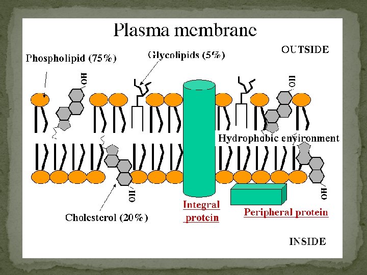
Makeup of the PM ① Phospholipids ② Cholesterol ③ Embedded Proteins ④ Glycoproteins ⑤ Glycolipids CARBOHYDRATES
 Phospholipids �most abundant lipid in the plasma membrane � amphipathic molecules, containing hydrophobic")
1) Phospholipids �most abundant lipid in the plasma membrane � amphipathic molecules, containing hydrophobic and hydrophilic regions � J. Singer & G. Nicolson (1972) proposed that the membrane is a mosaic of proteins dispersed within the bilayer, with only the hydrophilic regions exposed to water (fluid mosaic model) Plasma Membrane Video

Phospholipids Cont’d � Freeze-fracture studies of the plasma membrane supported the fluid mosaic model � Freeze-fracture is a specialized preparation technique that splits a membrane along the middle of the phospholipid bilayer TECHNIQUE RESULTS Extracellular layer Knife Plasma membrane Proteins Inside of extracellular layer Cytoplasmic layer Inside of cytoplasmic layer

Phospholipids Cont’d �Phospholipids can move within the bilayer �As temperatures cool, membranes become more solid �Membranes rich in unsaturated fatty acids are more fluid that those rich in saturated fatty acids �Membranes must be fluid to work properly; they are usually about as fluid as salad oil
 Cholesterol �Ring structure reduces membrane fluidity at moderate temperatures by reducing phospholipid movement")
2) Cholesterol �Ring structure reduces membrane fluidity at moderate temperatures by reducing phospholipid movement � low temperatures hinders solidification by disrupting the regular packing of phospholipids. � warm temperatures (such as 37°C), cholesterol restrains movement of phospholipids �Cholesterol is known as “temperature buffer”. Cholesterol (c) Cholesterol within the animal cell membrane
 Proteins �collage of different proteins embedded in the fluid matrix of the lipid")
3) Proteins �collage of different proteins embedded in the fluid matrix of the lipid bilayer � Proteins determine most of the membrane’s specific functions �The two sides of a membrane have different protein and lipid compositions. • Peripheral proteins are bound to the surface of the membrane Not embedded • Loosely bound to surface of membrane • Integral proteins penetrate the hydrophobic core • transmembrane proteins span the membrane • hydrophobic regions consist of one or more stretches of nonpolar amino acids, often coiled into alpha helices • Hydrophilic regions exposed to inside/outside of cell

Proteins Cont’d
 substances and ions across")
Proteins Cont’d �Transport proteins allow passage of large hydrophilic (polar) substances and ions across the membrane (FACILITATED DIFFUSION) 1) channel proteins have a hydrophilic channel that certain molecules or ions can use as a tunnel �Channel proteins called aquaporins facilitate the passage of water 2) carrier proteins bind to molecules and change shape to shuttle them across the membrane �A transport protein is specific for the substance it moves

The Permeability of the Lipid Bilayer �Small, uncharged polar and nonpolar molecules can freely pass through a cell membrane � Hydrophobic (nonpolar) molecules, such as hydrocarbons, can dissolve in the lipid bilayer and pass through the membrane rapidly �Hydrocarbons, CO 2 and O 2 � Polar molecules, such as sugars, do not cross the membrane easily �C 6 H 12 O 6, or charged molecules

Proteins Cont’d �Six major functions � Transport � Enzymatic activity � Signal transduction � Cell-cell recognition � Intercellular joining � Attachment to the cytoskeleton and extracellular matrix (ECM)

The Role of Membrane Carbohydrates in Cell-Cell Recognition �Cells recognize each other by binding to surface molecules, usually carbohydrates �Membrane carbohydrates may be covalently bonded to lipids (forming glycolipids) or more commonly to proteins (forming glycoproteins) �Carbohydrates on the external side of the plasma membrane vary among species, individuals, and even cell types in an individual Copyright © 2008 Pearson Education, Inc. , publishing as Pearson Benjamin Cummings
 Glycoproteins & 5) Glycolipids � Carbohydrates are third major component of plasma membranes")
4) Glycoproteins & 5) Glycolipids � Carbohydrates are third major component of plasma membranes � found on the exterior surface of cells � bound either to proteins (forming glycoproteins) � or bound to lipids (forming glycolipids) � Chains can be either straight or branched � Form specialized sites on the cell surface that allow cells to recognize each other. � unique patterns that allow the cell to be recognized � allows the immune system to differentiate between body cells (called “self”) and foreign cells or tissues (called “non-self”). � Similar types of glycoproteins and glycolipids are found on the surfaces of viruses and may change frequently, preventing immune cells from recognizing and attacking them.

Glycoproteins & Glycolipids Cont’d

Passive Transport: � Substances diffuse down their concentration gradient (the difference in concentration of a substance from one area to another) � From an area of high concentration to low concentration � No work must be done to move substances down the concentration gradient � O 2 gets into cells this way for cellular respiration � Diffusion is the tendency for molecules to spread out evenly into the available space � At dynamic equilibrium, as many molecules cross one way as cross in the other direction � This is not static – even at equilibrium molecules are still moving.
 WATER Net diffusion (a)")
Fig. 7 -11 a Molecules of dye Membrane (cross section) WATER Net diffusion (a) Diffusion of one solute Net diffusion Equilibrium
 Diffusion of two solutes The diffusion")
Fig. 7 -11 b Net diffusion Equilibrium (b) Diffusion of two solutes The diffusion of one solute is unaffected by the diffusion of another solute.

Passive Transport Cont’d �Osmosis is the diffusion of water across a selectively permeable membrane � The direction of osmosis is determined only by a difference in total solute concentration. �Water diffuses across a membrane from the region of lower solute concentration to the region of higher solute concentration � or you can think [high water] to [low water]
 Higher concentration of sugar H 2")
Fig. 7 -12 Lower concentration of solute (sugar) Higher concentration of sugar H 2 O Selectively permeable membrane Osmosis Same concentration of sugar

Passive Transport Cont’d �Tonicity is the ability of a solution to cause a cell to gain or lose water � Isotonic solution: Solute concentration is the same as that inside the cell; no net water movement across the plasma membrane � Hypertonic solution: Solute concentration is greater than that inside the cell; cell loses water � Hypotonic solution: Solute concentration is less than that inside the cell; cell gains water
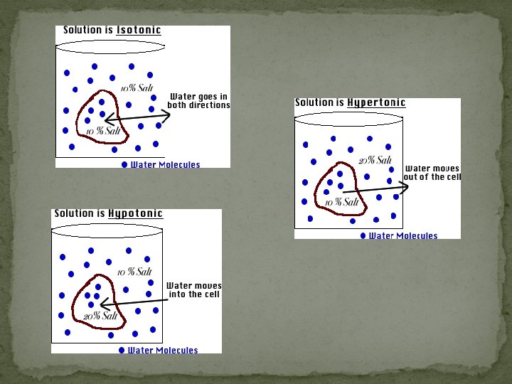

Fig. 7 -13 Hypotonic solution H 2 O Isotonic solution Hypertonic solution H 2 O (a) Animal cell Lysed H 2 O Normal H 2 O Shriveled H 2 O (b) Plant cell Turgid (normal) Flaccid Plasmolyzed

Passive Transport Cont’d �Hypertonic or hypotonic environments create osmotic problems for organisms �Osmoregulation, the control of water balance, is a necessary adaptation for life in such environments � Ex) The protist Paramecium, which is hypertonic to its pond water environment, has a contractile vacuole that acts as a pump

Passive Transport Cont’d �Cell walls of plants, bacteria, fungi, algae, and some archaea help maintain water balance � plant cell in a hypotonic solution cell swells � turgid (firm) � plant cell and its surroundings are isotonic �there is no net movement of water into the cell; the cell becomes flaccid (limp), and the plant may wilt � Plant cell in hypertonic environment � cells lose water; eventually, the membrane pulls away from the wall, a usually lethal effect called plasmolysis

Passive Transport Cont’d: Facilitated Diffusion � In facilitated diffusion, transport proteins speed the passive movement of molecules across the plasma membrane � Most transport proteins are very specific � 2 types of transport proteins �Channel proteins and carrier proteins

Passive Transport Cont’d: Facilitated Diffusion via Channel Proteins � Channel proteins provide corridors that allow a specific molecule or ion to cross the membrane � Aquaporins, for facilitated diffusion of water – speed up osmosis � Ion channels that open or close in response to a stimulus (gated channels)

Passive Transport Cont’d: Facilitated Diffusion via Carrier Proteins � Carrier proteins transport substances out of or into the cell by facilitated diffusion and active transport. � Each designed to recognize only ONE substance or ONE group of very similar substances. � Diffusion of sugars, amino acids, nucleoside. � Uptake of glucose. � Transportation of salts, glucose, and amino acids undergo a subtle change in shape that translocates the solute-binding site across the membrane

Active Transport �Substances diffuse against their concentration gradient � From an area of low concentration to high concentration �Work must be done to move substances from low to high � performed by specific proteins embedded in the membranes � Allows cells to maintain concentration gradients that differ from their surroundings � Ex) sodium-potassium pump

Figure 5 -14 Mechanism of the Na+-K+-ATPase 1 ECF ATP ADP 5 2 3 Na+ from ICF bind ICF ATPase is phosphorylated with Pi from ATP. 2 K+ released into ICF Protein changes conformation. 4 2 K+ from ECF bind Protein changes conformation. 3 3 Na+ released into ECF

Active Transport Cont’d: Ion Pumps �Membrane potential is the voltage difference across a membrane � Voltage is created by differences in the distribution of positive and negative ions �Cells have a net negative internal charge
")
Figure 5 -31 c Potassium equilibrium potential Concentration gradient Electrical gradient (c)

�Two combined forces, collectively called the electrochemical gradient, drive the diffusion of ions across a membrane: � A chemical force (the ion’s concentration gradient) � An electrical force (the effect of the membrane potential on the ion’s movement)

�An electrogenic pump is a transport protein that generates voltage across a membrane � EX: Sodium-Potassium pump – major electrogenic pump of animals � 3 Na+out 2 K+ in = overall 1 positive charge to the extracellular fluid �The main electrogenic pump of plants, fungi, and bacteria is a proton pump �These pumps generate voltage across membranes which stores energy for use for the cell

Active Transport Cont’d: Bulk transport: exocytosis and endocytosis �Large molecules cross the membrane in bulk via vesicles � polysaccharides and proteins �Bulk transport requires energy

Exocytosis �Transport vesicles migrate to the membrane, fuse with it, and release their contents �Many secretory cells use exocytosis to export their products � Pancreatic beta cells releasing insulin � Neurons releasing neurotransmiters � Plants making cell walls

Endocytosis �Cell takes in macromolecules by forming vesicles from the plasma membrane � reversal of exocytosis, involving different proteins �There are three types of endocytosis: � Phagocytosis (“cellular eating”) � Pinocytosis (“cellular drinking”) � Receptor-mediated endocytosis

Figure 5 -20 Phagocytosis Bacterium Phagocyte Lysosome 1 The phagocytic white blood cell encounters a bacterium that binds to the cell membrane. 2 The phagocyte uses its cytoskeleton to push its cell membrane around the bacterium, creating a large vesicle, the phagosome. 3 The phagosome containing the bacterium separates from the cell membrane and moves into the cytoplasm. 4 The phagosome fuses with lysosomes containing digestive enzymes. 5 The bacterium is killed and digested within the vesicle.
 a cell engulfs a particle in a vacuole �")
� In phagocytosis (cellular eating) a cell engulfs a particle in a vacuole � The vacuole fuses with a lysosome to digest the particle � In pinocytosis, ( cellular drinking) molecules are taken up when extracellular fluid is “gulped” into tiny vesicles � In receptor-mediated endocytosis, (picky eater) binding of ligands to receptors triggers vesicle formation � A ligand is any molecule that binds specifically to a receptor site of another molecule

Figure 5 -21 Receptor-mediated endocytosis and exocytosis 1 Ligand binds to membrane receptor. 9 Exocytosis Extracellular fluid 2 Receptor-ligand migrates to clathrin-coated pit. 8 Transport vesicle and cell membrane fuse (membrane recycling). Clathrin-coated pit 3 Endocytosis Receptor Clathrin 4 Vesicle loses clathrin coat. 7 Transport vesicle with receptors moves to the cell membrane. 5 Receptors and ligands separate. To lysosome or Golgi complex 6 Ligands go to lysosomes or Golgi for processing. Endosome Intracellular fluid

SUMMARY OF TRANSPORT

You should now be able to: Define the following terms: amphipathic molecules, aquaporins, diffusion 2. Explain how membrane fluidity is influenced by temperature and membrane composition 3. Distinguish between the following pairs or sets of terms: peripheral and integral membrane proteins; channel and carrier proteins; osmosis, facilitated diffusion, and active transport; hypertonic, hypotonic, and isotonic solutions 1.

You should now be able to: Explain how transport proteins facilitate diffusion 5. Explain how an electrogenic pump creates voltage across a membrane, and name two electrogenic pumps 6. Explain how large molecules are transported across a cell membrane 4.
- Slides: 43