Chapter 7 Membrane Structure and Function CELL MEMBRANE

Chapter 7 Membrane Structure and Function

CELL MEMBRANE: Basics: Regulates flow in and out of the cell Composed of phospholipids and proteins - some carbohydrates and lipids SELECTIVELY PERMEABLE - allows some things across more easily than others Structure varies from one cell type to another determines function of the membrane
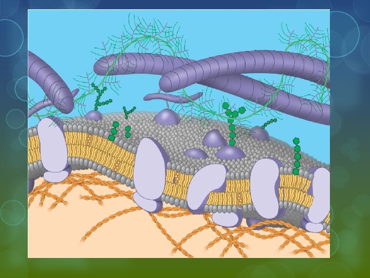

Membrane Structure § Cellular membranes are fluid mosaics of lipids and proteins § Phospholipids § Are the most abundant lipid in the plasma membrane § Are amphipathic, containing both hydrophobic and hydrophilic regions § Organized in a bilayer

WATER Hydrophilic head Hydrophobic tail WATER

Membrane Structure The membrane is a fluid structure with a “mosaic” of various proteins embedded in it Proteins are either integral or peripheral Integral – embedded in the membrane and traverse the phospholipid bilayer (transmembranal) Peripheral – on the surface - on both the inner and outer surfaces - surfaces differ – membrane is bifacial or asymmetric

Singer and Nicolson Model § Proposed that membrane proteins are dispersed and individually inserted into the phospholipid bilayer Hydrophobic region of protein Phospholipid bilayer Figure 7. 3 Hydrophobic region of protein

Freeze-fracture studies of the plasma membrane § Supported the fluid mosaic model of membrane structure • A cell is frozen and fractured with a knife. The fracture plane often follows the hydrophobic interior of a membrane, splitting the phospholipid bilayer into two separated layers. The membrane proteins go wholly with one of the layers. § Coat the split layers with a metal, usually gold or silver, and examine it under an electron scanning microscope

APPLICATION A cell membrane can be split into its two layers, revealing the ultrastructure of the membrane’s interior. TECHNIQUE Extracellular layer Proteins Knife Plasma membrane RESULTS Cytoplasmic layer These SEMs show membrane proteins (the “bumps”) in the two layers, demonstrating that proteins are embedded in the phospholipid bilayer.

Chemical Nature of Bilayer Amphipathic Parts Match Up - non-polar portions of proteins embed in the hydrophobic tails of the bilayer - polar portions of proteins embed in the hydrophilic heads or jut into the interior or exterior of the cell

The Fluidity of Membranes § Membrane parts are held together by hydrophobic interactions - weak bonds so phospholipids move around laterally (Side to side) § very rare for a phospholipid to flip-flop from one side of the membrane to the other
 (a) Movement of phospholipids")
The Fluidity of Membranes Lateral movement (~107 times per second) (a) Movement of phospholipids Figure 7. 5 A Flip-flop (~ once per month)

Movement of Molecules § phospholipids move quickly § Slows with temperature decrease until freezes § Unsaturated tails - more flowing § Saturated tails - more viscous
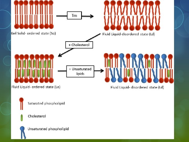

Role of cholesterol wedges between phospholipids and stabilizes structure § hinders close packing decreases freezing point of membrane Cholesterol (c) Cholesterol within the animal cell membrane Figure 7. 5

Proteins move slowly § larger § anchored to the cytoskeleton § some travel along the cytoskeleton § some immobile

Movement of Proteins in the Plasma Membrane EXPERIMENT Researchers labeled the plasma mambrane proteins of a mouse cell and a human cell with two different markers and fused the cells. Using a microscope, they observed the markers on the hybrid cell. RESULTS Membrane proteins + Mouse cell Human cell Hybrid cell Figure 7. 6 Mixed proteins after 1 hour CONCLUSION The mixing of the mouse and human membrane proteins indicates that at least some membrane proteins move sideways within the plane of the plasma membrane.
 Proteins: § Integral - transmembranal - interior")
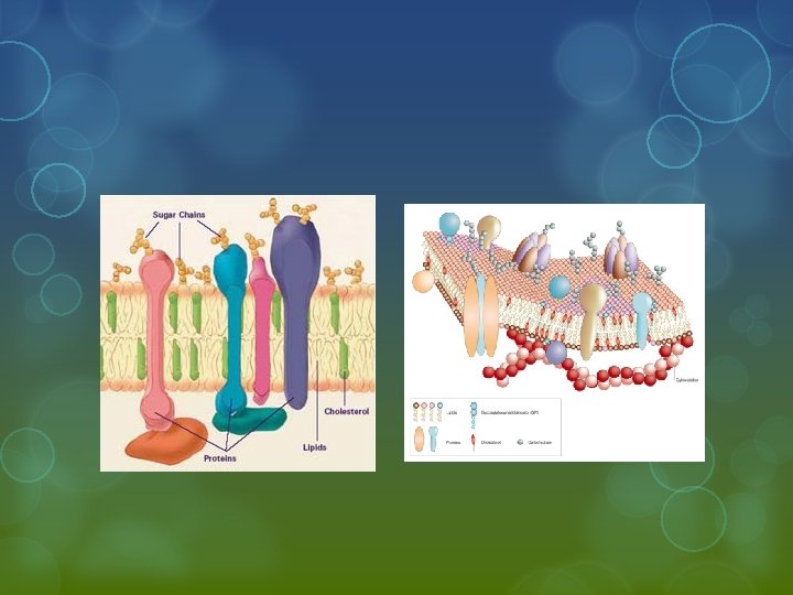
TYPES OF STRUCTURES ATTACHED TO MEMBRANES 1) Proteins: § Integral - transmembranal - interior and exterior end § Peripheral - surface - on outside attached to membrane but not imbedded § - attached to extracellular matrix and cytoskelton 2) Carbohydrates – § glycocalyx § glycoproteins § glycolipids FUNCTIONS: § § cell stickyness cell communication and recognition
 Glycoprotein Carbohydrate Glycolipid EXTRACELLULAR")
Membrane Proteins and Their Functions Fibers of extracellular matrix (ECM) Glycoprotein Carbohydrate Glycolipid EXTRACELLULAR SIDE OF MEMBRANE Microfilaments of cytoskeleton Figure 7. 7 Cholesterol Peripheral protein Integral CYTOPLASMIC SIDE protein OF MEMBRANE

Integral proteins § Penetrate the hydrophobic core of the lipid bilayer § Are often transmembrane proteins, completely spanning the membrane EXTRACELLULAR SIDE N-terminus C-terminus Figure 7. 8 a Helix CYTOPLASMIC SIDE

Peripheral proteins - appendages loosely bound to the surface of the membrane
 Transport. (left) A protein")
An overview of six major functions of membrane proteins (a) Transport. (left) A protein that spans the membrane may provide a hydrophilic channel across the membrane that is selective for a particular solute. (right) Other transport proteins shuttle a substance from one side to the other by changing shape. Some of these proteins hydrolyze ATP as an energy ssource to actively pump substances across the membrane. ATP (b) Enzymatic activity. A protein built into the membrane may be an enzyme with its active site exposed to substances in the adjacent solution. In some cases, several enzymes in a membrane are organized as a team that carries out sequential steps of a metabolic pathway. (c) Signal transduction. A membrane protein may have a binding site with a specific shape that fits the shape of a chemical messenger, such as a hormone. The external messenger (signal) may cause a conformational change in the protein (receptor) that relays the message to the inside of the cell. Figure 7. 9 Enzymes Signal Receptor
 Cell-cell recognition. Some glyco-proteins serve as identification tags that are specifically recognized by")
(d) Cell-cell recognition. Some glyco-proteins serve as identification tags that are specifically recognized by other cells. Glycoprotein (e) Intercellular joining. Membrane proteins of adjacent cells may hook together in various kinds of junctions, such as gap junctions or tight junctions (see Figure 6. 31). (f) Attachment to the cytoskeleton and extracellular matrix (ECM). Microfilaments or other elements of the cytoskeleton may be bonded to membrane proteins, a function that helps maintain cell shape and stabilizes the location of certain membrane proteins. Proteins that adhere to the ECM can coordinate extracellular and intracellular changes (see Figure 6. 29). Figure 7. 9

Synthesis and Sidedness of Membranes § Secreted proteins are on the inside of secretory vesicles § Proteins on the outside of the cell membrane are on the inside of a secretory vesicle. § Proteins on the inside of the cell membrane are on the outside of the secretory vesicle.

1 Transmembrane glycoproteins Secretory protein ER Glycolipid Golgi apparatus 2 Vesicle 3 Plasma membrane: Cytoplasmic face 4 Secreted protein Figure 7. 10 Extracellular face Transmembrane glycoprotein Membrane glycolipid

Traffic Across Membranes § movement of materials is dependent upon the make up of the membrane § certain substances move more easily § certain substances move more quickly

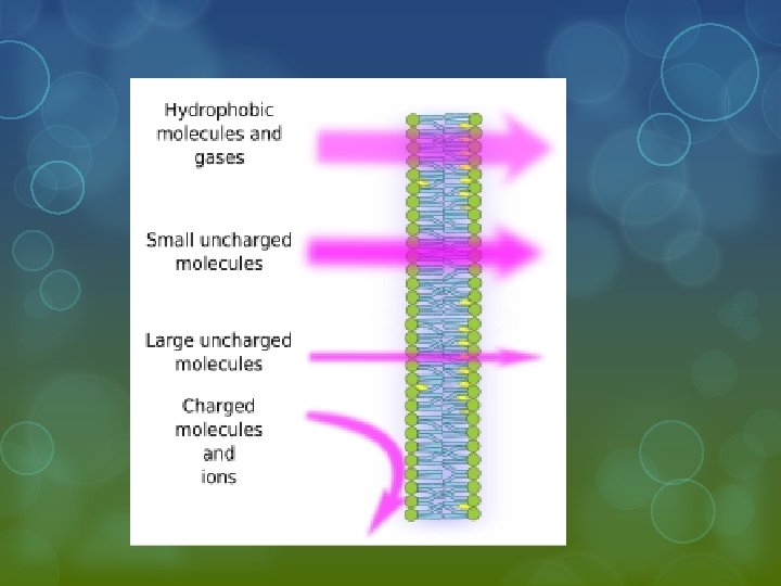
The Permeability of the Lipid Bilayer Hydrophobic molecules § Are lipid soluble and can pass through the membrane rapidly Polar molecules § Do not cross the membrane rapidly Transports: § hydrocarbons § carbon dioxide § oxygen § small polar molecules with a neutral charge (H 2 O) Impedes: § ions § polar molecule § large, uncharged polar molecules - glucose

Transport Proteins: § Create channels for ions and polar molecules to pass through the lipid bilayer § Selectively transport like enzymes - substrate specific § each protein transports one specific molecule § Methods: § hydrophilic channel § bind substance and move it across the bilayer

Types of Transport Across Membrane Passive vs. Active Passive: does not require energy § downhill process § moves from high concentration to low concentration with the concentration gradient (comparison of concentration levels across a membrane) Active: Requires energy § uphill process § moves from low concentration to high concentration § against the concentration gradient § requires transport proteins

Types of Passive Transport Diffusion Osmosis Facilitated Diffusion

Diffusion § based on thermal kinetics – particles move throughout a space until they are distributed evenly § movement of particles from high concentration to low concentration until net flow is zero § NET FLOW: change in concentrations due to flow across membrane § each substance moves independently of the others § continues until equilibrium is reached
 Diffusion of one solute. The membrane has pores large enough for molecules")
Diffusion (a) Diffusion of one solute. The membrane has pores large enough for molecules of dye to pass through. Random movement of dye molecules will cause some to pass through the pores; this will happen more often on the side with more molecules. The dye diffuses from where it is more concentrated to where it is less concentrated (called diffusing down a concentration gradient). This leads to a dynamic equilibrium: The solute molecules continue to cross the membrane, but at equal rates in both directions. Figure 7. 11 A Molecules of dye Membrane (cross section) Net diffusion Equilibrium
 Diffusion of two solutes. Solutions of two different dyes are separated by a")
(b) Diffusion of two solutes. Solutions of two different dyes are separated by a membrane that is permeable to both. Each dye diffuses down its own concentration gradient. There will be a net diffusion of the purple dye toward the left, even though the total solute concentration was initially greater on the left side. Net diffusion Figure 7. 11 B Net diffusion Equilibrium

Osmosis special case of diffusion - APPLIES TO WATER ONLY § movement of water across the membrane to balance solute concentration § movement of water continues directionally until solute concentrations are equal Based on relative comparisons of solute concentration (tonicity) § hypertonic: solution with more solute § hypotonic: solution with less solute § isotonic: solutions with equal concentrations Movement of water: 2 ways to say the same thing: § from least solute to higher solute § from high water concentration to low water concentration -

Net Flow

Water Balance of Cells Without Walls Isotonic § The concentration of solutes is the same as it is inside the cell § There will be no net movement of water

Hypertonic § The concentration of solutes is greater than it is inside the cell § The cell will lose water

Hypotonic § The concentration of solutes is less than it is inside the cell § The cell will gain water

. 13 Water balance in cells without walls Identify the Tonicity H 2 O Lysed H 2 O Normal H 2 O Shriveled

. 13 Water balance in cells without walls Hypotonic solution H 2 O Lysed Isotonic solution Hypertonic solution H 2 O Normal H 2 O Shriveled

Water Balance of Cells with Walls Hypertonic environment - More solute outside of cell - Cell loses water - Becomes plasmolyzed

Water Balance of Cells with Walls § Isotonic § Same level of solute § Net flow of water is zero § No internal pressure – plant cells have no support – flaccid § Plants wilt

Water Balance of Cells with Walls Hypotonic Environment More solute inside than out Positive flow of water into the cell § plant cell is turgid § healthy state in most plants § Cells don’t rupture because of strength of the cell wall § Combination of osmotic pressure and the pressure of the walls is called the cell’s water potential

. 13 Water balance in cells with walls Identify the Tonicity H 2 O

. 13 Water balance in cells with walls Hypotonic solution Isotonic solution H 2 O Turgid (normal) Hypertonic solution H 2 O Flaccid H 2 O Plasmolyzed

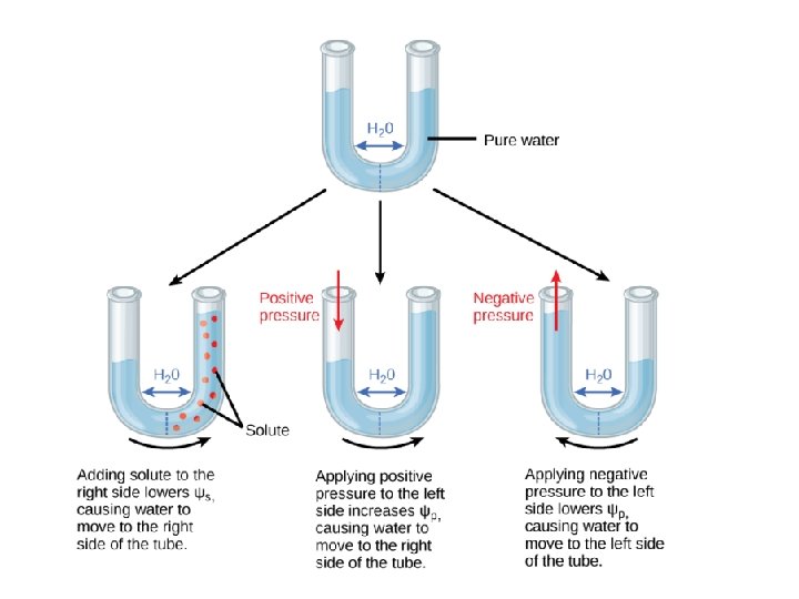
Water Potential § determines the flow of water § Water flows from high water potential to low water potential § High pressure from the walls raises the water potential (tendency to lose water) § High levels of solute in the cell lower water potential (tendency to gain water) § Water flows into/out of the cell until the water potential reaches zero – pressure from the cell walls prevents any more water from entering the cell.

Water Potential Equation § Ψ = ΨP + Ψπ ΨP = Pressure Potential § § Ψπ = Osmotic Potential § Ψπ = − Mi. RT or Ψπ = − Ci. RT § M or C = osmotic molar concentration § R = pressure constant (Gas Laws) § T = temperature (K) § i = ionization constant (with plants the solute is mainly sucrose which has an ionization constant of 1 because sucrose does not ionize)

Facilitated Diffusion: Passive Transport Aided by Proteins Transmembranal proteins speed the movement of molecules across the plasma membrane passive - no energy required - moves with the concentration gradient Regulation of Facilitated Diffusion: § protein saturation § can only transport so much at once § proteins are specialized for a specific substance § protein inhibition § similar molecules to normal transport substance bind to protein and block path of transport § THESE ARE ONLY PHYSICAL CHANGES NOT CATALYZED REACTIONS

Mechanism of Transport Conformational changes - binding and release of substance causes change in protein - moves substance across membrane § gated channels - stimulus causes protein to open and allow the flow of the substance - the stimulus is not the substance transported

Facilitated Diffusion

Active Transport § pumping of solutes against the concentration gradient § moving uphill - requires ATP or a membrane potential § membrane potential: electrical charge built up across the cell membrane due to unequal balance of anions and/or cations -

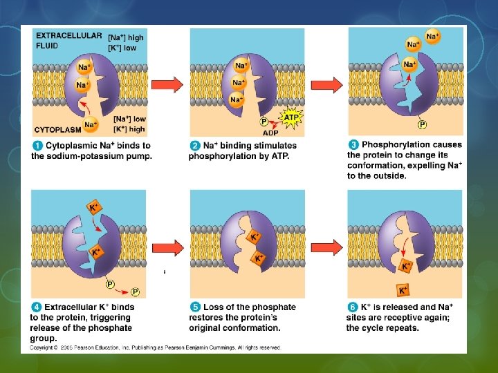
Types of Active Transport Use of ATP: § phosphorylation reaction - adds phosphate to transport molecule and causes a conformational change § EX: sodium-potassium pump

Na/K Ion Pump Generates an ELECTROCHEMICAL GRADIENT § Unbalanced concentration of ions § creates a new dynamic for diffusion ions now move from high concentration and high positive charge § important for Nerve Impulses

COTRANSPORT § movement of one type of ion out allows for the movement of another substance in when the ion diffuses back in through a cotransport protein ATP – + H+ + – H+ Proton pump H+ – + Sucrose-H+ cotransporter – – H+ H+ H+ Diffusion of H+ H+ + + Sucrose

Other Types of Active Transport § Use of Energized Electrons To Generate Chemical Gradients Ex: Kreb’s Cycle and Light Dependent Reaction of Photosynthesis – Drives the production of ATP

Electron Transport Chain In the Kreb’s Cycle and Photosynthesis

§ Passive and active transport compared Passive transport. Substances diffuse spontaneously down their concentration gradients, crossing a membrane with no expenditure of energy by the cell. The rate of diffusion can be greatly increased by transport proteins in the membrane. Active transport. Some transport proteins act as pumps, moving substances across a membrane against their concentration gradients. Energy for this work is usually supplied by ATP Figure 7. 17 Diffusion. Hydrophobic molecules and (at a slow rate) very small uncharged polar molecules can diffuse through the lipid bilayer. Facilitated diffusion. Many hydrophilic substances diffuse through membranes with the assistance of transport proteins, either channel or carrier proteins.

BULK MOVEMENT active transport that requires change in the cell membrane § movement of large molecules (proteins and polysaccharides)

Exocytosis - movement of materials out of the cell transport vesicle fuses with cell membrane dumping contents into extracellular fluid § Examples: insulin neurotransmitters glycocalyx

Endocytosis - § movement of materials into the cell results in the formation of a vacuole or vesicle Three types § Phagocytosis § Pinocytosis § Receptor Mediated Endocytosis
 Phagocytosis - cellular eating § cell membrane surrounds and engulfs large particles of")
1) Phagocytosis - cellular eating § cell membrane surrounds and engulfs large particles of food - becomes a food vacuole to be fused with lysosome
 Pinocytosis - cellular drinking § cell membrane draws in extra cellular fluid and")
2) Pinocytosis - cellular drinking § cell membrane draws in extra cellular fluid and forms a small vesicle – Non-discriminatory process

Three types of endocytosis In phagocytosis, a cell engulfs a particle by Wrapping pseudopodia around it and packaging it within a membraneenclosed sac large enough to be classified as a vacuole. The particle is digested after the vacuole fuses with a lysosome containing hydrolytic enzymes. PHAGOCYTOSIS EXTRACELLULAR CYTOPLASM FLUID Pseudopodium 1 µm Pseudopodium of amoeba “Food” or other particle Bacterium Food vacuole An amoeba engulfing a bacterium via phagocytosis (TEM). In pinocytosis, the cell “gulps” droplets of extracellular fluid into tiny vesicles. It is not the fluid itself that is needed by the cell, but the molecules dissolved in the droplet. Because any and all included solutes are taken into the cell, pinocytosis is nonspecific in the substances it transports. Figure 7. 20 PINOCYTOSIS 0. 5 µm Plasma membrane Pinocytosis vesicles forming (arrows) in a cell lining a small blood vessel (TEM). Vesicle
 Receptor Mediated Endocytosis specific contents are taken into cell regulated by surface proteins")
3) Receptor Mediated Endocytosis specific contents are taken into cell regulated by surface proteins that recognize specific substances called LIGANDS § receptor proteins usually gathered in one area of the cell membrane (coated pits) concentrates the gathering effort so cell can absorb concentrated amounts of a specific substance

§ Receptor Mediated Endocytosis Animation

RECEPTOR-MEDIATED ENDOCYTOSIS Receptor-mediated endocytosis enables the cell to acquire bulk quantities of specific substances, even though those substances may not be very concentrated in the extracellular fluid. Embedded in the membrane are proteins with specific receptor sites exposed to the extracellular fluid. The receptor proteins are usually already clustered in regions of the membrane called coated pits, which are lined on their cytoplasmic side by a fuzzy layer of coat proteins. Extracellular substances (ligands) bind to these receptors. When binding occurs, the coated pit forms a vesicle containing the ligand molecules. Notice that there are relatively more bound molecules (purple) inside the vesicle, other molecules (green) are also present. After this ingested material is liberated from the vesicle, the receptors are recycled to the plasma membrane by the same vesicle. Coat protein Receptor Coated vesicle Ligand Coated pit A coated pit and a coated vesicle formed during receptormediated endocytosis (TEMs). Coat protein Plasma membrane 0. 25 µm

CRASH COURSE
- Slides: 71