Chapter 7 Anatomy and Physiology 2009 Delmar Cengage


- Slides: 128

Chapter 7 Anatomy and Physiology © 2009 Delmar, Cengage Learning

7: 1 Basic Structure of the Human Body • The normal function of the human body is compared to an organized machine • The machine malfunctions, disease occurs • Anatomy: study of form and structure • Physiology: study of processes • Pathophysiology: study of how disease occurs and body’s response © 2009 Delmar, Cengage Learning

Protoplasm • Basic substance of life • Made of ordinary elements (e. g. , carbon, oxygen, hydrogen) • Scientists can combine these elements, but not create life © 2009 Delmar, Cengage Learning

Cells • • • Made of protoplasm Microscopic organisms Carry on all functions of life Body contains trillions of cells Vary in shape and size Perform different functions © 2009 Delmar, Cengage Learning

Basic Parts of Cells • • Cell membrane Cytoplasm Organelles Nucleus Nucleolus Chromatin Genome (continues) © 2009 Delmar, Cengage Learning

Basic Parts of Cells (continued) • • Centrosome Mitochondria Golgi apparatus Endoplasmic reticulum Vacuoles Lysosomes Pinocytic vesicles © 2009 Delmar, Cengage Learning

Mitosis • Asexual reproduction process used by most cells • Different types of cells reproduce at different rates • Process of mitosis—see Figure 7 -2 in text © 2009 Delmar, Cengage Learning

Meiosis • Process by which sex cells reproduce • Uses two separate cell divisions • Female cells (ova) and male cells (spermatozoa or sperm) divide to produce 23 chromosomes each • When ova and sperm combine, 46 chromosomes result to form zygote © 2009 Delmar, Cengage Learning

Tissues • Cells of same type joined together • 60%– 99% water • Groups of tissues – – Epithelial Connective Nerve Muscle © 2009 Delmar, Cengage Learning

Organs and Systems • Organs: two or more tissues joined together for a specific purpose • Systems: organs and other body parts joined together for a particular function © 2009 Delmar, Cengage Learning

Summary • • • Protoplasm is basic substance of life Protoplasm forms structural units called cells Cells combine to form tissue Tissues combine to form organs Organs and other parts combine to form systems • Systems work together to create miracle of human body © 2009 Delmar, Cengage Learning

7: 2 Body Planes/Directions/Cavities • Body planes: imaginary lines drawn through body at various levels to separate body into sections • Directional terms are created by planes • Transverse plane • Midsagittal or median plane • Frontal or coronal plane • Proximal and distal © 2009 Delmar, Cengage Learning

Cavities • Spaces within the body that contain vital organs • Dorsal or posterior cavity • Ventral or anterior cavities – Thoracic cavity – Abdominal cavity – Pelvic cavity • Three small cavities © 2009 Delmar, Cengage Learning

Abdominal Regions • Abdominal cavity is separated into regions or sections because it is so large • Quadrants – – RUQ LUQ RLQ LLQ (continues) © 2009 Delmar, Cengage Learning

Abdominal Regions (continued) • Regions – – – Epigastric Umbilical Hypogastric Hypochondriac Lumbar Iliac or inguinal © 2009 Delmar, Cengage Learning

7: 3 Integumentary System • Name for the skin and its structures • Called a membrane because it covers the body • Called an organ because it contains several kinds of tissues • Called a system because it has organs and other parts that work together for a particular function © 2009 Delmar, Cengage Learning

Layers of the Skin • Epidermis—outermost layer • Dermis—“true skin” • Subcutaneous fascia or hypodermis— the innermost layer © 2009 Delmar, Cengage Learning

Glands and Other Parts of the Skin • • Sudoriferous glands (sweat glands) Sebaceous glands (oil glands) Hair Nails © 2009 Delmar, Cengage Learning

Functions • • Protection Sensory perception Regulation of body temperature Storage Absorption Excretion Production © 2009 Delmar, Cengage Learning

Skin Color—Pigmentation • Skin color is inherited and is determined by pigments in the epidermis • Melanin • Carotene © 2009 Delmar, Cengage Learning

Skin Color—Albino • • Absence of skin pigments Skin has pinkish tint Hair is pale yellow or white Eyes are red in color and sensitive to light © 2009 Delmar, Cengage Learning

Skin Color—Abnormal • Erythema • Jaundice • Cyanosis © 2009 Delmar, Cengage Learning

Skin Eruptions • • Macules (macular rash) Papules (papular rash) Vesicles Pustules Crusts Wheals Ulcer © 2009 Delmar, Cengage Learning

Diseases and Abnormal Conditions • Acne vulgaris • Athlete’s foot • Skin cancer – Basal cell carcinoma – Squamous cell carcinoma – Melanoma (continues) © 2009 Delmar, Cengage Learning

Diseases and Abnormal Conditions (continued) • • • Dermatitis Eczema Impetigo Psoriasis Ringworm Verrucae/warts/plantar warts © 2009 Delmar, Cengage Learning

7: 4 Skeletal System • Made of organs called bones • Adult has 206 bones • Serves as framework for muscles, fat, and skin • Protects internal structures • Produces blood cells • Stores calcium, phosphorus, and fats © 2009 Delmar, Cengage Learning

Long Bones • • • Bones of the extremities Diaphysis Epiphysis Medullary canal Yellow marrow (continues) © 2009 Delmar, Cengage Learning

Long Bones (continued) • • Endosteum Red marrow Periosteum Articular cartilage © 2009 Delmar, Cengage Learning

Skeleton • Axial – Main trunk of body – Skull, spinal column, ribs, and sternum • Appendicular – Extremities – Shoulder girdle, arm bones, pelvic girdle, and leg bones © 2009 Delmar, Cengage Learning

Skull • • Cranial and facial bones Sutures Sinuses Foramina © 2009 Delmar, Cengage Learning

Cranial Bones • Eight bones of skull that surround and protect the brain • Frontal • Parietal (2) • Temporal (2) • Occipital • Ethmoid • Sphenoid © 2009 Delmar, Cengage Learning

Facial Bones • • 14 bones of skull that form facial features Mandible—lower jaw Maxilla (2)—upper jaw Zygomatic (2)—cheek Nasal (5)—upper part of nose Lacrimal (2)—inner aspect of eye Palatine (2)—hard palate (roof of mouth) © 2009 Delmar, Cengage Learning

Vertebrae • • Spinal column— 26 bones Protects the spinal cord Supports head and trunk Cervical (7)—neck Thoracic (12)—chest, attach to ribs Lumbar (5)—waist Sacrum (1)—back of pelvic girdle Coccyx (1)—tailbone © 2009 Delmar, Cengage Learning

Intervertebral Disks • Pads of cartilage separating vertebrae • Act as shock absorbers • Permit bending and twisting movements © 2009 Delmar, Cengage Learning

Ribs (costae) • • 12 pairs of long slender bones Attach to thoracic vertebrae True ribs—first 7 pairs; attach to sternum False ribs—last 5 pairs © 2009 Delmar, Cengage Learning

Sternum • • Breastbone Consists of 3 parts Two clavicles attach Ribs attach with cartilage © 2009 Delmar, Cengage Learning

Shoulder or Pectoral Girdle • 2 clavicles (collarbones) • 2 scapula (shoulder bones) • Upper arm bones attach to scapula © 2009 Delmar, Cengage Learning

Bones of the Arm • • • Humerus Radius Ulna Carpals Metacarpals Phalanges © 2009 Delmar, Cengage Learning

Bones of Pelvic Girdle • • Consists of 2 os coxae (coxal or hip bones) Symphysis pubis Ilium Ischium Pubis Acetabula Obturator foramen © 2009 Delmar, Cengage Learning

Bones of the Legs • • Femur Patella Tibia Fibula Tarsals Metatarsals Phalanges © 2009 Delmar, Cengage Learning

Joints • Where two or more bones join • Ligaments • Three types of joints – Diarthrosis or synovial – Amphiarthrosis – Synarthrosis © 2009 Delmar, Cengage Learning

Diseases and Abnormal Conditions • • • Arthritis Bursitis Fractures Dislocation Sprain Osteomyelitis (continues) © 2009 Delmar, Cengage Learning

Diseases and Abnormal Conditions (continued) • Osteoporosis • Ruptured disk • Abnormal curvature of spine – Kyphosis – Scoliosis – Lordosis © 2009 Delmar, Cengage Learning

7: 5 Muscular System • 600+ muscles in the body • Bundles of muscle fibers held together with connective tissue • Properties of muscles – – Excitability/irritability Contractibility Extensibility Elasticity © 2009 Delmar, Cengage Learning

Kinds of Muscles • Cardiac—involuntary • Visceral or smooth—involuntary • Skeletal—voluntary © 2009 Delmar, Cengage Learning

Functions of Muscles • • Attach to bones to provide movement Produce heat and energy Help maintain posture Protect internal organs © 2009 Delmar, Cengage Learning

Attachments to Bone • Tendon • Fascia • Origin and insertion © 2009 Delmar, Cengage Learning

Actions or Movements of Muscles • • • Adduction Abduction Flexion Extension Rotation Circumduction © 2009 Delmar, Cengage Learning

Muscle Tone • Partially contracted at all times • Muscle tone allows for state of readiness • Loss of muscle tone © 2009 Delmar, Cengage Learning

Diseases and Abnormal Conditions • • • Fibromyalgia Muscular dystrophy Duchenne’s dystrophy Myasthenia gravis Muscle spasms or cramps Strain © 2009 Delmar, Cengage Learning

7: 6 Nervous System • Complex and highly organized • Coordinates all of the many activities of the body • Allows the body to respond adapt to changes that occur both inside and outside the body © 2009 Delmar, Cengage Learning

Neuron • Neuron is also called a nerve cell • Basic structural unit of the nervous system • Parts of neuron – Cell body – Nucleus – Nerve fibers (dendrites, axon) © 2009 Delmar, Cengage Learning

Nerves • • • Combination of nerve fibers Located outside the brain and spinal cord Afferent—sensory nerves Efferent—motor nerves Associative—internuncial nerves © 2009 Delmar, Cengage Learning

Central Nervous System • Consists of two main divisions – – – Central nervous system (CNS) Brain and spinal cord Peripheral nervous system Somatic nervous system Autonomic nervous system © 2009 Delmar, Cengage Learning

Central Nervous System The Brain • • • Cerebrum Cerebellum Diencephalon Midbrain Pons Medulla oblongata © 2009 Delmar, Cengage Learning

Central Nervous System The Spinal Cord • • Continues down from medulla oblongata Surrounded and protected by the vertebrae Responsible for many reflex actions Carries sensory (afferent) messages to the brain • Carries motor (efferent) message from the brain © 2009 Delmar, Cengage Learning

Central Nervous System • • • Meninges Dura mater Arachnoid membrane Pia mater Ventricles © 2009 Delmar, Cengage Learning

Peripheral Nervous System • Cranial nerves • Spinal nerves • Autonomic nervous system – Sympathetic – Parasympathetic © 2009 Delmar, Cengage Learning

Diseases and Abnormal Conditions • • • Amyotrophic lateral sclerosis (ALS) Carpal tunnel syndrome Cerebral palsy Cerebrovascular accident (CVA) Encephalitis Epilepsy or seizure syndrome (continues) © 2009 Delmar, Cengage Learning

Diseases and Abnormal Conditions (continued) • • Hydrocephalus Meningitis Multiple sclerosis (MS) Neuralgia Paralysis Parkinson’s disease Shingles or herpes zoster © 2009 Delmar, Cengage Learning

7: 7 Special Senses • Senses allow body to react to the environment • See, hear, taste, smell, and to maintain balance • Body structures receive sensation, nerves carry to brain, brain interprets and responds to message © 2009 Delmar, Cengage Learning

Eye • • Sense of sight Light rays transmitted to the optic nerve Optic nerve relays information to brain Eye is well protected – – Bony socket Eyelids and eyelashes Lacrimal glands Conjunctiva © 2009 Delmar, Cengage Learning

Layers of the Eye • Sclera—outer • Choroid coat—middle • Retina—innermost © 2009 Delmar, Cengage Learning

Other Special Structures • • • Iris Pupil Lens Aqueous humor Vitreous humor Muscles © 2009 Delmar, Cengage Learning

Diseases and Abnormal Conditions • • • Amblyopia—lazy eye Astigmatism Cataract Conjuctivitis—pink eye Glaucoma (continues) © 2009 Delmar, Cengage Learning

Diseases and Abnormal Conditions (continued) • • • Hyperopia—farsightedness Myopia—nearsightedness Macular degeneration Presbyopia Strabismus © 2009 Delmar, Cengage Learning

Ear • Controls hearing and balance • Sound waves transmitted to the auditory nerve • Auditory nerve relays information to the brain for interpretation • Consists of the outer ear, middle ear, and inner ear © 2009 Delmar, Cengage Learning

Outer Ear • Pinna or auricle • Auditory canal • Tympanic membrane © 2009 Delmar, Cengage Learning

Middle Ear • • Malleus Incus Stapes Eustachian tube © 2009 Delmar, Cengage Learning

Inner Ear • • • Oval window Vestibule Cochlea Organ of Corti Semicircular canals © 2009 Delmar, Cengage Learning

Diseases and Abnormal Conditions • • • Hearing loss Meniere’s disease Otitis externa Otitis media Otosclerosis © 2009 Delmar, Cengage Learning

Sense of Taste • Taste receptors located on the tongue • Four main tastes – – Sweet Salty Sour Bitter © 2009 Delmar, Cengage Learning

Sense of Smell • Nose is the organ of smell • Olfactory receptors in nasal cavity • Impulses carried from the olfactory nerve to the brain for interpretation • Humans can detect over 6, 000 smells • Sense of taste and smell related © 2009 Delmar, Cengage Learning

Skin and General Senses • Sense receptors for pressure, heat, cold, touch, and pain located in the skin and connective tissue • Allows the human body to respond to its environment • Help body react to conditions that could cause injury © 2009 Delmar, Cengage Learning

7: 8 Circulatory System • Also known as the cardiovascular system • Consists of heart, blood vessels, blood • Transports oxygen and nutrients to all body cells • Transports carbon dioxide and metabolic materials away from the body cells © 2009 Delmar, Cengage Learning

Heart • • Muscular, hollow organ functions as pump Weight is less than one pound Location Three layers of tissue – Endocardium – Myocardium – Pericardium (continues) © 2009 Delmar, Cengage Learning

Heart (continued) • Septum • Heart chambers • Valves – – Tricuspid Pulmonary Mitral Aortic (continues) © 2009 Delmar, Cengage Learning

Heart (continued) • Cardiac cycle • Conductive pathways • Arrhythmias © 2009 Delmar, Cengage Learning

Blood Vessels • Blood is carried throughout the body in blood vessels • Arteries • Capillaries • Veins © 2009 Delmar, Cengage Learning

Blood • • Average adult: 4– 6 quarts Transports many substances Plasma Blood cells – Erythrocytes or red blood cells – Leukocytes or white blood cells – Thrombocytes © 2009 Delmar, Cengage Learning

Diseases and Abnormal Conditions • • • Anemia Aneurysm Arteriosclerosis Atherosclerosis Congestive heart failure (CHF) Embolus (continues) © 2009 Delmar, Cengage Learning

Diseases and Abnormal Conditions (continued) • • • Hemophilia Hypertension Leukemia Myocardial infarction—heart attack Phlebitis Varicose veins © 2009 Delmar, Cengage Learning

7: 9 Lymphatic System • Works with the circulatory system • Removes waste and excess fluids from the body tissues • Lymphatic vessels • Lymph nodes (glands) (continues) © 2009 Delmar, Cengage Learning

Lymphatic System (continued) • • Lymphatic ducts Lymph tissue Spleen Thymus © 2009 Delmar, Cengage Learning

Diseases and Abnormal Conditions • • • Adenitis Hodgkin’s disease Lymphangitis Splenomegaly Tonsillitis © 2009 Delmar, Cengage Learning

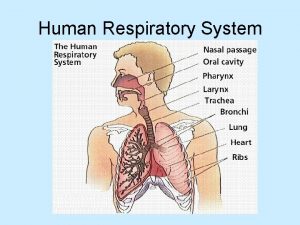
7: 10 Respiratory System • Lungs and air passages • Takes oxygen in and removes carbon dioxide • Works continuously or death occurs in 4– 6 minutes (continues) © 2009 Delmar, Cengage Learning

Respiratory System (continued) • • Nose Sinuses Pharynx—throat Larynx—voice box Trachea—windpipe Bronchi Alveoli Lungs © 2009 Delmar, Cengage Learning

Ventilation • • • Process of breathing Inspiration—inhalation Expiration—exhalation External respiration Internal respiration © 2009 Delmar, Cengage Learning

Diseases and Abnormal Conditions • • • Asthma Bronchitis Chronic obstructive pulmonary disease Emphysema Epistaxis—nosebleed (continues) © 2009 Delmar, Cengage Learning

Diseases and Abnormal Conditions (continued) • • • Influenza—flu Laryngitis Lung cancer Pleurisy Pneumonia (continues) © 2009 Delmar, Cengage Learning

Diseases and Abnormal Conditions (continued) • • • Rhinitis Sinusitis Sleep apnea Tuberculosis (TB) Upper respiratory infection (URI) © 2009 Delmar, Cengage Learning

7: 11 Digestive System • Physical and chemical breakdown of food for use by the body • System consists of the alimentary canal and the accessory organs © 2009 Delmar, Cengage Learning

Alimentary Canal • Long muscular tube • Begins at the mouth and ends at the anus • Accessory organs: salivary glands, tongue, teeth, liver, gallbladder, pancreas © 2009 Delmar, Cengage Learning

Mouth, Buccal, or Oral Cavity • • Receives food as it enters the body Actions in the mouth Teeth Tongue Hard palate Soft palate Salivary glands © 2009 Delmar, Cengage Learning

Pharynx or Throat • Carrier for both air and food • Carries food bolus to the esophagus • When bolus swallowed, epiglottis closes to prevent food from entering respiratory tract © 2009 Delmar, Cengage Learning

Esophagus • Muscular tube dorsal to the trachea • Carries bolus to stomach • Peristalsis moves food toward stomach © 2009 Delmar, Cengage Learning

Stomach • • • Receives food from esophagus Mucous membrane lining contains rugae Cardiac sphincter Pyloric sphincter Food remains in stomach about 1– 4 hours Gastric juices © 2009 Delmar, Cengage Learning

Small Intestine • About 20 feet long; 1 inch in diameter • Receives food from the stomach in the form of chyme • Small intestine – Duodenum – Jejunum – Ileum (continues) © 2009 Delmar, Cengage Learning

Small Intestine (continued) • • • Intestinal juices Bile Pancreatic juice Villi When food has finished its journey through the small intestine, only wastes, indigestible materials, and excess water remain © 2009 Delmar, Cengage Learning

Large Intestine • • • About 5 feet long; 2 inches in diameter Functions Cecum Colon Rectum © 2009 Delmar, Cengage Learning

Liver • • Largest gland in the body Accessory organ for digestive system Location Functions © 2009 Delmar, Cengage Learning

Gallbladder • • Small muscular sac Location Stores and concentrates bile Bile needed to emulsify fats © 2009 Delmar, Cengage Learning

Pancreas • Fish-shaped organ located behind the stomach • Produces pancreatic juices to digest food • Produces insulin which is secreted into the blood stream; regulates burning of carbohydrates to convert glucose to energy © 2009 Delmar, Cengage Learning

Diseases and Abnormal Conditions • • Appendicitis Cholecystitis Cirrhosis Constipation Diarrhea Diverticulitis Gastroenteritis (continues) © 2009 Delmar, Cengage Learning

Diseases and Abnormal Conditions (continued) • • Hemorrhoids Hepatitis Hernia or rupture Pancreatitis Peritonitis Ulcerative colitis © 2009 Delmar, Cengage Learning

7: 12 Urinary System • Excretory system • Removes certain wastes and excess water from the body • Maintains homeostasis • Maintains acid-base balance • 2 kidneys, 2 ureters, bladder, and urethra © 2009 Delmar, Cengage Learning

Kidneys • • Bean-shaped organs Location Protection Cortex Medulla Hilum Nephrons © 2009 Delmar, Cengage Learning

Ureters • Muscular tubes about 10– 12 inches long • Extend from renal pelvis of each kidney to bladder • Peristalsis moves urine through tube to bladder © 2009 Delmar, Cengage Learning

Bladder • • • Muscular sac Lined with mucous membranes Three layers of visceral muscle form walls Function Urge to void Circular sphincter muscles © 2009 Delmar, Cengage Learning

Urethra • • • Carries urine from bladder to the outside Urinary meatus Female and male systems Urine Conditions affecting urination © 2009 Delmar, Cengage Learning

Diseases and Abnormal Conditions • • Cystitis Glomerulonephritis or nephritis Pyelonephritis Renal calculus or urinary calculus Renal failure Chronic renal failure Uremia Urethritis © 2009 Delmar, Cengage Learning

7: 13 Endocrine System • Group of ductless (without tubes) glands • Secrete substances called hormones • Hormones that are secreted directly into bloodstream © 2009 Delmar, Cengage Learning

Pituitary Gland • • Master gland of the body Located at the base of the brain Anterior and posterior lobes Acromegaly Giantism Diabetes insipidus Dwarfism © 2009 Delmar, Cengage Learning

Thyroid Gland • • Regulates body’s metabolism Located in neck Requires iodine from food intake Goiter Hyperthyroidism Graves’ disease Hypothyroidism © 2009 Delmar, Cengage Learning

Parathyroid Glands • • Attached to thyroid glands Regulate amount of calcium in the blood Hyperparathyroidism Hypoparathyroidism © 2009 Delmar, Cengage Learning

Adrenal Glands • • • Located above the kidneys Cortex Medulla Addison’s disease Cushing’s syndrome © 2009 Delmar, Cengage Learning

Pancreas • Located behind the stomach • Both an exocrine and endocrine gland • Diabetes mellitus © 2009 Delmar, Cengage Learning

Other Endocrine Glands • Ovaries: female sex glands, located in the pelvis, secrete hormones that regulate menstruation and secondary sexual characteristics • Testes: male sex glands, located in the scrotal sac, produce hormones that regulate secondary sexual characteristics © 2009 Delmar, Cengage Learning

Thymus • • Located in the upper part of chest Active in early life Atrophies (wastes away) during puberty Produces thymosin © 2009 Delmar, Cengage Learning

Pineal Body • Located in the brain • Exact function unknown © 2009 Delmar, Cengage Learning

Placenta • Temporary endocrine gland produced during pregnancy • Functions • Expelled after the birth of the child © 2009 Delmar, Cengage Learning

7: 14 Reproductive System • Function is to produce life • Consists of gonads (sex glands) and accessory organs © 2009 Delmar, Cengage Learning

Male Reproductive System • • • Testes Scrotum Epididymis Vas deferens Seminal vesicles Ejaculatory ducts (continues) © 2009 Delmar, Cengage Learning

Male Reproductive System (continued) • • Prostate gland Cowper’s glands Urethra Penis © 2009 Delmar, Cengage Learning

Diseases and Abnormal Conditions Male • • Epididymitis Orchitis Prostatic hypertrophy or hyperplasia Testicular cancer © 2009 Delmar, Cengage Learning

Female Reproductive System • • Ovaries Fallopian tubes Uterus Vagina Bartholin’s glands Vulva Breasts or mammary glands © 2009 Delmar, Cengage Learning

Diseases and Abnormal Conditions Female • • • Breast tumors Cancer of the cervix and/or uterus Endometriosis Ovarian cancer Pelvic inflammatory disease (PID) Premenstrual syndrome (PMS) © 2009 Delmar, Cengage Learning

Sexually Transmitted Diseases (STDs) • • AIDS Chlamydia Gonorrhea Herpes Pubic lice Syphilis Trichomonas vaginalis © 2009 Delmar, Cengage Learning
 Delmar cengage learning medical terminology
 2009 delmar cengage learning
 1/52 medical terminology
 2009 delmar cengage learning
 2009 delmar cengage learning
 Chapter 13 medical math
 Chapter 10 cultural diversity
 Cengage anatomy and physiology
 Delmar cengage learning instructor resources
 Chapter 14 anatomy and physiology
 Chapter 1 introduction to human anatomy and physiology
 Anatomy and physiology chapter 8 special senses
 Chapter 13 anatomy and physiology of pregnancy
 Anatomy and physiology chapter 2
 Chapter 7 anatomy and physiology
 Art labeling activity: figure 14.1 (3 of 3)
 Chapter 10 blood anatomy and physiology
 Anatomy and physiology chapter 15
 Necessary life functions anatomy and physiology
 Holes anatomy and physiology chapter 1
 Anatomy and physiology chapter 15
 Distal and proximal
 Chapter 2 human reproductive anatomy and physiology
 Appendicular skeleton pectoral girdle
 Chapter 6 general anatomy and physiology
 Chapter 1 an introduction to anatomy and physiology
 Upper respiratory tract
 Tattoo anatomy and physiology
 Anatomy science olympiad
 Crown plants examples
 Bone metabolism
 Anatomy and physiology of peptic ulcer
 Liver anatomy and physiology
 Hypogastric region
 Epigastric region
 The blood anatomy and physiology
 3 layers of muscle
 Http://anatomy and physiology
 Appendectomy anatomy and physiology
 Aohs foundations of anatomy and physiology 1
 Aohs foundations of anatomy and physiology 1
 Anatomy and physiology of swine
 Unit 26 agriscience
 Science olympiad anatomy and physiology 2020 cheat sheet
 Physiology of stomach ppt
 Pancreas anatomy and physiology
 Aohs foundations of anatomy and physiology 1
 Aohs foundations of anatomy and physiology 1
 Anatomy and physiology
 Cornell notes for anatomy and physiology
 Human anatomy & physiology edition 9
 Holes essential of human anatomy and physiology
 Anatomy and physiology unit 7 cardiovascular system
 Anatomy and physiology
 Aohs foundations of anatomy and physiology 1
 Aohs foundations of anatomy and physiology 1
 Human physiology exam 1
 Welcome to anatomy and physiology
 Physiology of the foot and ankle
 Integumentary system psoriasis
 Pancreas anatomy and physiology
 Anatomy and physiology vocabulary
 Anatomy and physiology
 Biceps muscle names
 Anatomy and physiology
 Body landmarks diagram
 Anatomy and physiology
 Anatomy and physiology
 Anatomy and physiology
 Anatomy and physiology
 Figure 11-8 arteries
 Anatomy and physiology
 Anatomy and physiology
 Anatomy and physiology
 Human anatomy and physiology 10th edition
 Brain histology labeled
 Anatomy and physiology of the eye
 Principles of anatomy and physiology tortora
 Irn.org anatomy and physiology
 Anatomy and physiology body parts
 Unit 26 animal anatomy physiology and nutrition
 Figure 14-1 digestive system
 Anatomy and physiology of the retina
 Mucous connective tissue
 Anatomy and physiology of meningitis ppt
 Jeopardy anatomy and physiology game
 What is homeostasis
 Anatomy and physiology
 Respiration
 Anatomy and physiology trivia
 Superior mouth fish
 Brain anatomy and physiology
 Anatomy and physiology
 Horizontal anatomical plane
 2012 pearson education inc anatomy and physiology
 Layers of esophagus
 Fish anatomy and physiology
 Pse4u
Pse4u Delmar isotonic
 Delmar tsi
 Delmar customs broker windsor
 Delmar thomson learning
 Borgify
 Chapter 7:10 respitory system
 Cengage chapter 7
 Accounting chapter 13 study guide
 Copyright cengage learning. powered by cognero
 Cengage learning heart diagram
 Chapter 8 the digestive system labeling exercises
 Knowledge of medical terminology
 Matching muscle directions and positions
 Cengage medical terminology chapter 1
 Geometric sequence function
 Kramer formulasi
 Cengage differential equations
 Bank reconciliation cengage
 Separation of variables differential equations
 Cengage learning heart diagram
 Cengage
 Cengage
 Cengage
 South-western cengage learning
 Cengage
 Cengage
 Cengage
 Cengage learning heart diagram
 Cengage learning australia
 Whille
 Cengage learning
