CHAPTER 6 THE SKELETAL SYSTEM FUN FACTS Babies

CHAPTER 6 THE SKELETAL SYSTEM

FUN FACTS Babies have ~ 300 bones Adults have ~ 206 bones Humans and Giraffes have the same # of bones in the neck Longest bone = femur Smallest bone = inner ear

Functions of the skeletal system 1. 2. 3. 4. 5. Support – structural framework Protection of internal organs Blood Cell Production (hematopoiesis) Movement – skeletal muscle attaches to bone Mineral Homeostasis (calcium and phosphorous)

Organization of the Skeletal System 2 main divisions of the skeletal system 1. Axial skeleton (head, neck, trunk) – 80 bones 2. Appendicular skeleton (limbs) – 126 bones
")
Classification of Bones 1. 2. 3. 4. Long bones (bones of arms and legs) Short bones (bones of wrist and ankle) Flat bones (skull, ribs, shoulder blades) Irregular bones (vertebrae)

Classification of Bones

Bone Anatomy Bone Tissue types 1. Compact Bone ▪ Solid ▪ Forms the outer layer of bones ▪ Contains yellow marrow (produces fat, cartilage and bone) 2. Spongy Bone (Cancellous bone) ▪ Resembles a sponge ▪ Fills the inner layer of bones ▪ Contains red marrow (produces blood cells and platelets) ▪ Reduces the weight of the skeleton

Bone Anatomy Coverings 1. Periosteum ▪ Outer surface of bone ▪ Tendons and ligaments fuse to connect muscles and bones ▪ Isolates bone from other bone ▪ Provides route for vessels and nerves ▪ Participates in growth and repair 2. Endostium ▪ Lines marrow cavity ▪ Active only during growth and repair

Structure of a Long Bone Cartilage Epiphysis Epiphyseal Line Diaphysis: central shaft Spongy Bone Epiphysis: forms joint with another bone Medullary Cavity Epiphyseal line = growth plate Articular (Hyaline) Cartilage Medullary cavity: Hollow chamber filled with bone marrow Periosteum: Covers outside of bone Endosteum: Lines medullary cavity Diaphysis Compact Bone Endosteum Periosteum Cartilage Epiphysis

Bone Anatomy Cells 1. Osteocytes ▪ mature bone cells ▪ Exchange nutrients and wastes with blood 2. Osteoclasts – (clast = break) ▪ Release enzymes to dissolve bone matrix 3. Osteoblasts (blast = precursor) ▪ Produce new bone
 1. Organic Components (33. 3%) ▪ Collagen")
Bone Anatomy Bone Matrix (secreted by osteocytes) 1. Organic Components (33. 3%) ▪ Collagen fibers ▪ provide resilience against stretching and twisting 2. Inorganic components (66. 7%) ▪ Mg, F, Na, Ca, P (minerals) ▪ Provide strength and resist compression
 ▪ Basic functional")
Bone Anatomy Features specific to compact bone 1. Osteon (Haversion System) ▪ Basic functional unit of compact bone ▪ Osteocytes arranged in concentric layers 2. Central canal ▪ Houses blood vessels 3. Perforating Channels (Volkmann’s Canals) ▪ Link blood vessels of the central canal with the periosteum and marrow cavity

Bone Anatomy Features specific to compact bone 4. Lacunae ▪ Small pockets which house osteocytes 5. Lamellae = “thin plates” ▪ Sheets of calcified matrix 6. Canaliculi = “small channels” ▪ Connect lacunae with blood vessels

Bone Anatomy Features specific to spongy bone 1. trabeculae ▪ Spiny bone processes ▪ Provide support and protection of the red bone marrow

Bone Histology COMPACT BONE Lamella Spongy Bone osteon Blood vessels Periosteum Volkmann’s Canal Haversion Canal

Bone Histology Lamellae Osteocyte Lacuna Canaliculi Haversion Canal

Bone Histology

Bone Histology SPONGY BONE

Bone Formation
 Ossification = bone formation Calcification = the deposition of calcium salts")
Bone Growth (osteogenesis) Ossification = bone formation Calcification = the deposition of calcium salts Occurs during ossification but can also occur in other tissues besides bone
 ▪")
Bone Growth During Development 1. Intramembranous ossification (complete by the end of adolescence) ▪ Occurs in flat bones of the skull, lower jaw and collar bones ▪ Bone develops from dense connective tissue ▪ Ossification center = where bone growth begins ▪ Growth is outward from the center

Bone Growth During Development 2. Endochondral ossification – lasts into adulthood ▪ “inside” “cartilage” – bone develops from hyaline cartilage ▪ Most bones of the body grow this way 1. Primary ossification center in diaphysis; bone grows in both directions 2. Epiphyseal cartilage grows at same rate 3. Secondary ossification center at epiphysis

Endochondral Ossification

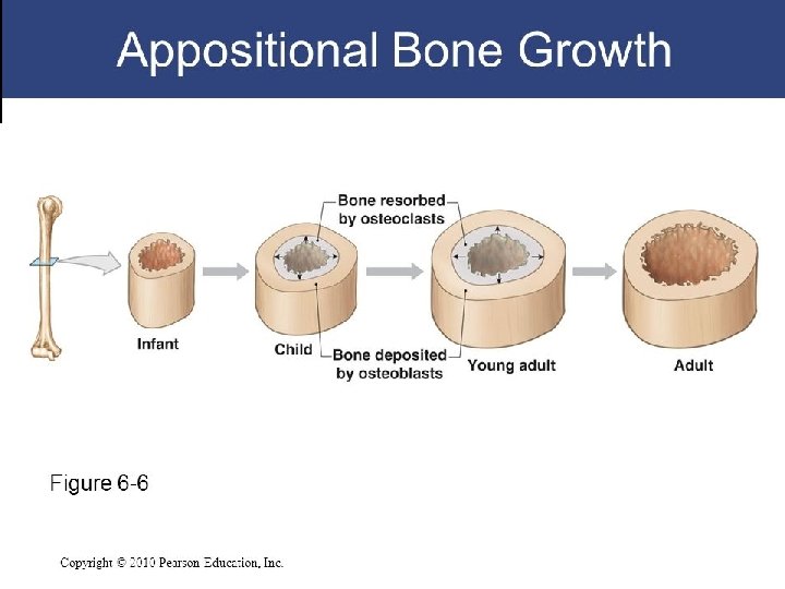
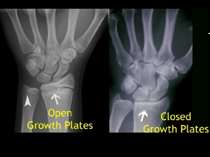
Bone Growth During Development Growth During puberty: Sex hormones speed up osteoblast activity = rapid growth Epiphyseal cartilage growth stops Epiphyseal line = former location of epiphyseal cartilage; still evident by X-Ray through adulthood Bone continues to grow in diameter (appositional growth)

Requirements for normal bone growth Minerals, especially calcium and phosphate Vitamin D 3 stimulates absorption of calcium vitamin D 3 deficiency – causes bone to soften = Rickett’s Vitamin A stimulates osteoblasts Vitamin C synthesis of collagen vitamin - C deficiency = scurvy

Calcium Homeostasis The role of the skeleton as a mineral reserve

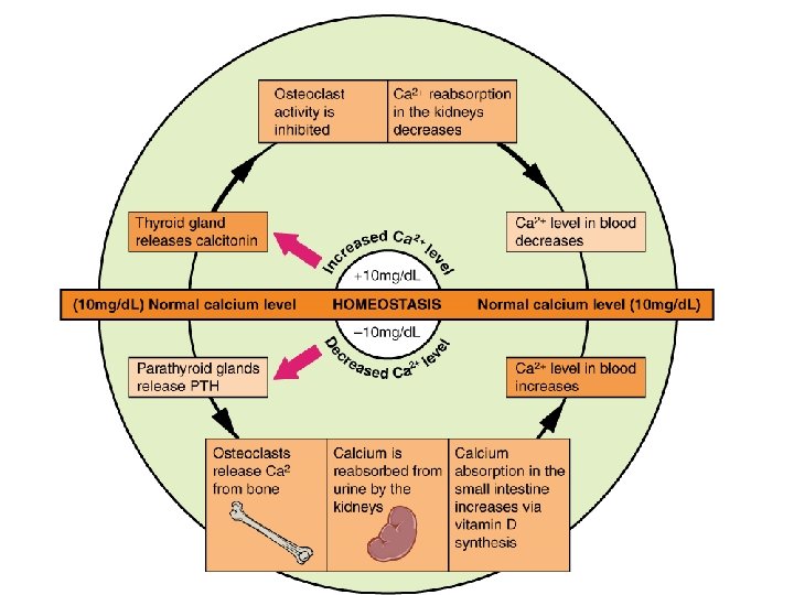
Calcium Homeostasis Calcium = most abundant mineral in the body Stored in skeleton (bone matrix) Necessary for muscle and nerve function Blood calcium levels are tightly regulated
 released Osteoclast activity increases")
Calcium Homeostasis When blood calcium is low Parathyroid hormone (PTH) released Osteoclast activity increases ▪ Bone resorption = calcium released from bone Calcium reabsorption from urine in the kidney Calcium absorption from small intestine Overall response = elevate blood Ca 2+
 is released Osteoblast activity increases")
Calcium Homeostasis When blood calcium is high Calcitonin (CT) is released Osteoblast activity increases ▪ Bone formation; calcium deposited in bone Overall response: decrease blood Ca 2+

Bone Remodeling

Bone Remodeling minerals and tissues are added and removed from bone Occurs throughout the human lifetime as a result of coupled activity of osteoblasts and osteoclasts Imbalance in activity of either cell type disease

Bone Remodeling Activation Phase 1. Pre-osteoclasts are attracted to remodeling sites 2. Pre-osteoclasts fuse to form multi-nucleate osteoclasts

Bone Remodeling Resorption 3. Osteoclasts dig out a cavity called a resorption pit 4. Calcium is released into bloodstream 5. Osteoclasts disappear
 appear along the resorption pit")
Bone Remodeling Reversal 6. Stem cells (pre-cursors to osteoblasts) appear along the resorption pit 7. Osteoblasts increase in numbers and change into preosteoblasts

Bone Remodeling Formation 8. pre-osteoblasts mature 9. Osteoblasts release osteoid at the site, forming a new soft non-mineralized matrix

Bone Remodeling Quiescence 10. The new matrix is mineralized with calcium and phosphorous 11. Site remains dormant until next cycle of remodeling

Abnormal Bone Remodeling Decreased “blast” activity, normal “clast” Aging, alcohol Increased “clast” activity, normal “blast” Osteoporosis, menopause, anorexia

FRACTURES

Bone Repair after Fracture 1. Fracture hematoma – blood leaks from broken blood vessels in bone

Bone Repair after Fracture 2. Callus Formation – fibroblasts invade the tissue and produce collagen fibers

Bone Repair after Fracture 3. Endochondral ossification – cartilage replaced with bone

Bone Repair after Fracture 1. Fracture hematoma – blood leaks from broken blood vessels in bone 2. Callus Formation – fibroblasts invade the tissue and produce collagen fibers 3. Endochondral ossification – cartilage replaced with bone

Fracture and Repair

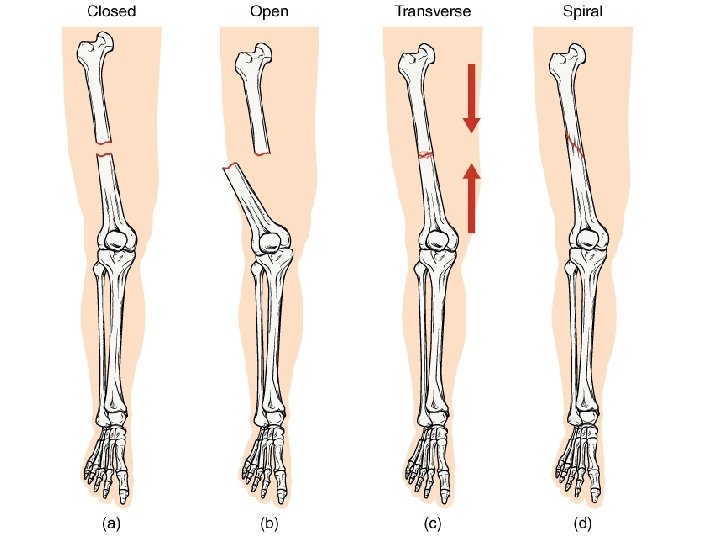
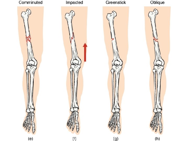
Types of Fractures Transverse occurs at an exact 90 degree horizontal angle Caused by bending force that causes the bone to snap (like a carrot)

Types of Fractures Oblique occurs at an angle that is not 90 degrees Caused by trapping of one bone while the other twists over it (i. e. foot caught between two rocks as the leg twists)

Types of Fractures Spiral fracture spirals around the bone Caused by twisting force

Types of Fractures Comminuted fracture breaks into multiple pieces Caused by crushing force

Types of Fractures Avulsion ligament or tendon pulls away from its attachment on the bone and a fragment breaks off with it Caused by muscle contraction or stretch that is stronger than the force that holds the tendon/ligament to the bone.

Types of Fractures Impacted one fragment is driven into itself and buckles Caused by compression of the bone from end to end

Types of Fractures Fissure or Hairline incomplete fracture; multiple small lines visible but do not pass through entire bone Caused by any force that is not great enough to cause complete fracture
")
Types of Fractures Greenstick bone bends rather than breaks (like a green tree branch) Occurs most often in children in bones that have not completely ossified and still contain cartilage
 = broken bone breaks through skin Closed (simple) =")
Types of Fractures Open (compound) = broken bone breaks through skin Closed (simple) = skin remains intact video

Fracture and Repair

BONE MARKINGS

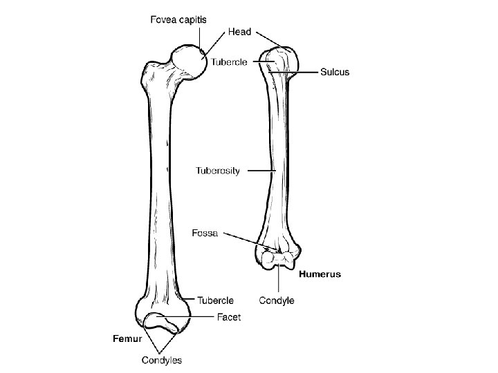
Elevations and Projections Process = prominent feature Ramus = curved portion of a bone (ram’s horn)

Processes formed where tendons and ligaments attach Trochanter = large rough projection Tuberosity = small rough surface Tubercle = small rounded surface Crest = ridge Line = elongated ridge Spine = sharp process

Processes formed for articulation with other bones Head = epiphysis Neck = connection between epiphysis and diaphysis Condyle = rounded surface Trochlea = small, grooved pulley shaped process Facet = flat surface

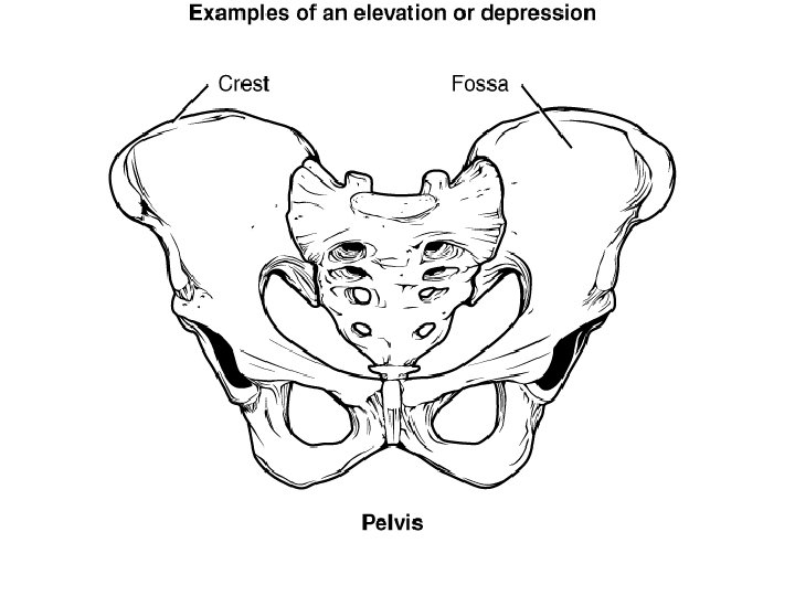
Depressions Fossa = elongated basin Sulcus = narrow grove

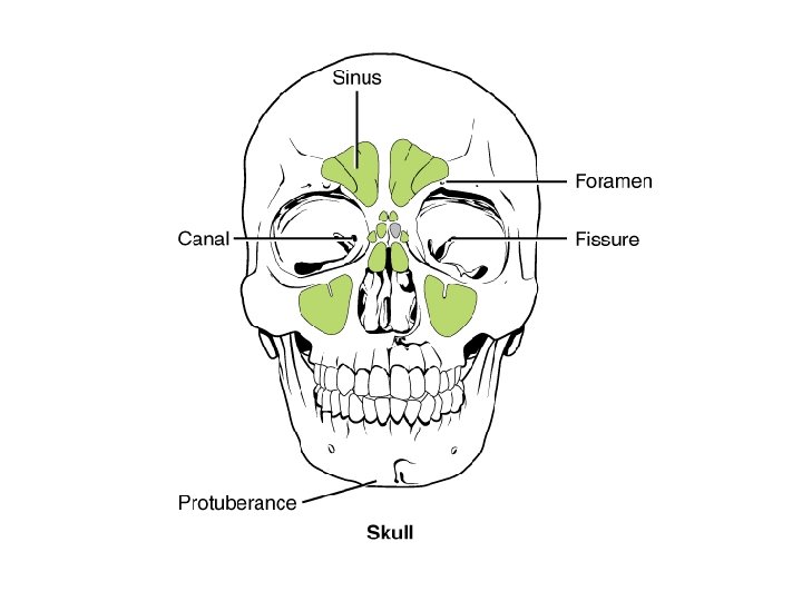
Openings Foramen = hole in bone Canal = passage in bone Meatus = opening into canal Fissure = slit Sinus = air=filled space
- Slides: 67