Chapter 6 Muscular System Types of Muscles Skeletal


 • Each muscle cell is surrounded by")
 and actin (thin). •")


- Slides: 21
Chapter 6 Muscular System
Types of Muscles • Skeletal – striated & voluntary • Smooth - involuntary • Cardiac – heart • Striated means it appears striped
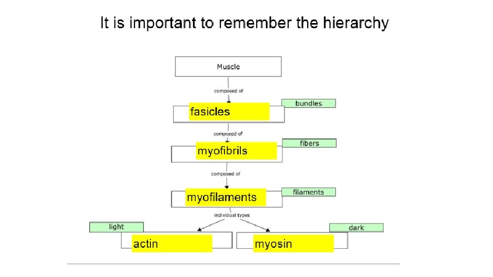
Structure of a Muscle • Muscles are composed of many FIBERS that are arranged in bundles called FASCICLES. • Muscles are separated from each other by FASCIA, which form the APONEUROSES and TENDONS that connect the muscles to bones. • Muscle Connective Tissue: • Epimysium – outermost layer that surrounds the entire muscle • Perimysium – separates and surrounds fascicles (bundles of muscle fibers) • Endomysium – surrounds each individual muscle fiber
Muscle Functions • The SOLE function of Muscular Tissue is to contract or shorten. As it contracts it: • Produces Movement • Maintains Posture • Stabilizes Joints • Generates Heat
Structure of a Muscle Fiber (muscle cells) • Each muscle cell is surrounded by a specialized cell membrane called the SARCOLEMMA. • The cytoplasm is called the SARCOPLASM. • Each muscle cell is filled with MYOFIBRILS which in turn are composed of MYOFILAMENTS.
Myofibril Structure • Myofibrils – contain myofilaments of myosin (thick) and actin (thin). • • • The myofilaments overlap to form I and A bands on the fiber: DARK BANDS – A bands - myosin LIGHT BANDS – I bands – actin In the middle of each I band is a dark line called a Z line. One Z line to the next is a SARCOMERE.
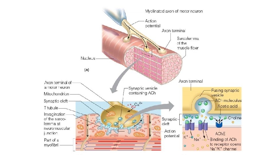
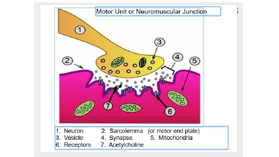
Neuromuscular Junction • The muscle fiber and the motor neuron make up the neuromuscular junction. • Motor end plate – folded area where the muscle and the neuron communicate • Synaptic cleft – the gap between the neuron and the motor end plate • Synaptic vesicles – where the neurotransmitters are stored
Neuromuscular Junction • The neurotransmitter that crosses the gap is ACETYLCHOLINE • ACH is broken down by CHOLINESTERASE.
Neuromuscular Junction • The theory of how a muscle contracts is the SLIDING FILAMENT THEORY. • The contraction occurs when the THICK filament (myosin) slides past the THIN filament (actin). • The sliding filament theory involves 5 different molecules plus CALCIUM 1. 2. 3. 4. 5. ATP Myosin Actin Acetylcholine Cholinesterase
Energy Source • The energy for muscle contraction comes from ATP provided by the process of CELLULAR RESPIRATION. • The molecule CREATINE PHOSPHATE helps with the regeneration of ATP. • Much of our body’s heat is produced by this process, which in turns helps maintain homeostasis.
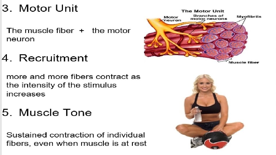
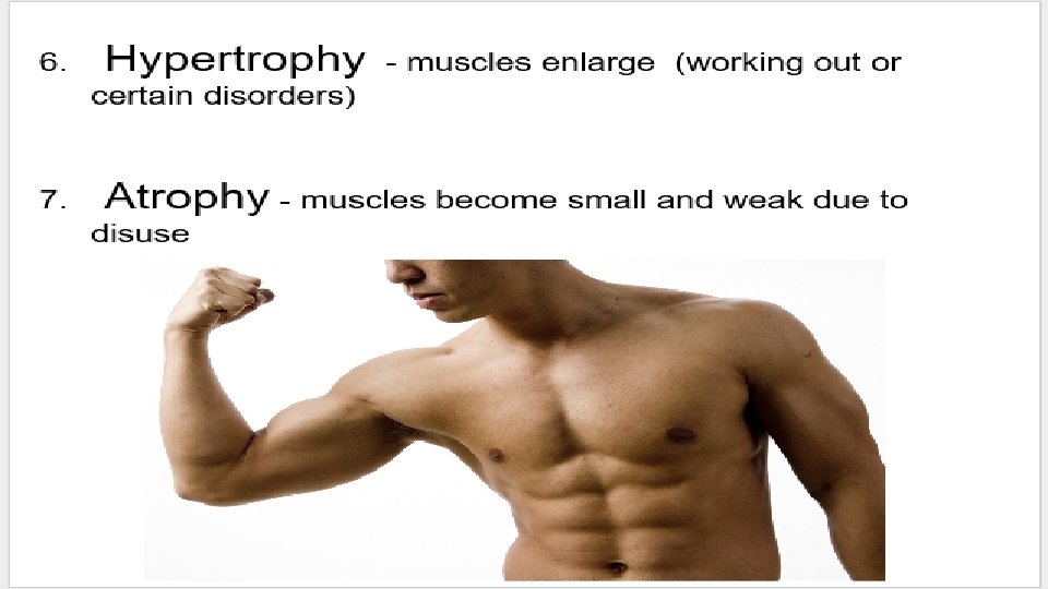
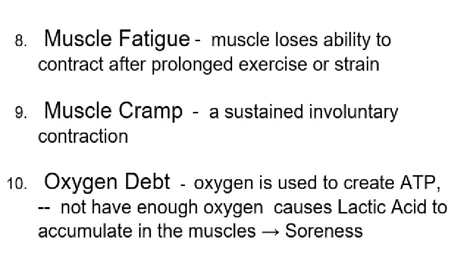
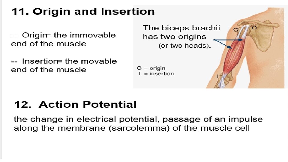
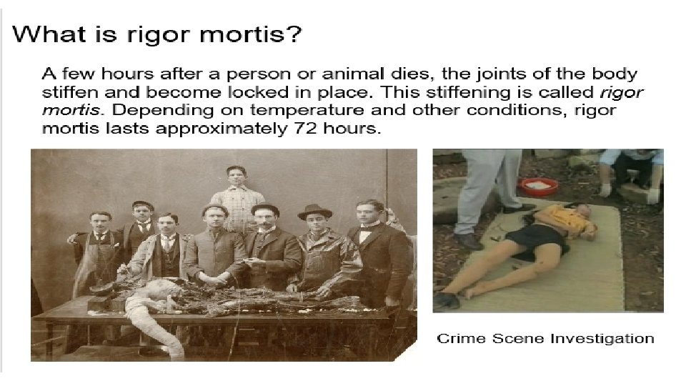
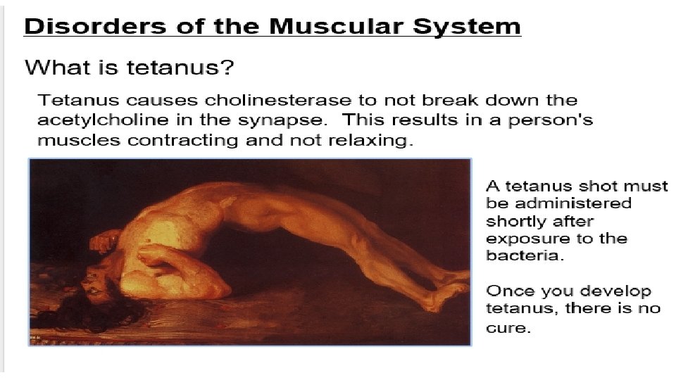
Terms related to Muscle Contraction 1. Threshold stimulus 2. All or none response 3. Motor Unit 4. Recruitment 5. Muscle tone 6. Muscle Hypertrophy 7. Muscle Atrophy 8. Muscle fatigue 9. Muscle cramp 10. Oxygen debt 11. Origin and insertion 12. Tetanus 13. Rigor mortis
1. Threshold Stimulus Minimal strength to cause a contraction Motor neuron releases enough Acetylcholine to reach threshold 2. All-or-None Response Fibers don’t contract partially, they either do or they don’t