Chapter 6 Chromosomes and Cell Reproduction Section 6


 contains")


 • 2. Synthesis Phase (S) •")

 • Cell’s DNA is copied • Each chromosome consists of 2 chromatids")


 move toward opposite poles")


- Slides: 55
Chapter 6 Chromosomes and Cell Reproduction
Section 6. 1 Chromosomes
Objectives • Define the importance of cell division. • Explain the difference between prokaryotic and eukaryotic cell reproduction. • Define the components that make up a chromosome.
Cell Division – Formation of New Cells • 2 trillion cells produced by an adult human daily – 25 million new cells per second – Formed when older cells divide • Cell division (Cell Reproduction) – Occurs in humans an other organisms at different times in their life – Type of division differs depending on organisms – No matter what type of division, all info. in DNA must be present in new cells
Gametes • An organisms reproductive cells, such as sperm or egg cells
Prokaryotic Cell Reproduction • Binary fission – Form of asexual reproduction that produces identical offspring • Single parent passes exact copies of all DNA to offspring • Prokaryote DNA? – Single, circular
Binary Fission • 2 stages: – 1. DNA is copied – 2. Cell Division • New membrane is formed • Cell pinches itself in half
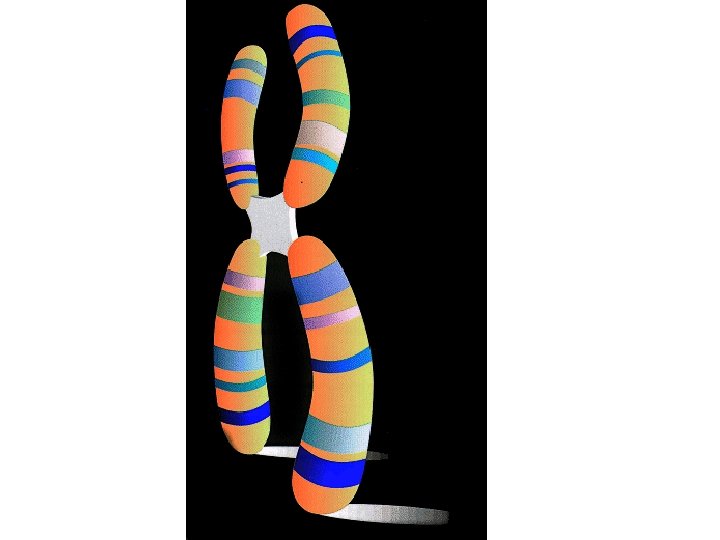
Eukaryotes • Gene – A segment of DNA that is located in a chromosome and that codes for a specific heredity trait • Chromosome – Structures in the nucleus that are made up of DNA and protein • Chromatid – One of the two strands of a chromosome that become visible during meiosis or mitosis • Centromere – Region of the chromosome that holds the 2 sister chromatids together during mitosis
Objectives • Define haploid and diploid. • Explain the difference between autosomes and sex chromosomes. • Describe what happens when chromosome numbers are changed.
Human Chromosome Number • Somatic Cells – any cell other than sperm or egg cells – Normally has 2 copies of 23 different chromosomes – Total = 46 – Chromosomes differ in size, shape, and set of genes
Sets of Chromosomes • Homologous Chromosomes – Each pair is the same – Similar in size, shape, and genetic content – Each one comes from one of the two parents
Sets of Chromosomes • Diploid – When a cell (usually a somatic cell) contains 2 sets of chromosomes • Haploid – When a cell (usually a gamete) contains 1 set of chromosomes • Zygote – A fertilized egg cell, the first cell of a new individual – Fusion of 2 haploid gametes
Chromosome Number and Complexity • Does having more chromosomes make an organism more complex? – p. 121
Sex Chromosomes • Autosomes – Chromosomes that are not directly related to determining the sex (gender) of an individual. – 22 of the 23 pairs • Sex Chromosomes – Chromosomes that determine the sex of an individual – 1 of the 23 pairs – X = female – Y = male
Karyotype • A photo of the chromosomes in a dividing cell that shows the chromosomes arranged by size
Abnormal Chromosome Numbers What happens when chromosome numbers change?
Prenatal Testing • Amniocentesis – Sample of amniotic fluid – Analyze fetal cells – Can be used to determine abnormalities in chromosomes • Chorionic Villi Sampling – Tissue sample of placenta – Can also be used to detect abnormalities
Mutations • Change in chromosome structure: – Deletion – Inversion – Duplication – Translocation
Modeling Mutations • You will be modeling the four types of mutations using the materials on the side counter. You will glue each chromatid you design to the white paper. • You must start by modeling your original chromosome, then modify it appropriately according to the different mutations we discussed. • You may choose the number of genes on your original chromatid, but you must have AT LEAST 5. • Cut the colored strips into smaller pieces to represent your genes. • Rearrange them to model the mutations. As you model each mutation, glue them to the white paper (be sure they are correct before you glue them down. • Cut the black pieces into circles to represent the centromere.
Objectives • Identify the 5 major events that occur during the cell cycle.
6. 2 The Cell Cycle
Division of Eukaryotic Cells • More complex than bacteria because… – Cytoplasm divides – Chromosomes divide inside the nucleus – Many internal organelles must be correctly copied and rearranged • …to form 2 functional cells
The Cell Cycle • A repeating sequence of cellular growth and division during the life of an organism • First 3 stages called INTERPHASE – Cell spends 90% of its time in interphase • Mitosis – When the nucleus of a cell is divided into 2 nuclei • Cytokinesis – When the cytoplasm divides
Stages • 1. First Growth Phase (G 1) • 2. Synthesis Phase (S) • 3. Second Growth Phase (G 2) • 4. Mitosis • 5. Cytokinesis
G 1 • Cell grows functions normally • Major portion of cell’s life • Non-dividing cells
Synthesis (S) • Cell’s DNA is copied • Each chromosome consists of 2 chromatids attaches at centromere
G 2 • Nucleus prepares to divide • Microtubules rearranged for mitosis
Mitosis • Nucleus is divided into 2 nuclei • Each nucleus has same # and kinds of chromosomes as original cell
Ctyokinesis • Cytoplasm divides • 2 new cells are formed
Mitosis and Cytokinesis • Responsible for producing new cells that are identical to the original cells • Allow organism to grow • Replace damaged tissues • Responsible for asexual reproduction – In some organisms
Objectives • Describe how eukaryotic cells control the cell cycle.
Control of Cell Cycle • Controlled by proteins at 3 checkpoints: – 1. G 1 Checkpoint – 2. G 2 Checkpoint – 3. Mitosis Checkpoint
G 1 Checkpoint • Determines whether cell will divide • If cell is ready proteins will tell the copy DNA • If cell is not ready it will enter a resting period • Some muscle and nerve cells never divide
G 2 Checkpoint • DNA checked by repair enzymes • If checkpoint is passed, proteins trigger mitosis
Mitosis Checkpoint • Triggers end of mitosis • Signals beginning of G 1 phase
Cancer • Uncontrolled growth of cells • Disorder of cell division • Cancer cells don’t respond to normally to body’s control mechanisms
Bell-Ringer • List the 5 stages of the cell cycle. • List the 3 checkpoints of cell division.
Stages of Mitosis
Stages of Mitosis
Stages of Mitosis
Stages of Mitosis
Bell Ringer • List the 4 stages of mitosis. • Describe what is occurring during each stage.
Section 6. 3 Mitosis and Cytokinesis
Objectives • Describe the structure and function of the spindle during mitosis.
Chromatid Separation in Mitosis • Spindles – Cell structures made of microtubules that move chromosomes during cell division • Spindles pull chromosomes apart
Forming the Spindle • Centrosomes organize the formation of the spindle
Separation of Chromatids by Attaching Spindle Fibers • Spindle fibers made of microtubules • Microtubules attach to centromeres and poles • Microtubules break down and pull chromosomes toward poles
Mitosis and Cytokinesis • PMAT – Prophase – Metaphase – Anaphase – Telophase • Cytokinesis
Prophase • Chromosomes become visible • Nuclear envelope dissolves • Spindle forms
Metaphase • Chromosomes line up at equator
Anaphase • Centromeres divide • Chromatids (now called chromosomes) move toward opposite poles
Telophase • • Nuclear envelope forms at each pole Chromosomes uncoil Spindle dissolves Cytokinesis begins
Cytokinesis • Cytoplasm of cell divides • Forms 2 genetically identical cells • Cell pinched in half by protein threads
Review Challenge • Teams of 4 students – If you are unable to form groups of 4 I will choose for you • You may use your book, notes, and the Test Prep Pretest that you received at the beginning of the chapter to answer the review questions. • You must have your answer on your whiteboard within the given time limit. • Scores will be kept as running totals. For the last question you can risk up to as many points as you currently have to achieve the highest team score. • The winning team will earn 3 extra credit points towards the test tomorrow.