
Chapter 6 by Tian Keli Dept of Biochemistry
Chapter 6 by Tian Keli (田克立) Dept. of Biochemistry and Molecular Biology Tel: 88382346 – 0 (O), Room: 6426 E-mail: tiankeli@sdu. edu. cn 所有图片均来自互联网
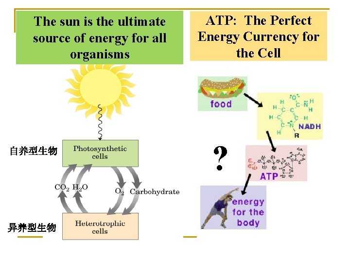
The sun is the ultimate source of energy for all organisms 自养型生物 异养型生物 ATP: The Perfect Energy Currency for the Cell ?
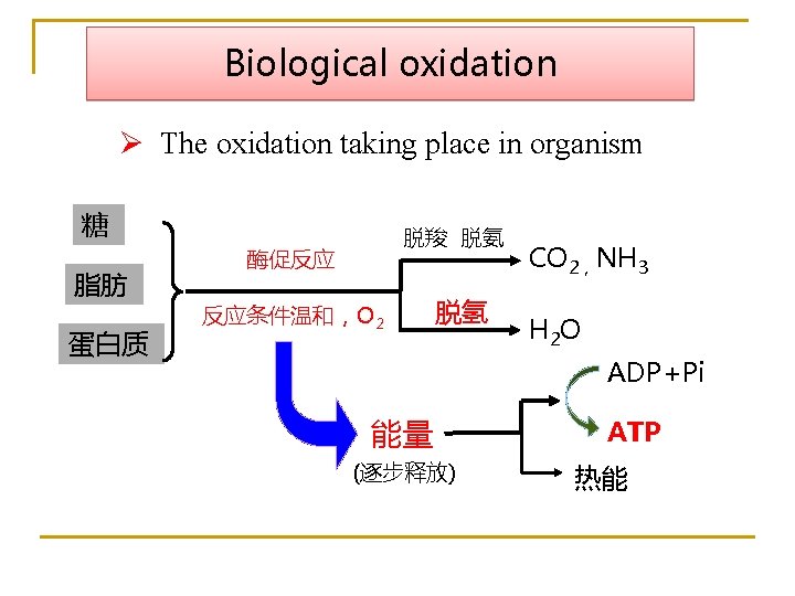
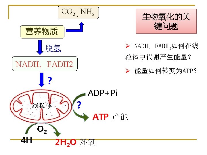
Biological oxidation Ø The oxidation taking place in organism 糖 脂肪 蛋白质 脱羧 脱氨 酶促反应 反应条件温和,O 2 脱氢 CO 2,NH 3 H 2 O ADP+Pi 能量 (逐步释放) ATP 热能
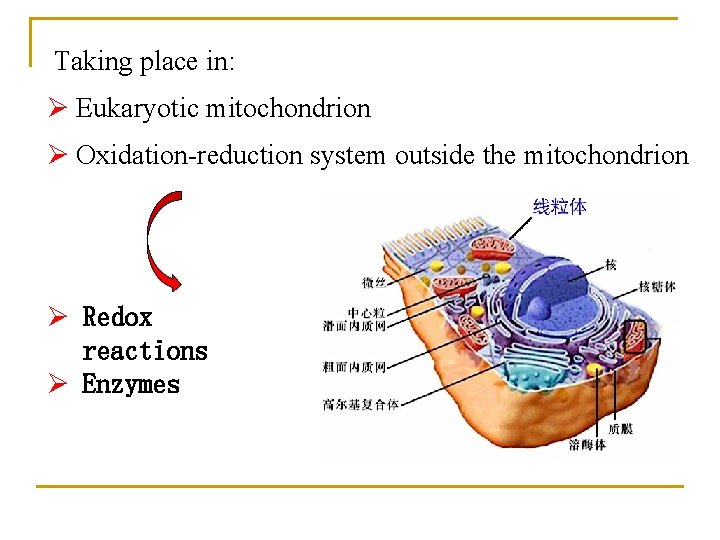
Taking place in: Ø Eukaryotic mitochondrion Ø Oxidation-reduction system outside the mitochondrion Ø Redox reactions Ø Enzymes
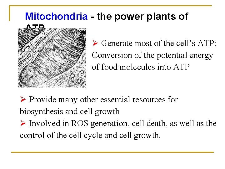
Mitochondria - the power plants of ATP Ø Generate most of the cell’s ATP: Conversion of the potential energy of food molecules into ATP Ø Provide many other essential resources for biosynthesis and cell growth Ø Involved in ROS generation, cell death, as well as the control of the cell cycle and cell growth.
Section 1 Characteristics of the mitochondrion
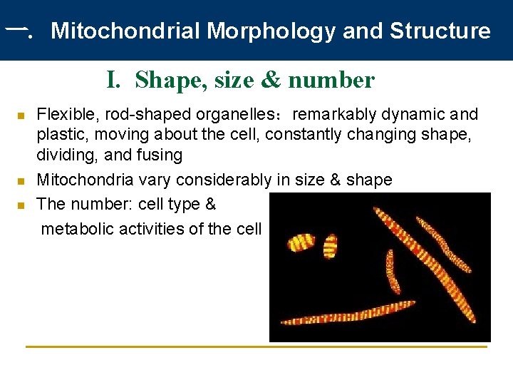
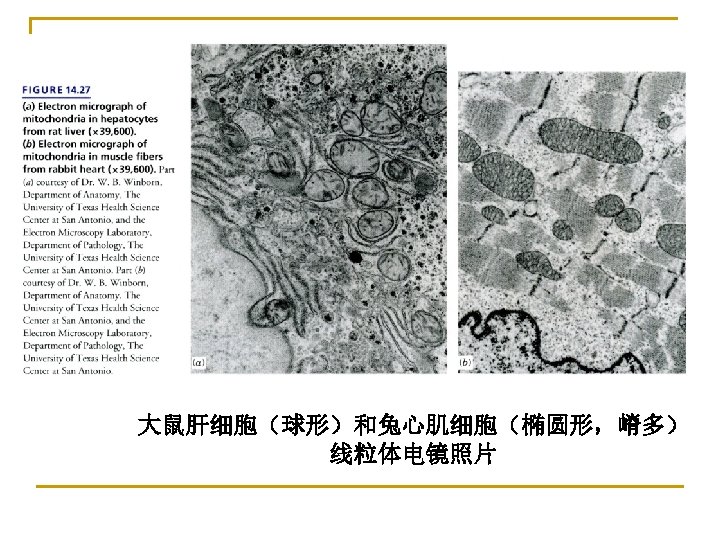
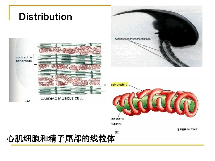
一. Mitochondrial Morphology and Structure I. Shape, size & number n n n Flexible, rod-shaped organelles:remarkably dynamic and plastic, moving about the cell, constantly changing shape, dividing, and fusing Mitochondria vary considerably in size & shape The number: cell type & metabolic activities of the cell
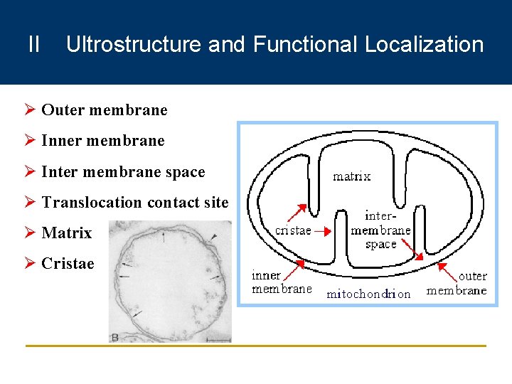
II Ultrostructure and Functional Localization Ø Outer membrane Ø Inner membrane Ø Inter membrane space Ø Translocation contact site Ø Matrix Ø Cristae
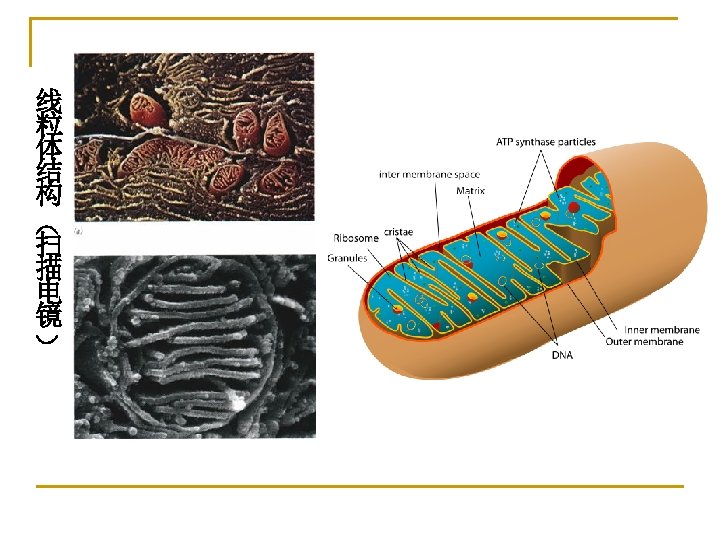
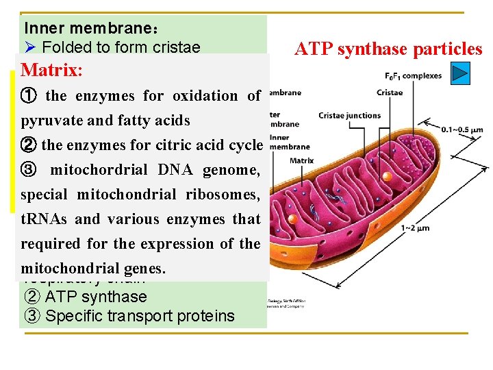
Inner membrane: Ø Folded to form cristae Ø Rich in cardiolipin, Matrix: Impermeable to most charged Outer ① the membrane: enzymes for oxidation of molecules, like ions, electrons, Ø Protein-topyruvate and fatty acids and protons Ø phospholipid Very high protein-toratio: ② the enzymes for citric acid cycle phospholipid ratio (about 4: 1 by ③ mitochordrial DNA genome, about 1: 1 by weight) special mitochondrial ribosomes, Ø Ø Porins(孔蛋白) Contains three major types t. RNAs and various enzymes that of proteins: ① Those that carry out the required for the expression of the oxidation reactions of the mitochondrial genes. respiratory chain ② ATP synthase ③ Specific transport proteins ATP synthase particles

The structure of cardiolipin (心磷脂) Ø Cardiolipin consists of two covalently linked phospholipid units, with a total of four rather than the usual two fatty acid chains. Ø Cardiolipin is produced in the mitochondrial inner membrane, where it interacts closely with membrane proteins involved in oxidative phosphorylation and ATP transport. Its two juxtaposed phosphate groups may act as a local proton trap on the membrane surface.
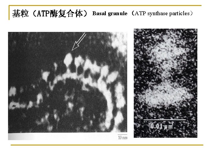
基粒(ATP酶复合体) Basal granule (ATP synthase particles)
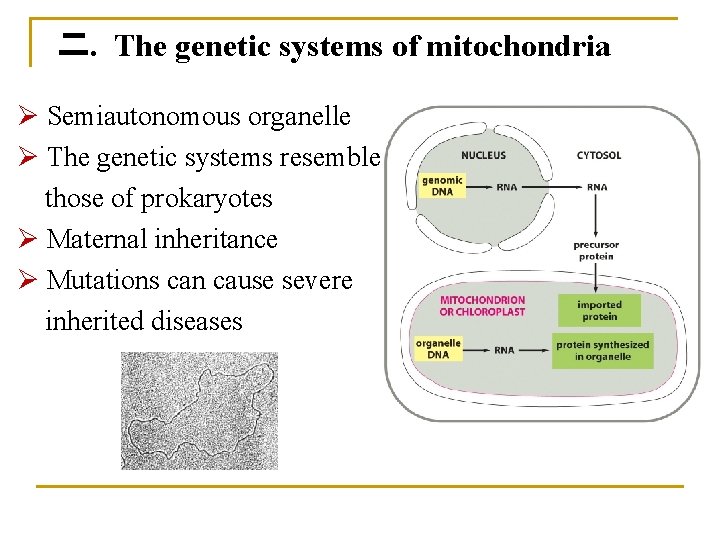
二. The genetic systems of mitochondria Ø Semiautonomous organelle Ø The genetic systems resemble those of prokaryotes Ø Maternal inheritance Ø Mutations can cause severe inherited diseases
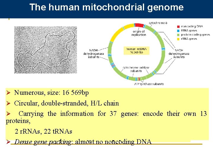
The human mitochondrial genome Ø Numerous, size: 16 569 bp Ø The mitochondrial DNA is organized into Ø Circular, double-stranded, H/L chain compact bodies— the nucleoids —by Ø Carrying the information for 37 genes: encode their own 13 proteins, special scaffolding proteins that also function 2 r. RNAs, 22 t. RNAs as transcription regulatory proteins. Ø Dense gene packing: almost no noncoding DNA
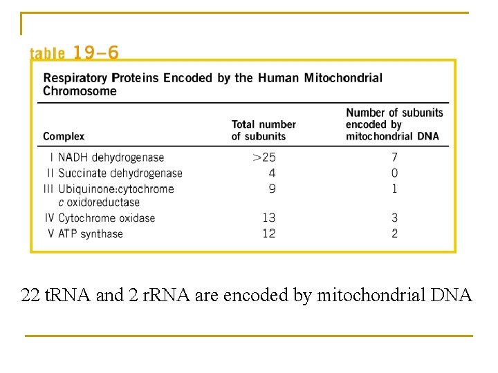
22 t. RNA and 2 r. RNA are encoded by mitochondrial DNA
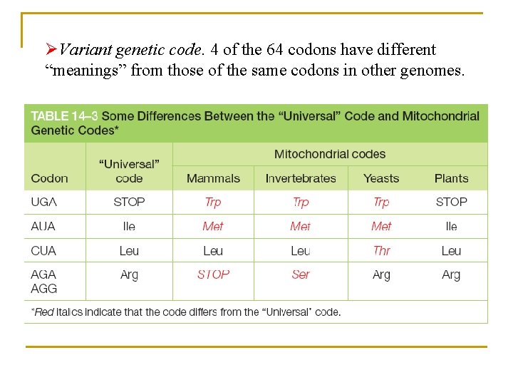
ØVariant genetic code. 4 of the 64 codons have different “meanings” from those of the same codons in other genomes.
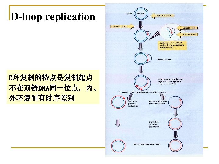
Transformation of mitochondrial Genetic information Ø Resemble those of prokaryotes Ø Replicate throughout all cell cycle, not just in S phase Ø Two replication origins: one for each strand of the mt. DNA: D-loop replication Ø Two transcriptional promoters: one for each strand of the mt. DNA Ø Lower fidelity of mitochondrial DNA replication, inefficient DNA repair: mitochondria have a high mutation ratio
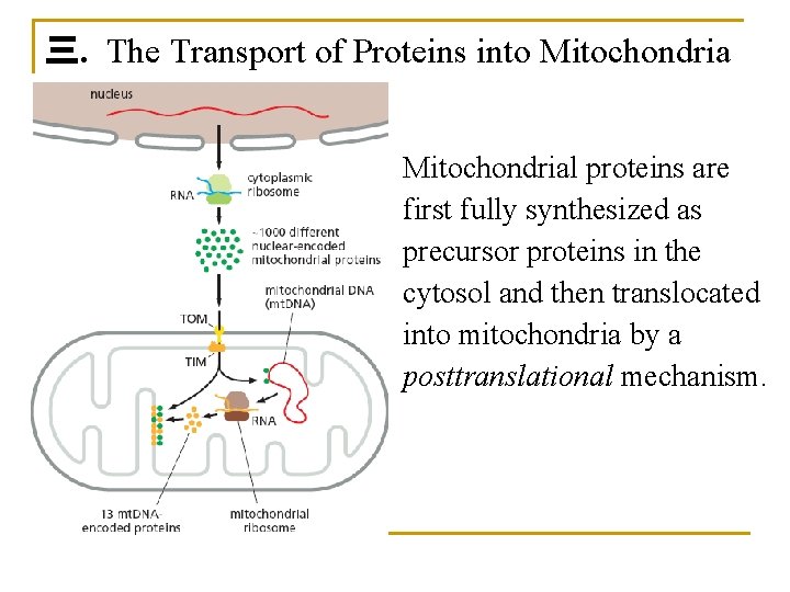
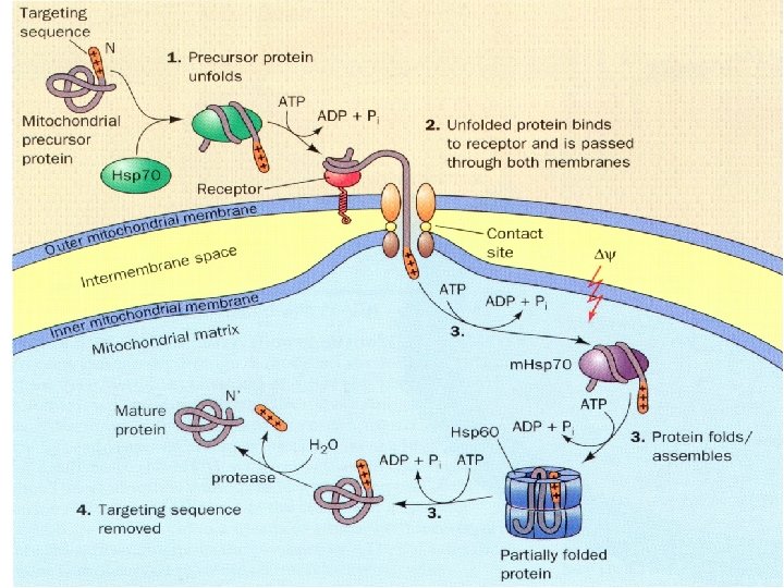
三. The Transport of Proteins into Mitochondrial proteins are first fully synthesized as precursor proteins in the cytosol and then translocated into mitochondria by a posttranslational mechanism.
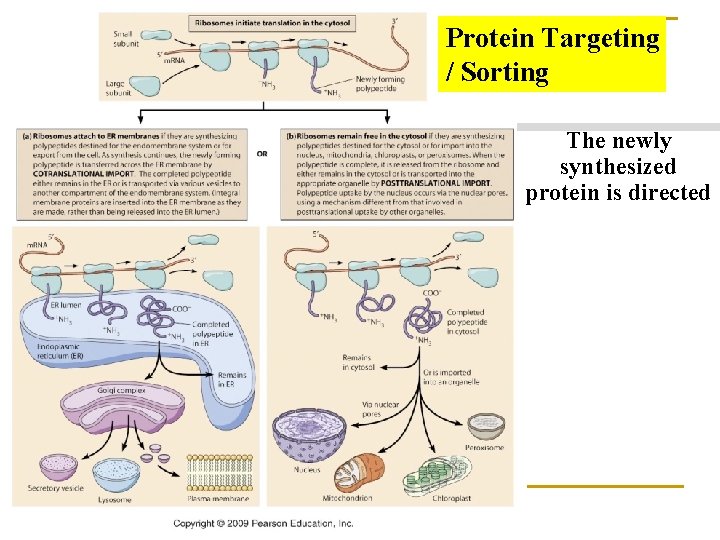
Protein Targeting / Sorting The newly synthesized protein is directed
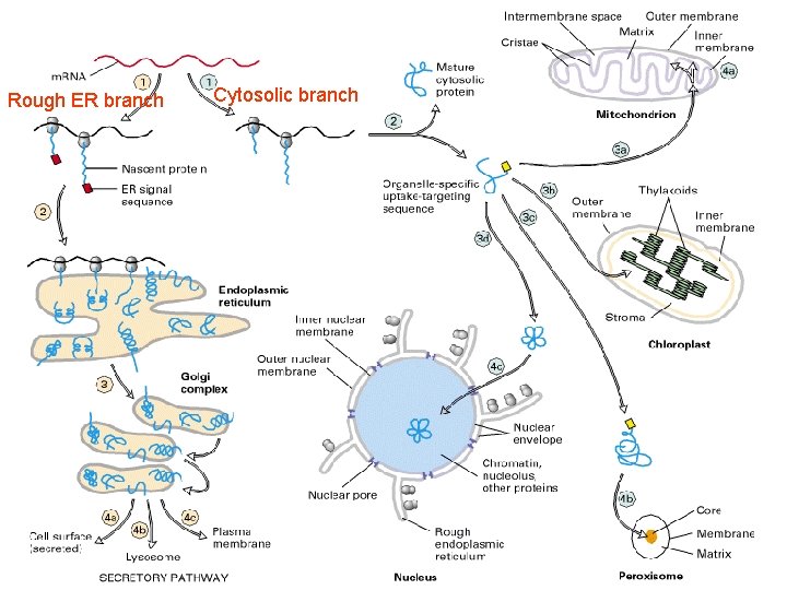
Rough ER branch Cytosolic branch
1. Mitochondrial Precursor Proteins Are Imported as Unfolded Polypeptide Chains. n Mitochondrial precursor proteins do not fold into their native structures after they are synthesized; instead, they remain unfolded through interactions with other proteins in the cytosol (hsp 70).
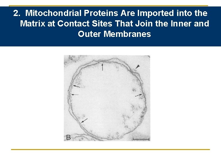
2. Mitochondrial Proteins Are Imported into the Matrix at Contact Sites That Join the Inner and Outer Membranes
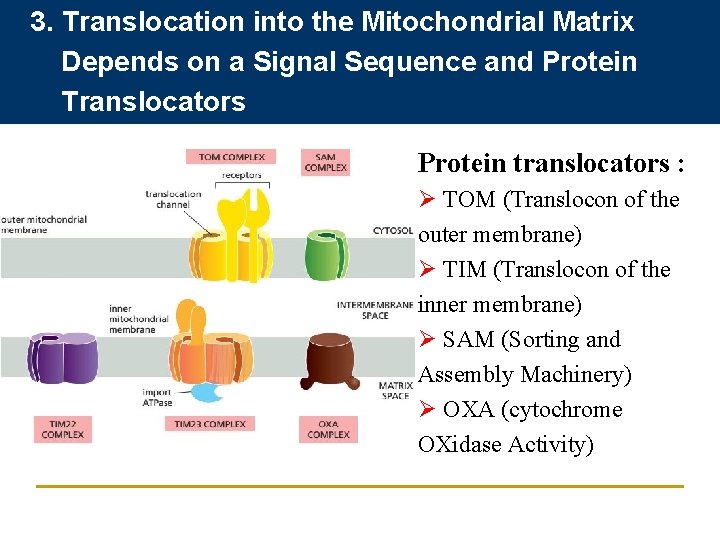
3. Translocation into the Mitochondrial Matrix Depends on a Signal Sequence and Protein Translocators Protein translocators : Ø TOM (Translocon of the outer membrane) Ø TIM (Translocon of the inner membrane) Ø SAM (Sorting and Assembly Machinery) Ø OXA (cytochrome OXidase Activity)
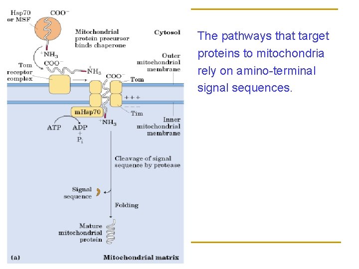
The pathways that target proteins to mitochondria rely on amino-terminal signal sequences.
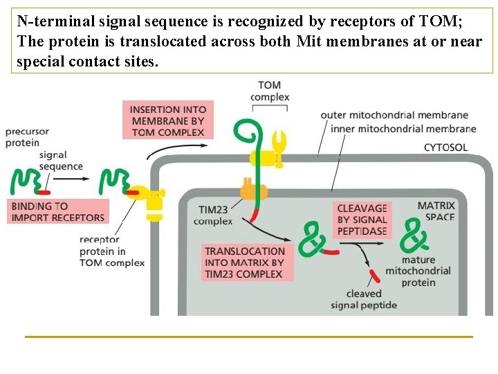
N-terminal signal sequence is recognized by receptors of TOM; The protein is translocated across both Mit membranes at or near special contact sites.
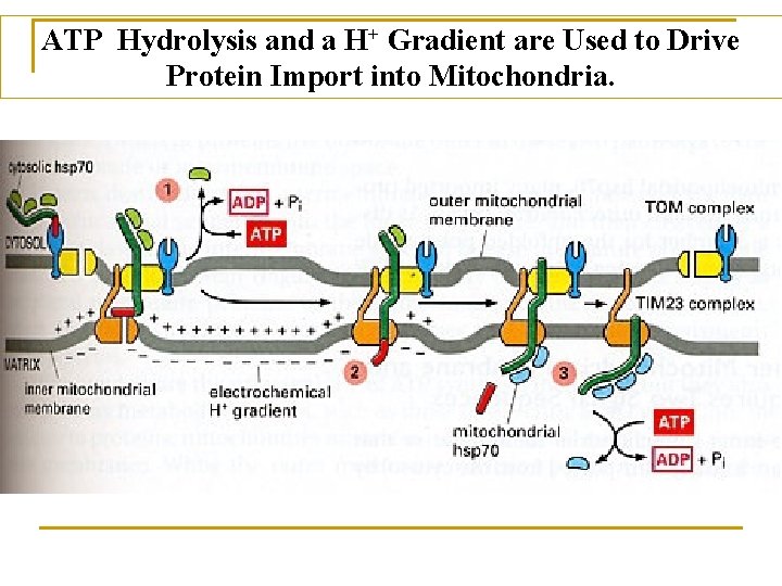
ATP Hydrolysis and a H+ Gradient are Used to Drive Protein Import into Mitochondria.
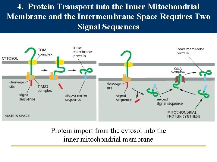
4. Protein Transport into the Inner Mitochondrial Membrane and the Intermembrane Space Requires Two Signal Sequences Protein import from the cytosol into the inner mitochondrial membrane
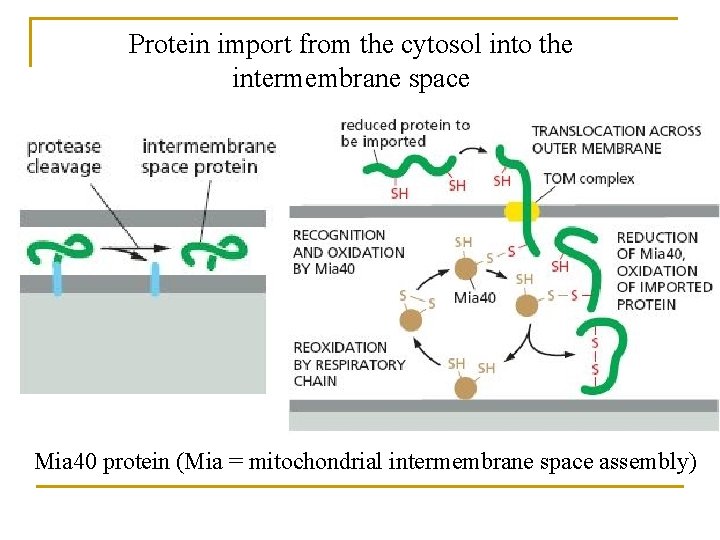
Protein import from the cytosol into the intermembrane space Mia 40 protein (Mia = mitochondrial intermembrane space assembly)
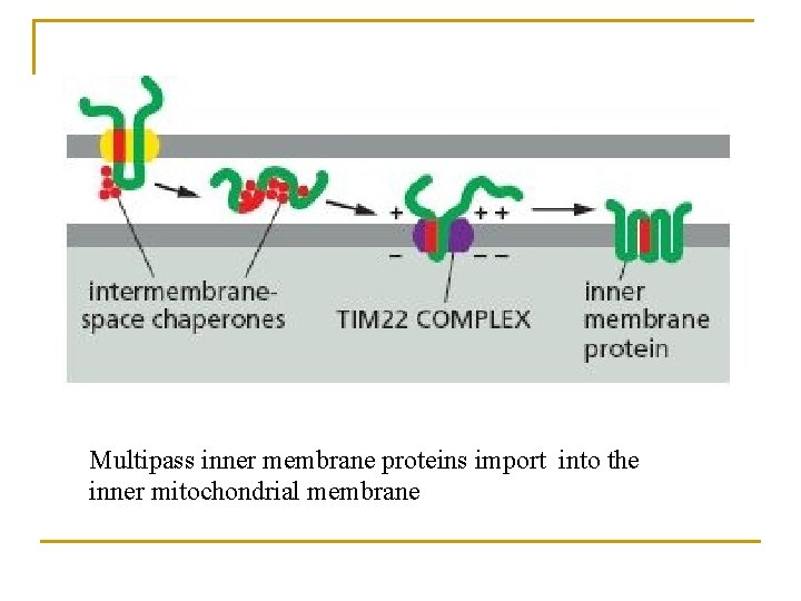
Multipass inner membrane proteins import into the inner mitochondrial membrane
5. Proper folding ensure the maturation of mitochondria proteins After the initial interaction with mitochondrial hsp 70, many imported proteins are passed on to another chaperone protein, mitochondrial hsp 60 that facilitates its folding by binding and releasing it through cycles of ATP hydrolysis.
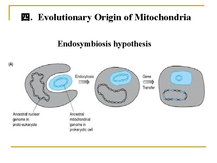
四. Evolutionary Origin of Mitochondria Endosymbiosis hypothesis
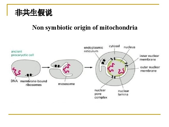
非共生假说 Non symbiotic origin of mitochondria
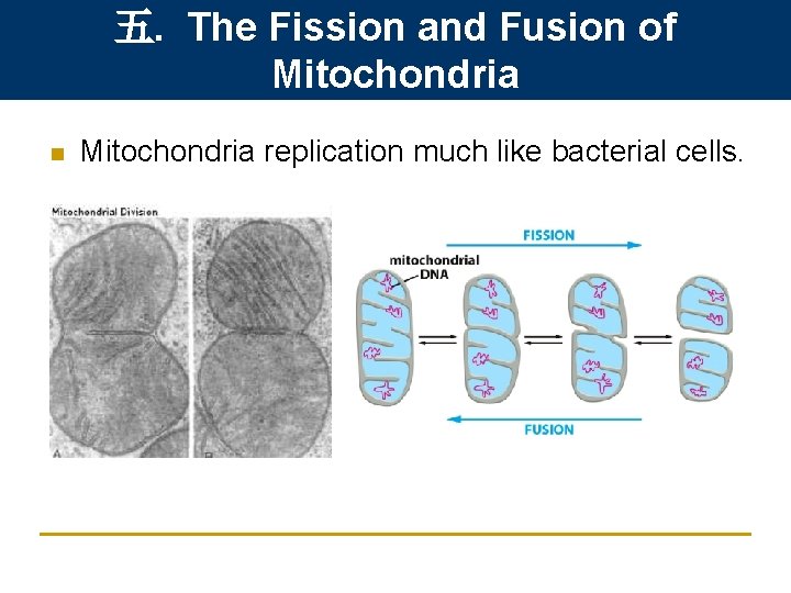
五. The Fission and Fusion of Mitochondria n Mitochondria replication much like bacterial cells.
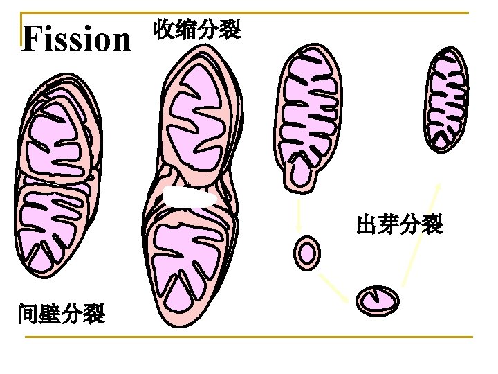
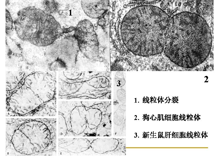
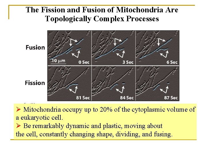
The Fission and Fusion of Mitochondria Are Topologically Complex Processes Ø Mitochondria occupy up to 20% of the cytoplasmic volume of a eukaryotic cell. Ø Be remarkably dynamic and plastic, moving about the cell, constantly changing shape, dividing, and fusing.
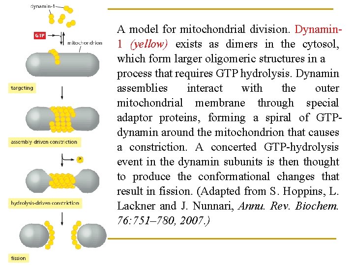
A model for mitochondrial division. Dynamin 1 (yellow) exists as dimers in the cytosol, which form larger oligomeric structures in a process that requires GTP hydrolysis. Dynamin assemblies interact with the outer mitochondrial membrane through special adaptor proteins, forming a spiral of GTPdynamin around the mitochondrion that causes a constriction. A concerted GTP-hydrolysis event in the dynamin subunits is then thought to produce the conformational changes that result in fission. (Adapted from S. Hoppins, L. Lackner and J. Nunnari, Annu. Rev. Biochem. 76: 751– 780, 2007. )
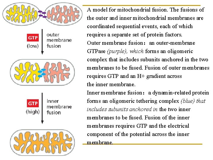
A model for mitochondrial fusion. The fusions of the outer and inner mitochondrial membranes are coordinated sequential events, each of which requires a separate set of protein factors. Outer membrane fusion:an outer-membrane GTPase (purple), which forms an oligomeric complex that includes subunits anchored in the two membranes to be fused. Fusion of outer membranes requires GTP and an H+ gradient across the inner membrane. Inner membrane fusion:a dynamin-related protein forms an oligomeric tethering complex (blue) that includes subunits anchored in the two inner membranes to be fused. Fusion of the inner membranes requires GTP and the electrical component of the potential across the inner membrane.
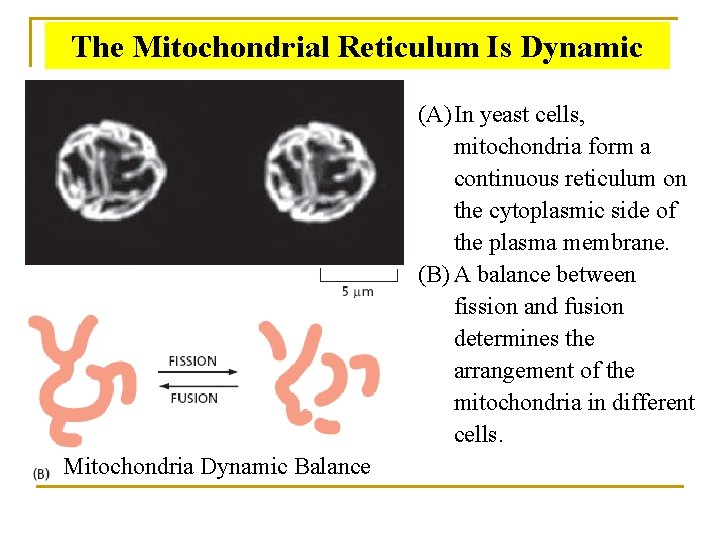
The Mitochondrial Reticulum Is Dynamic (A) In yeast cells, mitochondria form a continuous reticulum on the cytoplasmic side of the plasma membrane. (B) A balance between fission and fusion determines the arrangement of the mitochondria in different cells. Mitochondria Dynamic Balance
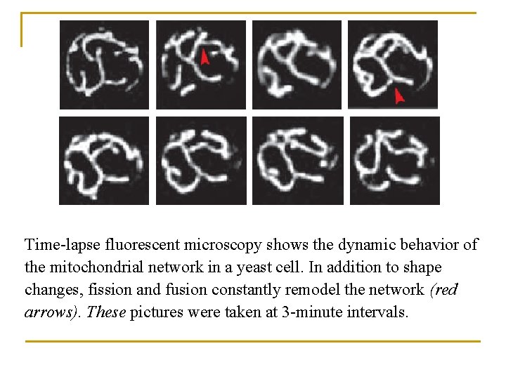
Time-lapse fluorescent microscopy shows the dynamic behavior of the mitochondrial network in a yeast cell. In addition to shape changes, fission and fusion constantly remodel the network (red arrows). These pictures were taken at 3 -minute intervals.
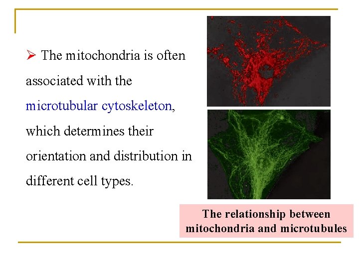
Ø The mitochondria is often associated with the microtubular cytoskeleton, which determines their orientation and distribution in different cell types. The relationship between mitochondria and microtubules
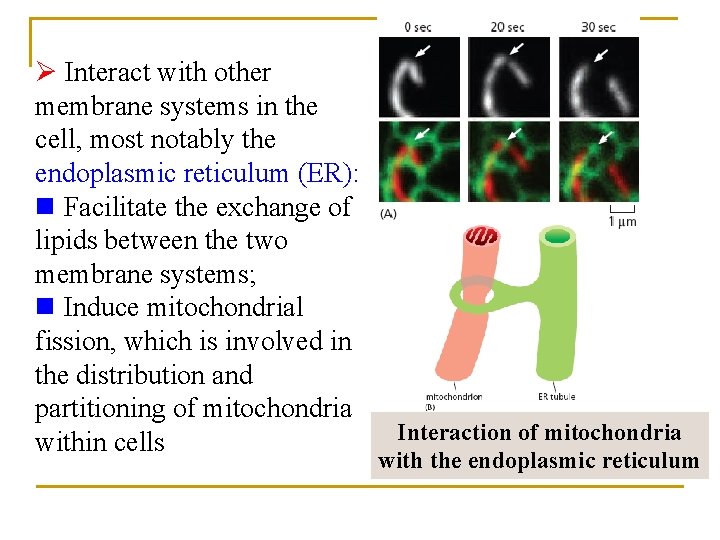
Ø Interact with other membrane systems in the cell, most notably the endoplasmic reticulum (ER): n Facilitate the exchange of lipids between the two membrane systems; n Induce mitochondrial fission, which is involved in the distribution and partitioning of mitochondria within cells Interaction of mitochondria with the endoplasmic reticulum
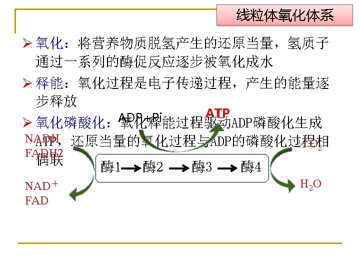
Section 2 Cell respiration and energy conversion Ø Cellular respiration The process of oxidizing food molecules to carbon dioxide and water. The energy released is trapped in the form of ATP for use by all the energy-consuming activities of the cell.
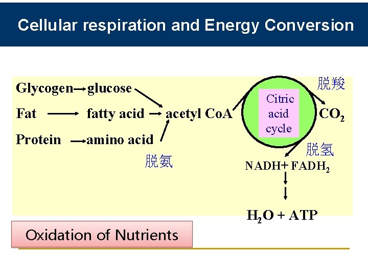
Cellular respiration and Energy Conversion 脱羧 Glycogen glucose Citric acid Fat fatty acid acetyl Co. A CO 2 cycle Protein amino acid 脱氢 脱氨 NADH+ FADH 2 H 2 O + ATP Oxidation of Nutrients
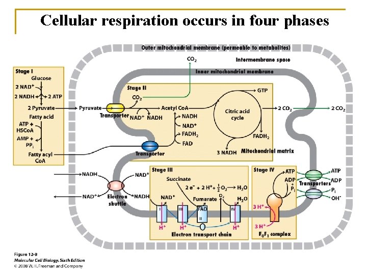
Cellular respiration occurs in four phases
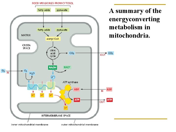
A summary of the energyconverting metabolism in mitochondria.
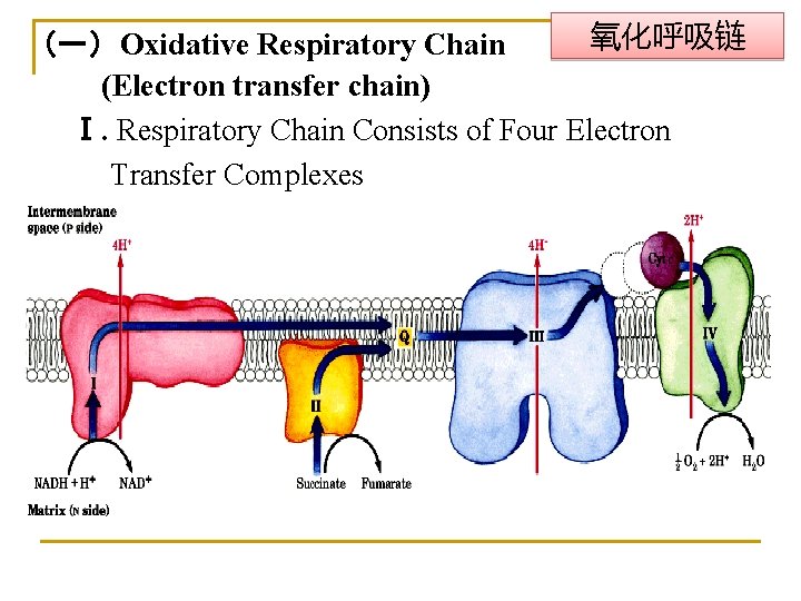
氧化呼吸链 (一)Oxidative Respiratory Chain (Electron transfer chain) Ⅰ. Respiratory Chain Consists of Four Electron Transfer Complexes
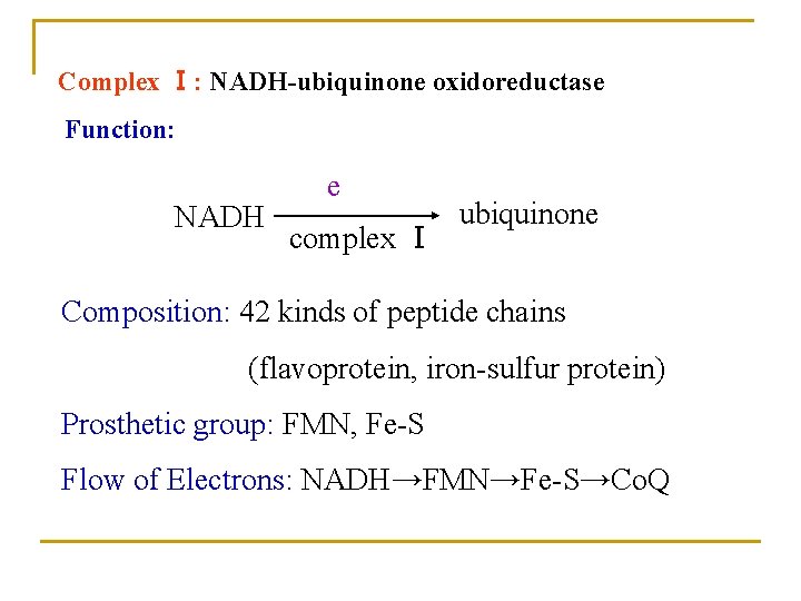
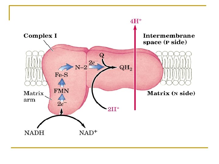
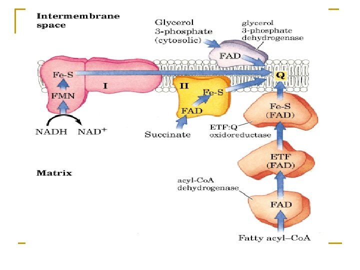
Complex Ⅰ: NADH-ubiquinone oxidoreductase Function: NADH e complex Ⅰ ubiquinone Composition: 42 kinds of peptide chains (flavoprotein, iron-sulfur protein) Prosthetic group: FMN, Fe-S Flow of Electrons: NADH→FMN→Fe-S→Co. Q
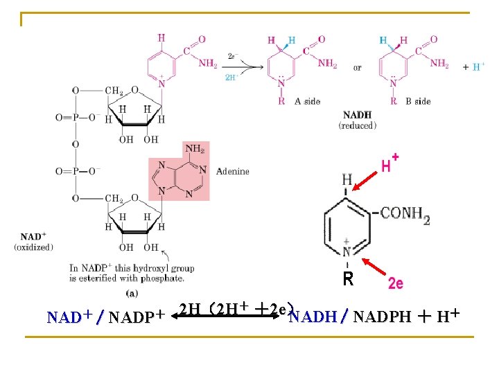
NAD+ / NADP+ 2 H(2 H+ +2 e) NADH / NADPH + H+
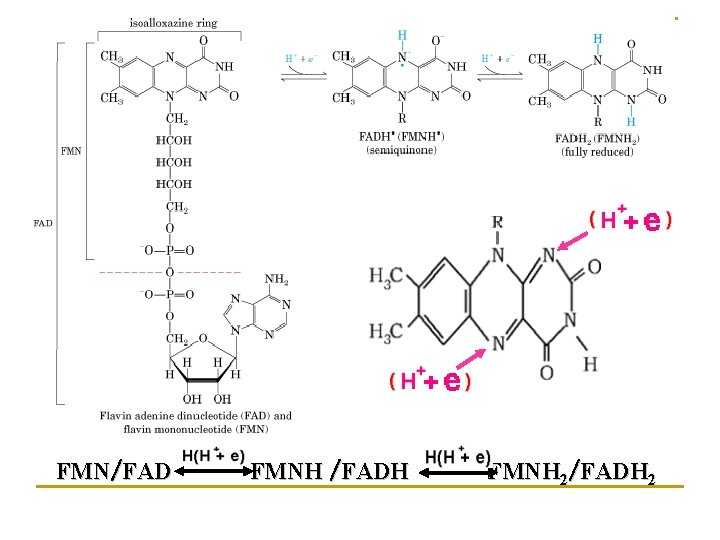
FMN/FAD FMNH /FADH FMNH 2/FADH 2
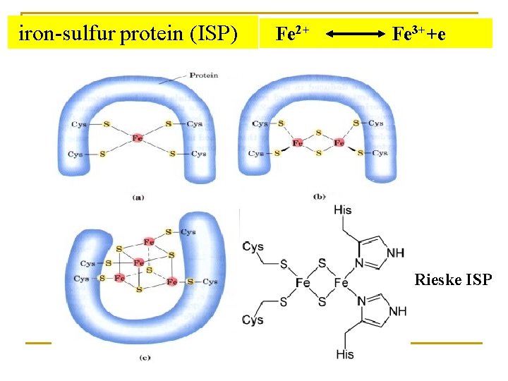
iron-sulfur protein (ISP) Fe 2+ Fe 3++e Rieske ISP
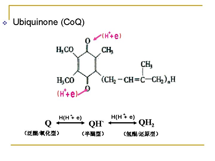
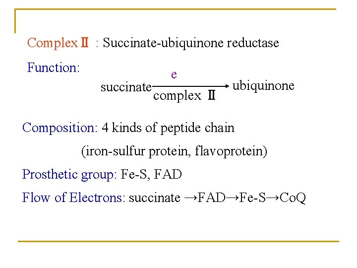
ComplexⅡ : Succinate-ubiquinone reductase Function: succinate e complex Ⅱ ubiquinone Composition: 4 kinds of peptide chain (iron-sulfur protein, flavoprotein) Prosthetic group: Fe-S, FAD Flow of Electrons: succinate →FAD→Fe-S→Co. Q
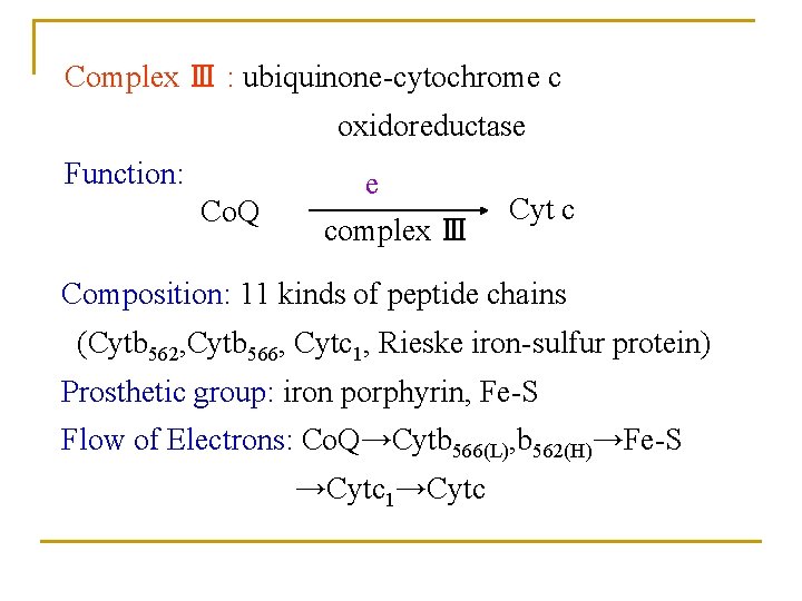
Complex Ⅲ : ubiquinone-cytochrome c oxidoreductase Function: Co. Q e complex Ⅲ Cyt c Composition: 11 kinds of peptide chains (Cytb 562, Cytb 566, Cytc 1, Rieske iron-sulfur protein) Prosthetic group: iron porphyrin, Fe-S Flow of Electrons: Co. Q→Cytb 566(L), b 562(H)→Fe-S →Cytc 1→Cytc
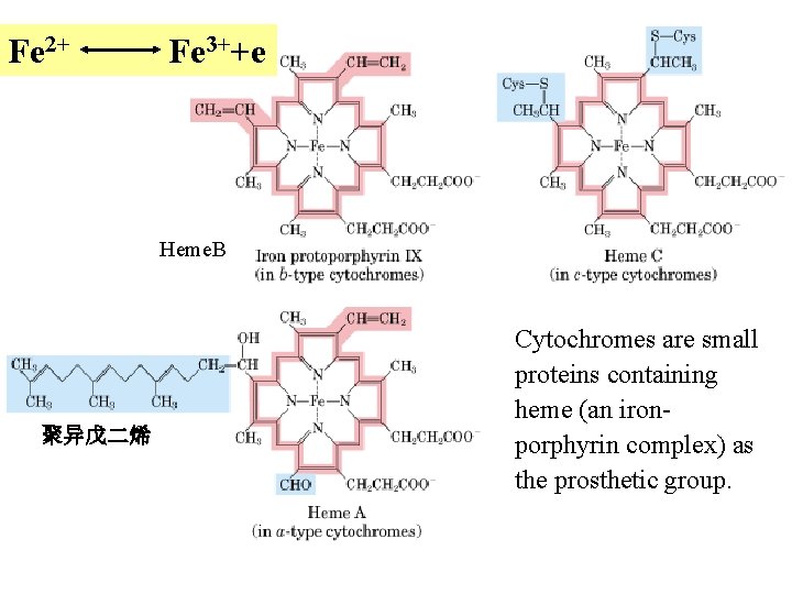
Fe 2+ Fe 3++e Heme. B 聚异戊二烯 Cytochromes are small proteins containing heme (an ironporphyrin complex) as the prosthetic group.
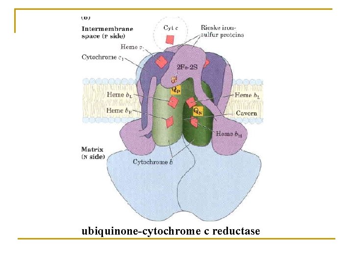
ubiquinone-cytochrome c reductase
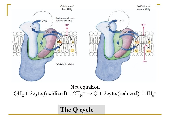
Net equation QH 2 + 2 cytc 1(oxidized) + 2 HN+ → Q + 2 cytc 1(reduced) + 4 Hp+ The Q cycle
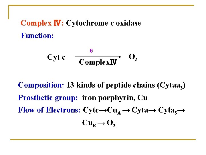
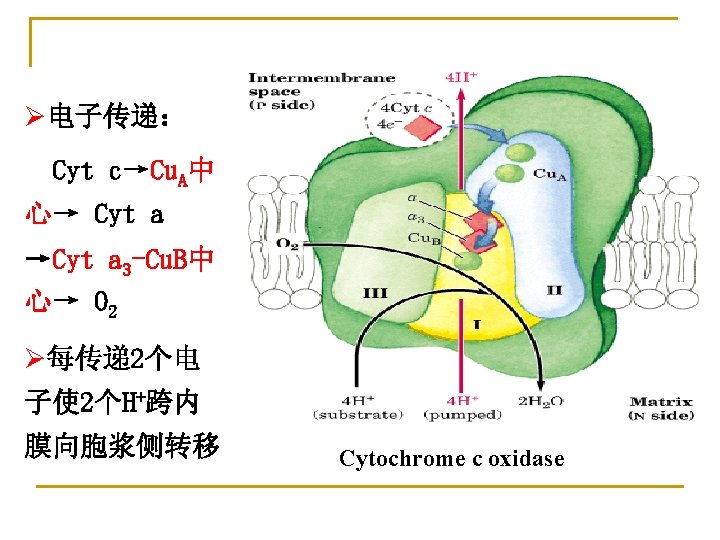
Complex Ⅳ: Cytochrome c oxidase Function: Cyt c e ComplexⅣ O 2 Composition: 13 kinds of peptide chains (Cytaa 3) Prosthetic group: iron porphyrin, Cu Flow of Electrons: Cytc→Cu. A → Cyta 3→ Cu. B → O 2
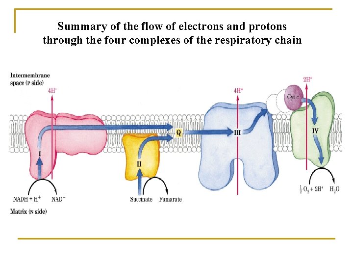
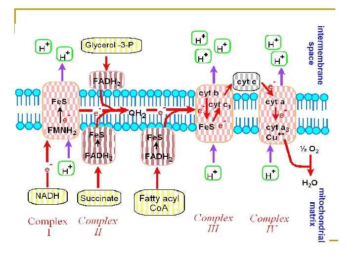
Summary of the flow of electrons and protons through the four complexes of the respiratory chain
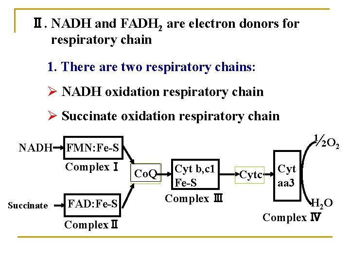
Ⅱ. NADH and FADH 2 are electron donors for respiratory chain 1. There are two respiratory chains: Ø NADH oxidation respiratory chain Ø Succinate oxidation respiratory chain ½O 2 NADH FMN: Fe-S ComplexⅠ Co. Q Succinate FAD: Fe-S ComplexⅡ Cyt b, c 1 Fe-S Complex Ⅲ Cytc Cyt aa 3 H 2 O Complex Ⅳ
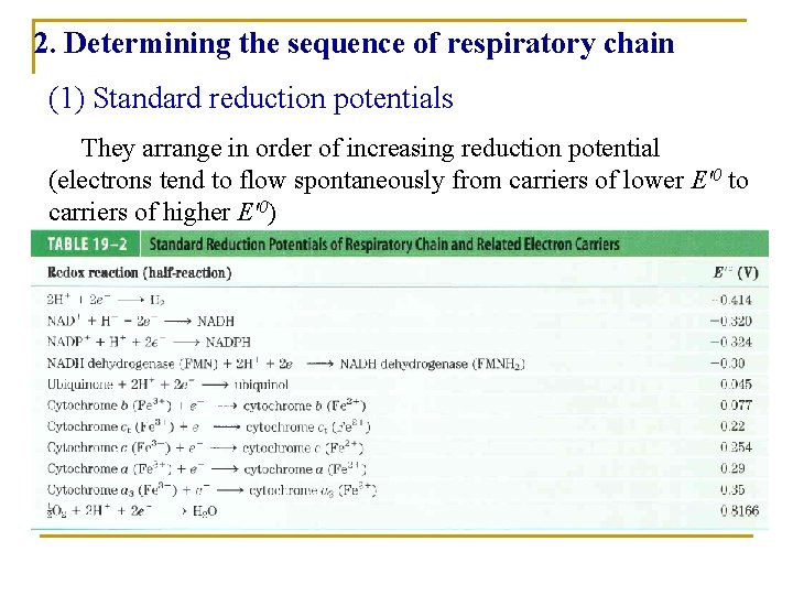
2. Determining the sequence of respiratory chain (1) Standard reduction potentials They arrange in order of increasing reduction potential (electrons tend to flow spontaneously from carriers of lower E'0 to carriers of higher E'0)
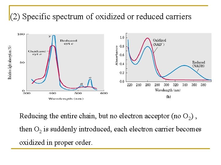
(2) Specific spectrum of oxidized or reduced carriers Reducing the entire chain, but no electron acceptor (no O 2) , then O 2 is suddenly introduced, each electron carrier becomes oxidized in proper order.
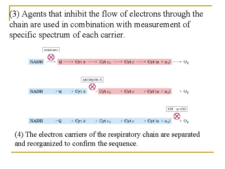
(3) Agents that inhibit the flow of electrons through the chain are used in combination with measurement of specific spectrum of each carrier. (4) The electron carriers of the respiratory chain are separated and reorganized to confirm the sequence.
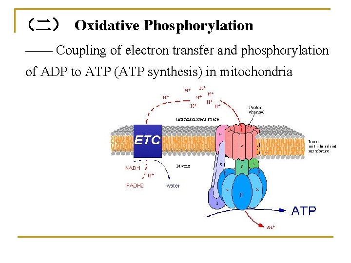
(二) Oxidative Phosphorylation —— Coupling of electron transfer and phosphorylation of ADP to ATP (ATP synthesis) in mitochondria
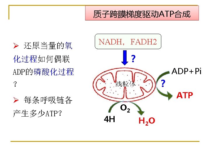
I. Coupling Sites of Oxidative Phosphorylation 1. P/O ratio The ratio of phosphate radicals esterified (forming ATP from ADP) to atoms of oxygen consumed by mitochondria. (The amount of ATP produced from the movement of two electrons through a defined electron transport chain, donated by reduction of an oxygen atom. )
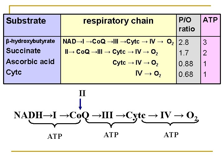
Substrate β-hydroxybutyrate Succinate Ascorbic acid Cytc respiratory chain P/O ratio ATP NAD→I →Co. Q →III →Cytc → IV → O 2 2. 8 1. 7 0. 88 0. 68 3 2 1 1 II→ Co. Q →III → Cytc → IV → O 2 II NADH→I →Co. Q →III →Cytc → IV → O 2 ATP ATP
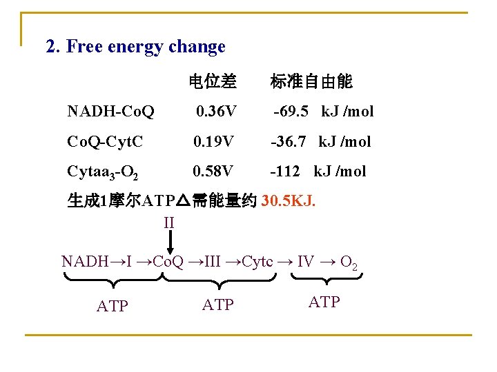
2. Free energy change 电位差 标准自由能 NADH-Co. Q 0. 36 V -69. 5 k. J /mol Co. Q-Cyt. C 0. 19 V -36. 7 k. J /mol Cytaa 3 -O 2 0. 58 V -112 k. J /mol 生成 1摩尔ATP△需能量约 30. 5 KJ. II NADH→I →Co. Q →III →Cytc → IV → O 2 ATP ATP
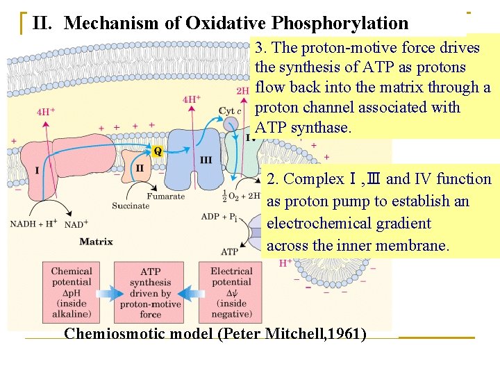
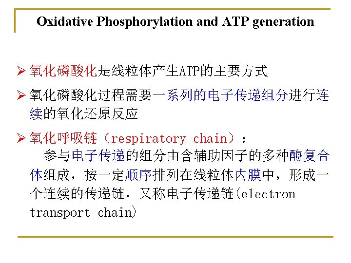
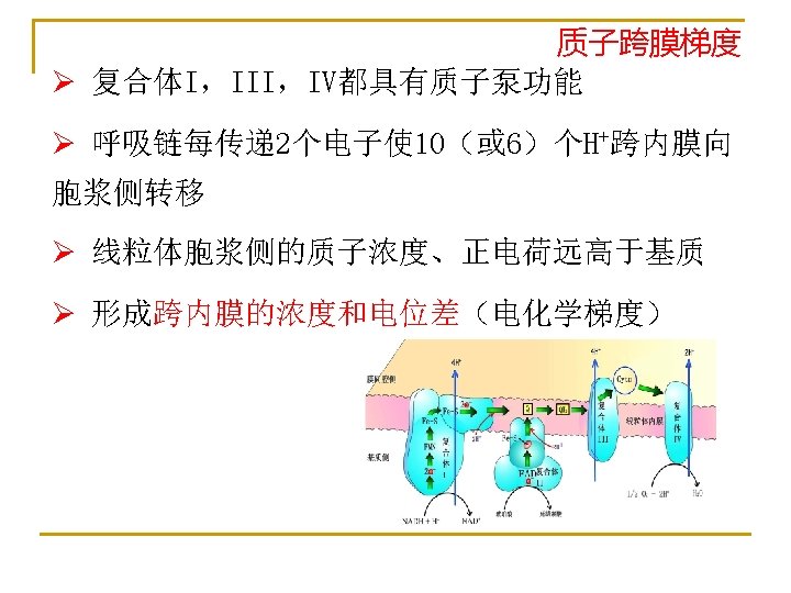
II. Mechanism of Oxidative Phosphorylation 3. The proton-motive force drives 1. Oxidative phosphorylation the synthesis of ATP as protons requires an intact inner flow back into the matrix through a membrane, which is proton channel associated with impermeable to protons. ATP synthase. 2. ComplexⅠ, Ⅲ and IV function as proton pump to establish an electrochemical gradient across the inner membrane. Chemiosmotic model (Peter Mitchell, 1961)
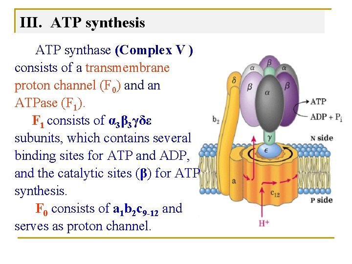
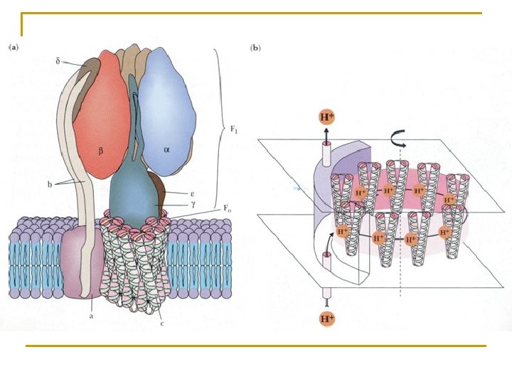
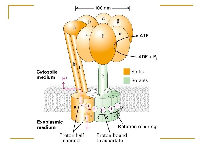
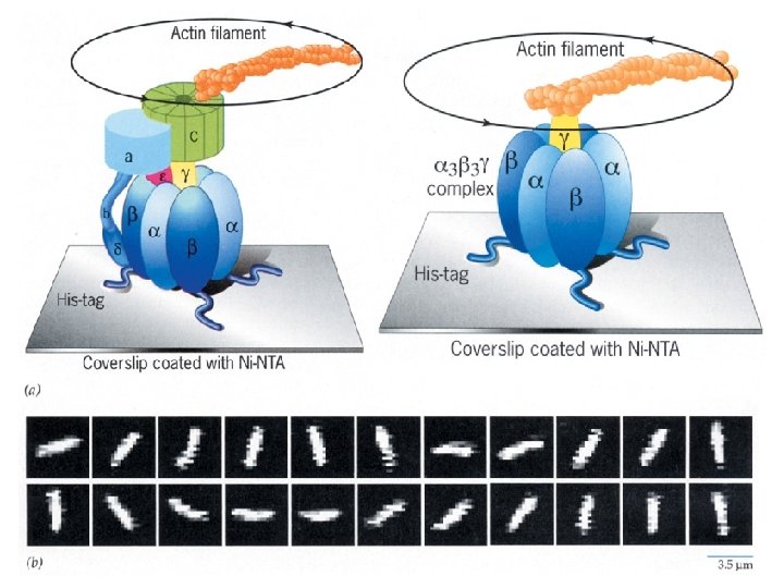
III. ATP synthesis ATP synthase (Complex V ) consists of a transmembrane proton channel (F 0) and an ATPase (F 1). F 1 consists of α 3β 3γδε subunits, which contains several binding sites for ATP and ADP, and the catalytic sites (β) for ATP synthesis. F 0 consists of a 1 b 2 c 9 -12 and serves as proton channel.
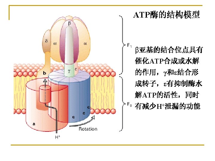
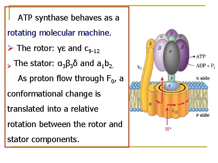
ATP synthase behaves as a rotating molecular machine. Ø The rotor: γε and c 9 -12 Ø The stator: α 3β 3δ and a 1 b 2. As proton flow through F 0, a conformational change is translated into a relative rotation between the rotor and stator components.
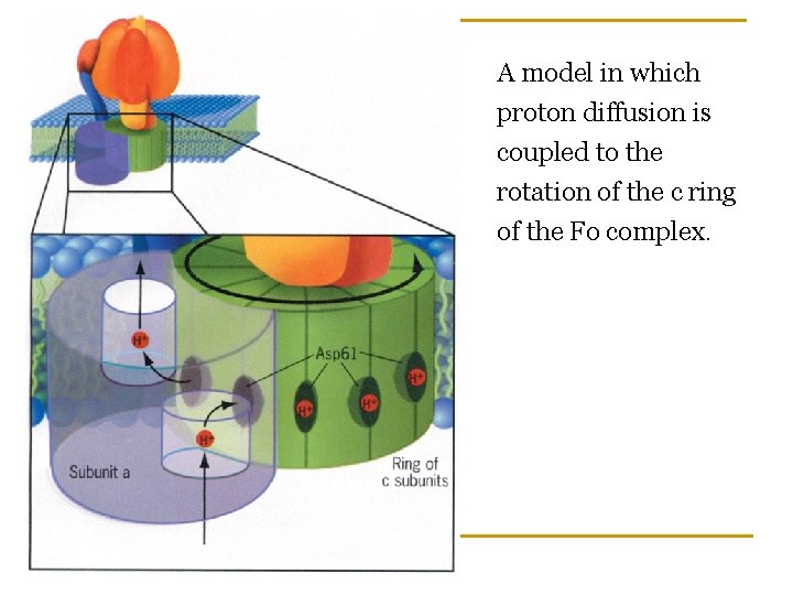
A model in which proton diffusion is coupled to the rotation of the c ring of the Fo complex.
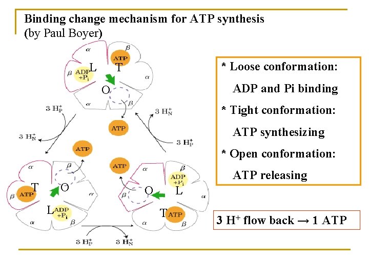
Binding change mechanism for ATP synthesis (by Paul Boyer) L T * Loose conformation: O ADP and Pi binding * Tight conformation: ATP synthesizing * Open conformation: ATP releasing T O O L L T 3 H+ flow back → 1 ATP
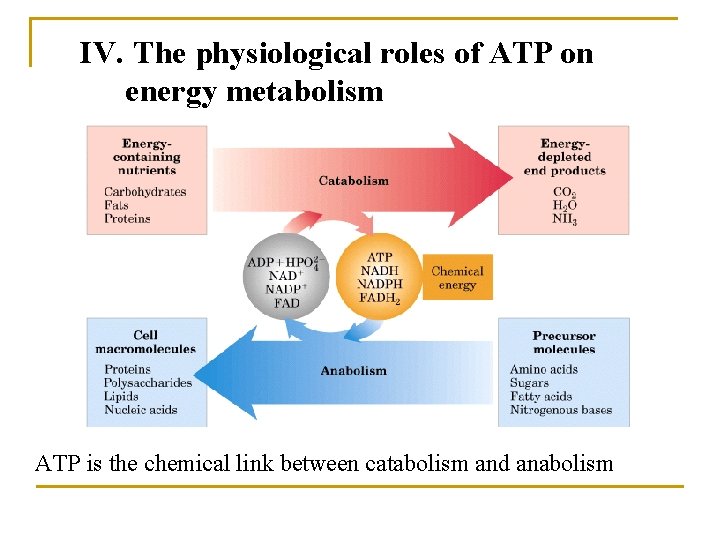
IV. The physiological roles of ATP on energy metabolism ATP is the chemical link between catabolism and anabolism
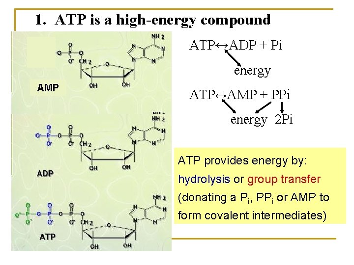
1. ATP is a high-energy compound ATP↔ADP + Pi energy AMP ATP↔AMP + PPi energy 2 Pi ATP provides energy by: hydrolysis or group transfer (donating a Pi, PPi or AMP to form covalent intermediates)
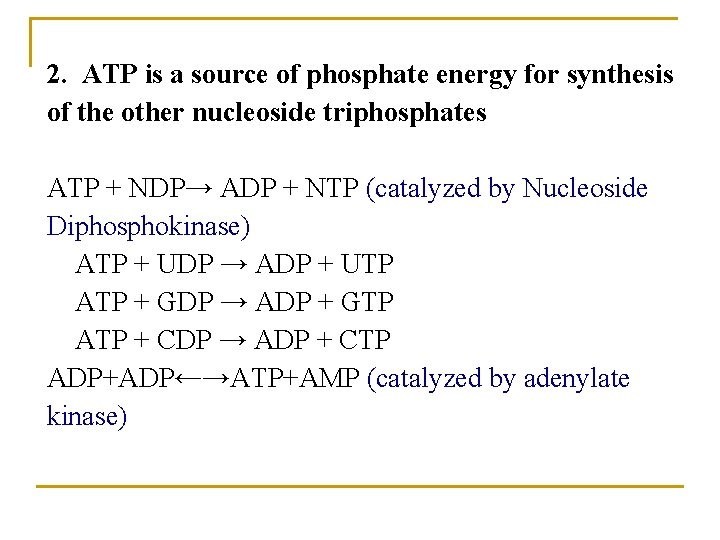
2. ATP is a source of phosphate energy for synthesis of the other nucleoside triphosphates ATP + NDP→ ADP + NTP (catalyzed by Nucleoside Diphosphokinase) ATP + UDP → ADP + UTP ATP + GDP → ADP + GTP ATP + CDP → ADP + CTP ADP+ADP←→ATP+AMP (catalyzed by adenylate kinase)
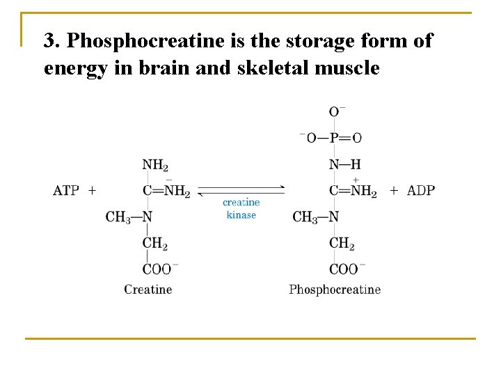
3. Phosphocreatine is the storage form of energy in brain and skeletal muscle
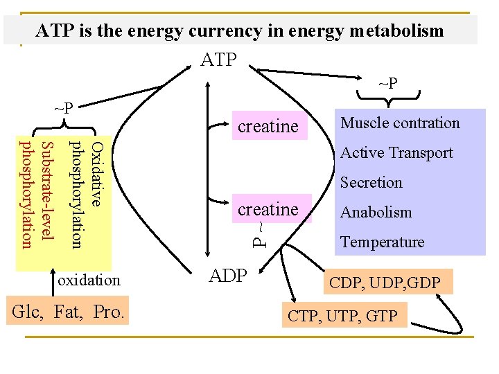
ATP is the energy currency in energy metabolism ATP ~P Oxidative phosphorylation Substrate-level phosphorylation oxidation Glc, Fat, Pro. creatine Muscle contration Active Transport Secretion creatine P ~ ~P ADP Anabolism Temperature CDP, UDP, GDP CTP, UTP, GTP
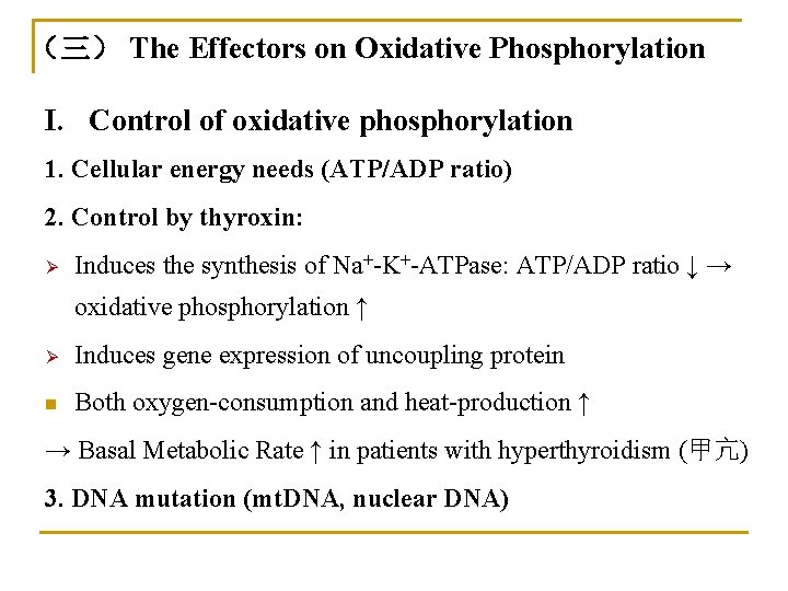
(三) The Effectors on Oxidative Phosphorylation I. Control of oxidative phosphorylation 1. Cellular energy needs (ATP/ADP ratio) 2. Control by thyroxin: Ø Induces the synthesis of Na+-K+-ATPase: ATP/ADP ratio ↓ → oxidative phosphorylation ↑ Ø Induces gene expression of uncoupling protein n Both oxygen-consumption and heat-production ↑ → Basal Metabolic Rate ↑ in patients with hyperthyroidism (甲亢) 3. DNA mutation (mt. DNA, nuclear DNA)
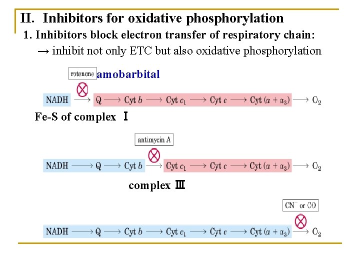
II. Inhibitors for oxidative phosphorylation 1. Inhibitors block electron transfer of respiratory chain: → inhibit not only ETC but also oxidative phosphorylation amobarbital Fe-S of complex Ⅰ complex Ⅲ
2. Uncouplers: * dissipate the proton gradient on both side of membrane * allow electron transfer to continue without ATP synthesis. Ø Dinitrophenol (DNP): * Hydrophobic * Picking up protons on one side and release then on the other.
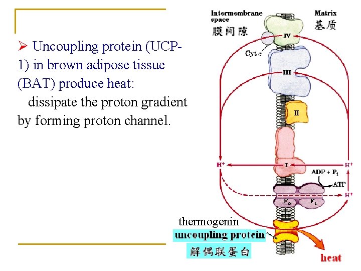
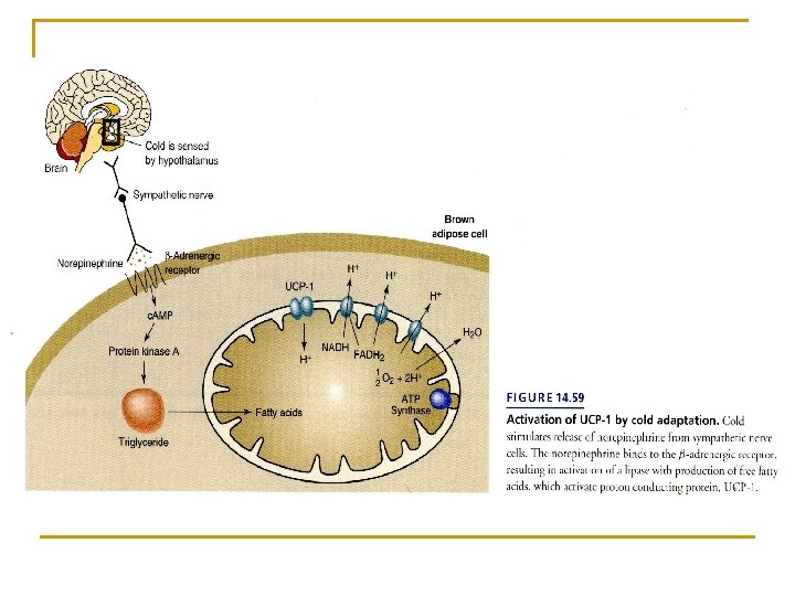
Ø Uncoupling protein (UCP 1) in brown adipose tissue (BAT) produce heat: dissipate the proton gradient by forming proton channel. thermogenin
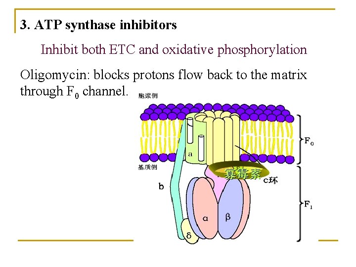
3. ATP synthase inhibitors Inhibit both ETC and oxidative phosphorylation Oligomycin: blocks protons flow back to the matrix through F 0 channel.
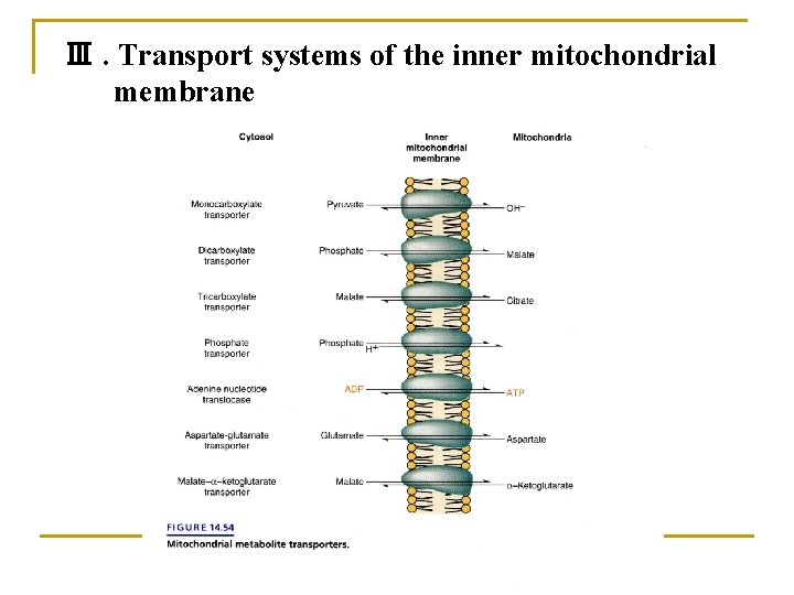
Ⅲ. Transport systems of the inner mitochondrial membrane
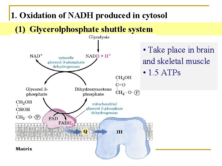
1. Oxidation of NADH produced in cytosol (1) Glycerolphosphate shuttle system • Take place in brain and skeletal muscle • 1. 5 ATPs
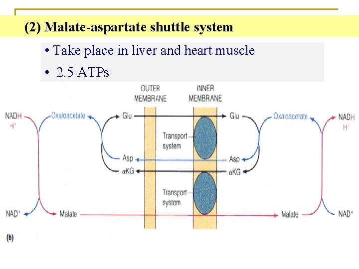
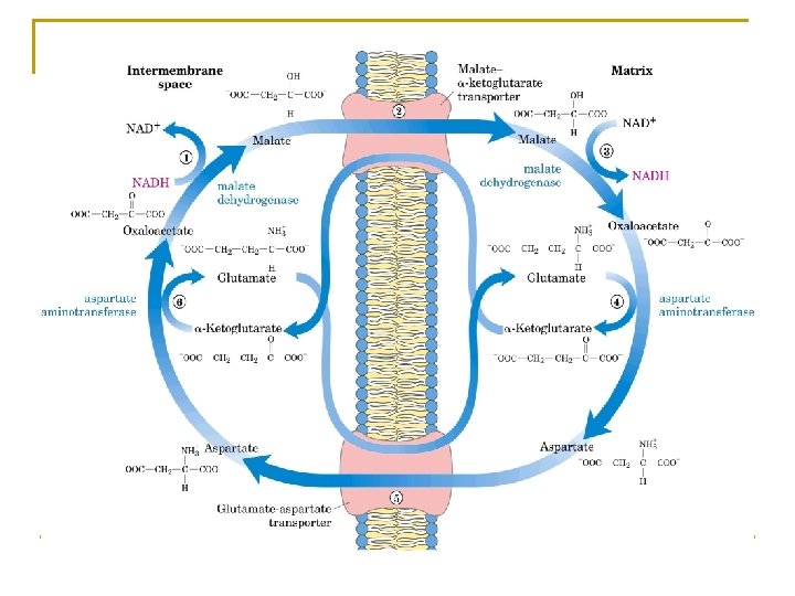
(2) Malate-aspartate shuttle system • Take place in liver and heart muscle • 2. 5 ATPs
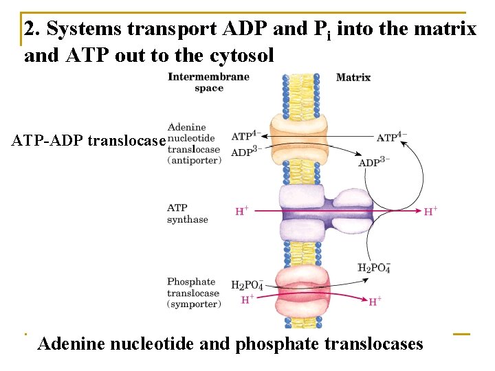
2. Systems transport ADP and Pi into the matrix and ATP out to the cytosol ATP-ADP translocase Adenine nucleotide and phosphate translocases
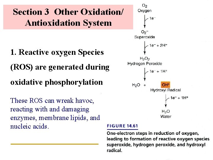
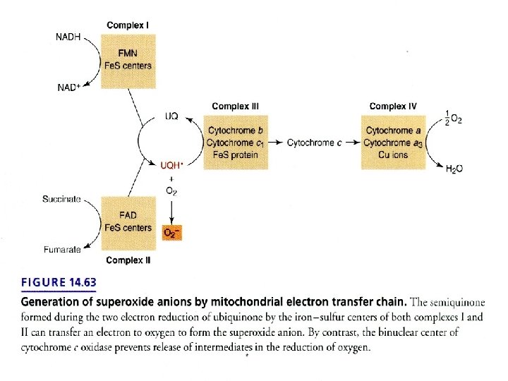
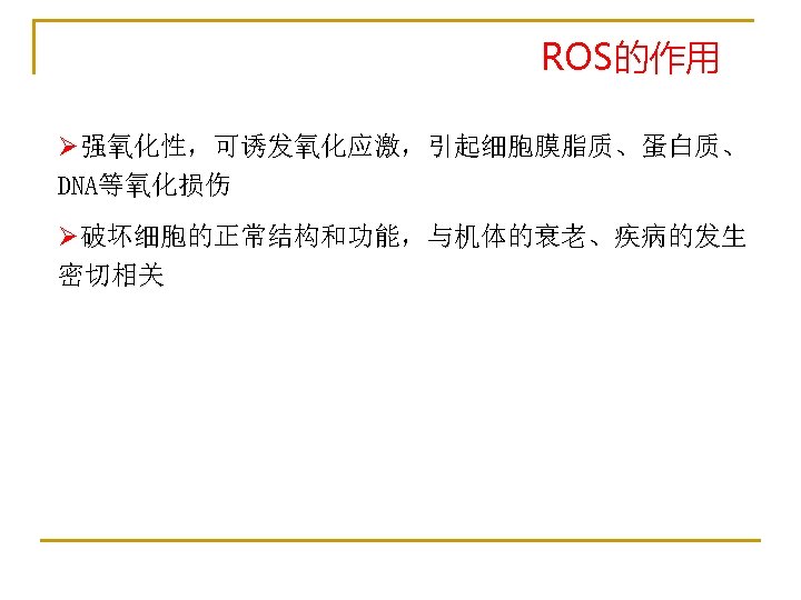
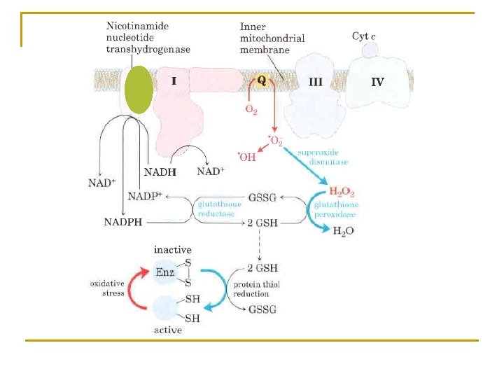
Section 3 Other Oxidation/ Antioxidation System 1. Reactive oxygen Species (ROS) are generated during oxidative phosphorylation These ROS can wreak havoc, reacting with and damaging enzymes, membrane lipids, and nucleic acids.
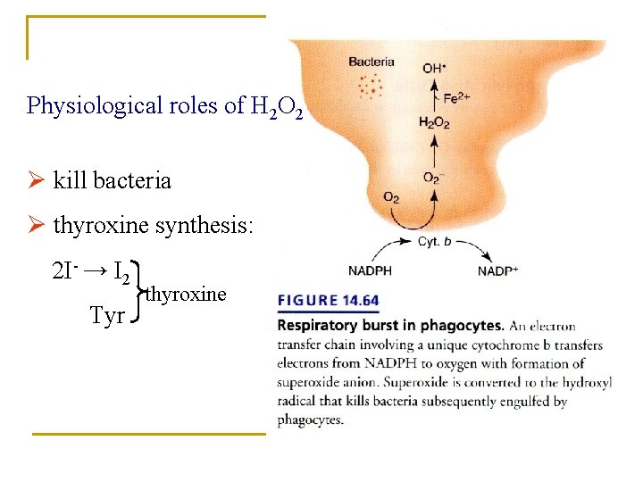
Physiological roles of H 2 O 2 Ø kill bacteria Ø thyroxine synthesis: 2 I- → I 2 Tyr thyroxine
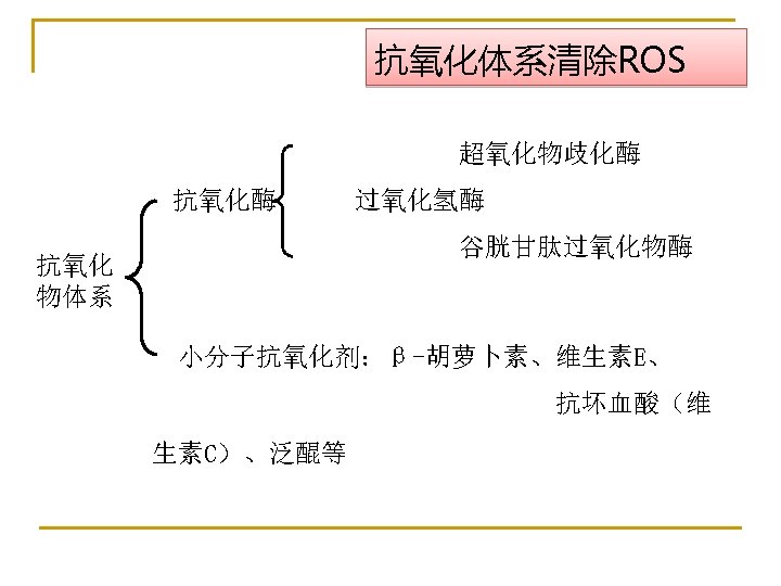
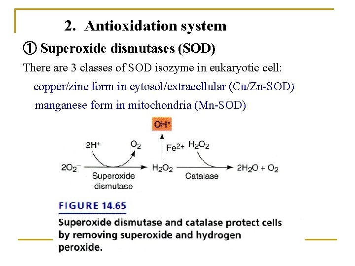
2. Antioxidation system ① Superoxide dismutases (SOD) There are 3 classes of SOD isozyme in eukaryotic cell: copper/zinc form in cytosol/extracellular (Cu/Zn-SOD) manganese form in mitochondria (Mn-SOD)
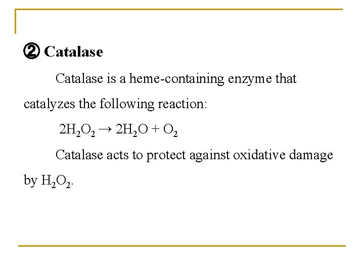
② Catalase is a heme-containing enzyme that catalyzes the following reaction: 2 H 2 O 2 → 2 H 2 O + O 2 Catalase acts to protect against oxidative damage by H 2 O 2.
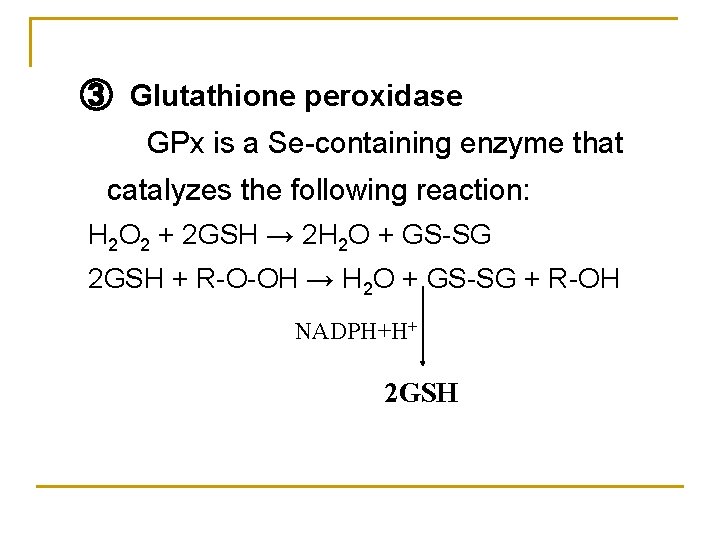
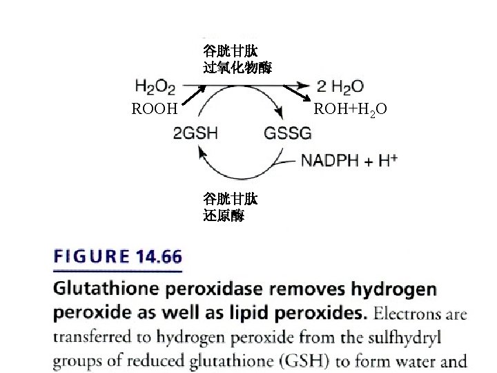
③ Glutathione peroxidase GPx is a Se-containing enzyme that catalyzes the following reaction: H 2 O 2 + 2 GSH → 2 H 2 O + GS-SG 2 GSH + R-O-OH → H 2 O + GS-SG + R-OH NADPH+H+ 2 GSH
3. Cytochrome P 450 Monooxygenase (mixed-function oxidase, or hydroxylase) in microsome Monooxygenases incorporate one atom from O 2 into a product and reduce the other atom to water. RH + NADPH+H+ +O 2 → R-OH + NADP+ + H 2 O This type of enzyme is used in hydroxylation of steroid hormones, bile acid, and biotransformation in liver, etc.
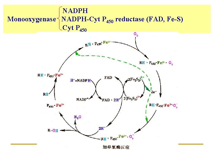
NADPH Monooxygenase NADPH-Cyt P 450 reductase (FAD, Fe-S) Cyt P 450
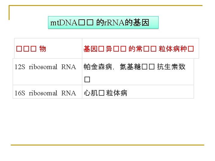
Section 4 Mitochondria and Disease Ø Mitochondrial have a high ratio of mutations: Lower fidelity of mitochondrial DNA replication / Inefficient DNA repair Ø Mutations in mitochondrial DNA accumulate throughout the life of the organism. Ø Mutations in mitochondrial DNA can cause a variety of human inherited diseases, which often most severely affect muscle, nervous system…
Ø Aging: The accumulation of mitochondrial DNA mutations is a contributor to aging Ø Mutations in nuclear-encoded mitochondrial proteins can also cause mitochondrial diseases, which are inherited in the regular, Mendelian fashion.
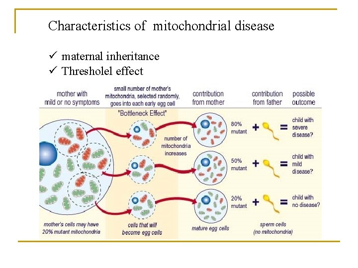
Characteristics of mitochondrial disease ü maternal inheritance ü Thresholel effect
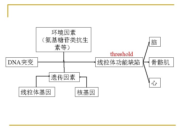
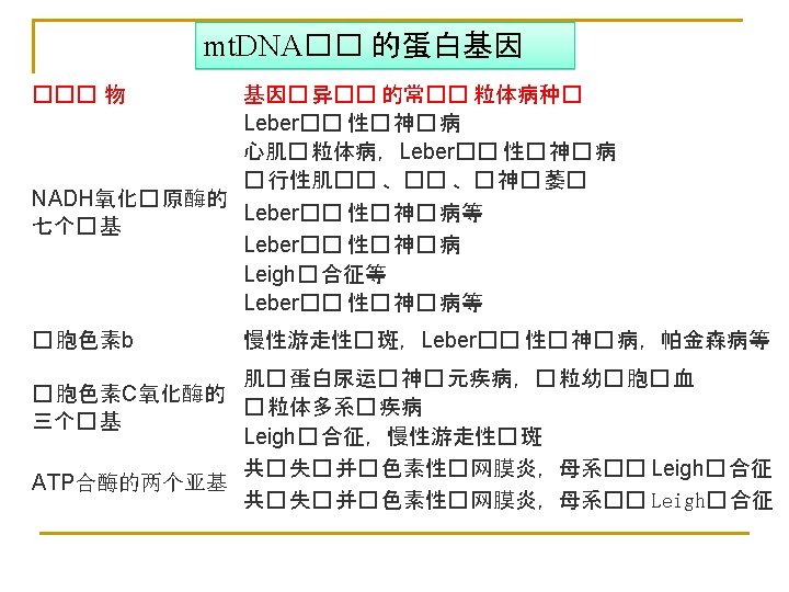

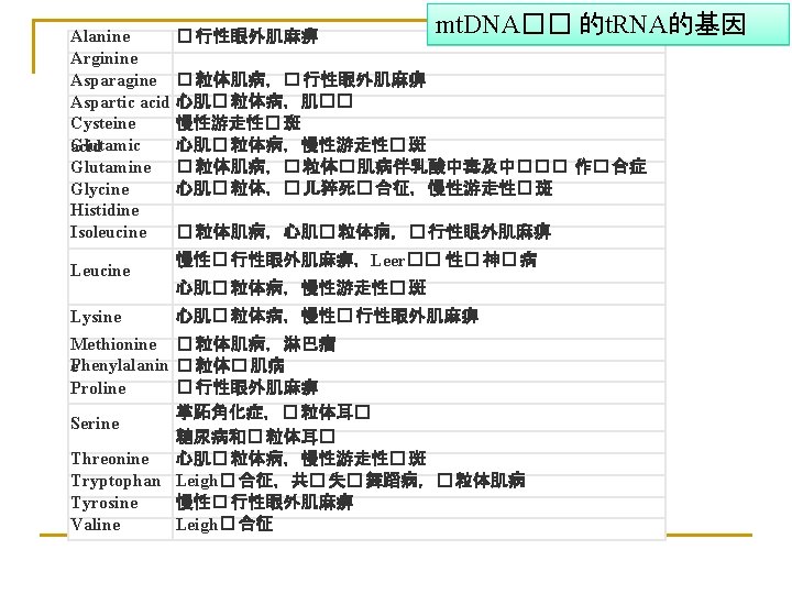
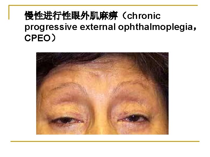
慢性进行性眼外肌麻痹(chronic progressive external ophthalmoplegia, CPEO)

Thank You Any questions?
- Slides: 130