Chapter 6 Bone Tissue Function of Bone Tissue

Chapter 6 Bone Tissue

� Function of Bone Tissue 1. Support of soft tissues and attachment of Tendons and skeletal muscle. 2. Protection of internal organs 3. Movement (skeletal muscle contraction) 4. Mineral storage 5. Blood cell production in red marrow 6. Fat storage in yellow marrow

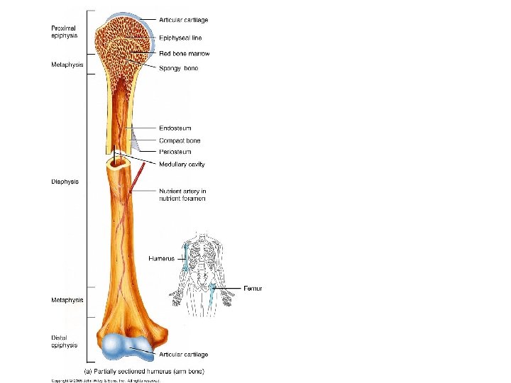
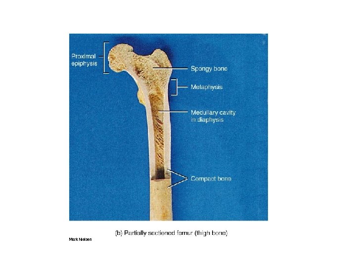
Structure of Bone-long bones 1. Diaphysis-bone shaft 2. Epiphysis-distal and proximal ends of bone 3. Metaphyses- regions where diaphysis joins epiphyses (growing-epiphyseal plate) 4. Articular cartilage-hyaline cartilage covering epiphysis at articulation 5. Periosteum-connective tissue sheath surrounding bone not covered by articular cartilage-attached to bone via perforating fibers • Medullary cavity-space within diaphysis containing yellow marrow 7. Endosteum-thin membrane lining medullary cavity-contains bone-forming cells

Types of cells in bone tissue 1. Osteogenic-stem cells from mesenchyme-only bone cells that undergo cell division 2. Osteoblasts-synthesize and secrete collagen for the extracellular matrix, initiate calcification 3. Osteocytes-mature bone cells, exchange nutrients and waste with blood, maintain the bone tissue. 4. Osteoclasts-large cells, found in endosteum. Secretes lysosomal enzymes for bone resorption, regulates blood Ca 2+
 2. Strength vs.")
Compact vs. Spongy Bone 1. Osteons vs. trabeculae (protect red marrow) 2. Strength vs. light weight 3. (although osteons and trabeculae are oriented 4. along stress lines) 5. 3. Diaphyses vs short flat irregularly shaped bones and epiphyses

Histology of Bone Calcified extracellular matrix Lacunae-small spaces

Histology of Spongy Bone trabeculae Section of a trabeculae Lacunae-small spaces contain osteocytes

Blood Supply of Bone 1. Periosteal arteries-enter diaphysis through perforating canals and supply periosteum and outer part of compact bone 2. Nutrient artery-passes through a hole in compact bone (foramen) 3. Metaphyseal arteries-supply red marrow and metaphyses Veins 1. Nutrient veins-run with nutrient artery in diaphysis 2. Epiphyseal veins and metaphyseal veins-run with their arteries 3. in the epiphyses 3. Periosteal veins-run with their artieries in the perosteum Nerves run with blood supplies to transmit pain especially in the periosteum (fractures, biopsy)

Blood and Nerve Supply of Bone

Bone Formation Ossification-process of bone formation Two methods of ossification: 1. Intramembrane ossification-bone forms within mesenchyme 2. Endochondral ossification-bone forms within hyaline cartilage Intramembrane Ossification 1. Ossification Center-mesenchyme (tissue from which 2. other tissues are derived) differentiates 3. 2. Calcification-osteocytes extend processes into canaliculi 4. 3. Formation of Trabeculae-extracellular matrix forms spongy bone 4. Development of Periosteum-mesenchyme condenses
![Intramembraneous Ossification Mesenchyme differentiates (flat skull bones [fontanels] and mandible) 1 2 3 4](http://slidetodoc.com/presentation_image_h2/a41647579d7a424fa2719c771ed3254b/image-13.jpg "Intramembraneous Ossification Mesenchyme differentiates (flat skull bones [fontanels] and mandible) 1 2 3 4")
Intramembraneous Ossification Mesenchyme differentiates (flat skull bones [fontanels] and mandible) 1 2 3 4

Endochondral Ossification Replacement of cartilage by bone-most bones formed this way. 1. Development of Cartilage Model-hyaline cartilage is surrounded by perichondrium membrane-chondroblasts secrete extracellular matrix become buried-called chondrocytes) 2. Growth of the Cartilage Model-interstitial growth 3. 1. (increases length-chondrocytes), appositional growth (increa thickness-chondroblasts) 3. Development of Ossification Center-inward growth from external surface • Development of medullary cavities-cavity formed by osteoclasts 5. Development of secondary ossification centersproceeds outward from epiphysis center 2. 6. Formation of articular cartilage/ephyseal plate 1. hyaline cartilage covering epiphyses becomes articular cartila
 1 2 4 3 5 /medullary cavity")
Endochondral Ossification (replacement of cartilage with bone) 1 2 4 3 5 /medullary cavity

Bone Growth-length Epiphyseal plate • Zone of resting cartilage-nearest epiphysis, chondrocytes, anchors epiphyseal plate to bone epiphysis. • Zone of proliferating cartilage-larger chondrocytes arranged in stacks, divide to replace dead cells at diaphyseal side of epiphyseal plate. • Zone of hypertrophic cartilage-large chondrocytes arranged in columns • Zone of calcified cartilage-dead chondrocytes surrounded by calcified extracellular matrix. Osteoclasts dissolve cartilage, area invaded by osteoblasts and capillaries from the diaphysis and produce bone extracellular matrix-becomes “new disphysis” cemented to disphysis of bone

Bone growth-epiphyseal plate

Bone growth-thickness 1. Periosteum differentiates into osteoblasts that secrete extracellular matrix and become osteocytes. Formation of bone ridges/grooves for periosteal blood supply. 2. The ridge/groove folds and fuses enclosing the blood vessels. Periosteum becomes endosteum. 3. Osteoblasts in endosteum deposit extracellular matrix forming concentric lamellae. Lamellae formation proceeds out-to-in (inward). 4. Osteoblasts deposit new outer cercumferential lamellae increasing bone thickness and enclosing additional blood vessels. 5. As new bone is deposited on the outer surface, the medullary cavity is being destroyed by osteoclasts-therefore the medullary cavity increases in size as the bone gets thicker.

Bone growth-thickness

Bone remodeling-replacement of old tissue by new tissue. Processes Involved are bone resorption and deposition Compact bone=4% per year Spongy bone=20% per year Why? 1. Weight stress=thicker bone 2. Shape can be altered based on need (e. g. orthodontics). Regulation 1. Minerals required 2. Vitamins: C (collagen synthesis), 3. K and B 12 (protein synthesis), A (osteoblasts) 3. Hormones: IGF stimulated by h. GH (stimulates osteoblast division), Sex hormones (estrogen terminates growth at epiphyseal plates).

Types of Bone Fractures 1. 2. 3. 4. 5. 6. 6. 1. Open fracture-bone protrudes through skin Comminuted-fragments between ends Greenstick-only in children; bend/break Impacted-one end inside other Pott’s-distal end of lateral fibula, injury to tibial articulation Colles-distal end of radius with posterior displacement

Bone fracture repair 1. 3. +6 h-3 w 2. ~3 w 4. >3 m

Negative feedback for blood calcium Blood calcium required for: 1. Nerve and muscle cell function 2. Blood clotting 3. Enzyme cofactor 4. Bone buffers Ca 2+ Blood plasma Ca 2+ (9 -11 mg/100 ml) 1. Parathyroid Hormone (PTH) 2. increases blood Ca 2+ 3. 2. Decrease in blood Ca 2+ causes: 4. increase in c. AMP which c increase in PTH increase in osteoclasts retention of Ca 2+ by kidneys

Osteoporosis Calcium depletion Bone resorption > Bone deposition Result: depletion of bone mass leading to fractures, height loss, hunched back, pain

Chapter 6 END
- Slides: 25