Chapter 6 A Tour of the Cell Recall


 • Sacs not interconnected •")
 1 Vesicles move 2 Vesicles")


 – Support, adhesion, protection – glycoproteins, fibronectin,")


- Slides: 49
Chapter 6 A Tour of the Cell
Recall: Molecules and atoms from the environment are necessary to build new molecules. • Carbon moves from the environment to organisms where it is used to build carbohydrate, proteins, lipids or nucleic acids. • Carbon is used in storage compounds and cell formation in all organisms. • Nitrogen moves from the environment to organisms where it is used in building proteins and nucleic acids. • Phosphorus moves from the environment to organisms where it is used in nucleic acids and certain lipids.
A Tour of the Cell • SA to V ratio • Introduction to the cell – prokaryotic and eukaryotic • Structure and Function: – Ribosome, SER, RER, Golgi, Mitochondria, Lysosome, Vacuole, Chloroplast
What is useful to know about the different types of microscopes…
Main ideas of the cell theory • Scientists, using microscopes, found cells in every organism they examined. This led to the cell theory— 1. All living things are composed of cells 2. Cells are the basic unit of structure and function in living things. 3. All cells come from pre-existing cells. 5
Most Cells are Microscopic • Most plant/animal cells are 10 - 100 mm – 10 x larger than bacterial cells! • Cells are small because they need a high surface area / volume ratio
Surface Area to Volume Ratio • affects the ability to get resources and wastes throughout the cell • increase volume, decrease surface area, demand for resources go up and need more cell structures to exchange materials • smaller cells have a more • root hairs, alveoli, villi, microvilli favorable SA to V ratio • plasma membrane is very efficient at exchanging materials
Eukaryotic cells have internal membranes that compartmentalize their functions • Two types of cells make up every organism – Prokaryotic – Eukaryotic • All cells have several basic features in common -plasma membrane -cytosol -chromosomes -ribosomes
• The plasma membrane – Functions as a selective barrier – Allows sufficient passage of nutrients and waste – Cells that exchange a lot of material with their surroundings may have long, thin projections from the cell (microvilli) Carbohydrate side chain Outside of cell Hydrophilic region Inside of cell 0. 1 µm (a) TEM of a plasma membrane. The plasma membrane, here in a red blood cell, appears as a pair of dark bands Hydrophobic region Hydrophilic region Phospholipid Proteins (b) Structure of the plasma membrane 9
Prokaryotic Cells Simple • Plasma membrane, cytoplasm, DNA, ribosomes, cell wall • No membrane bound organelles – DNA in nucleoid (NO nucleus) • Most are 1 - 10 mm in length • Cell wall: chemically complex • Some have capsule, fimbriae, flagella
Prokaryotic Cells are Simple Pili: attachment structures on the surface of some prokaryotes Nucleoid: region where the cell’s DNA is located (not enclosed by a membrane) Ribosomes: organelles that synthesize proteins Bacterial chromosome (a) A typical rod-shaped bacterium Plasma membrane: membrane enclosing the cytoplasm Cell wall: rigid structure outside the plasma membrane Capsule: jelly-like outer coating of many prokaryotes 0. 5 µm Flagella: locomotion organelles of some bacteria (b) A thin section through the bacterium Bacillus coagulans (TEM)
Eukaryotic Cells are Complex • Generally quite bigger than prokaryotic cells • Plasma membrane, cytoplasm, DNA, ribosomoes • Contains membrane bounded organelles – – Compartmentalizes the cells Allows differing environments within one cell Site of cellular metabolism reactions Greatly increase membrane area (reduces that SA / volume ratio problem associated with increased size) • Non-organelle structures: – Centriole and cytoskeleton – Ribosomes in cytoplasm and attached to organelles
Main Differences Animal cilia contractile vacuole centrioles Plant cell wall central vacuole chloroplast 13
Animal Cells
Plant Cells
The Endomembrane System • Some membranes are physically connected • Some are connected through vesicles (membrane bubbles) • Many organelles function together to synthesize, store, and export molecules • all have interchangeable membranes, they are all made of a phospholipid bilayer and are fusible with one another. The endomembrane system’s interconnectedness.
Eukaryotic Makeup of Endomembrane System • In eukaryotes the organelles of the endomembrane system include: the nuclear envelope, the endoplasmic reticulum, the Golgi apparatus, lysosomes, vacuoles, vesicles, endosomes and the cell membrane.
• Relationships among organelles of the endomembrane system 1 Nucleus Nuclear envelope is connected to rough ER, which is also continuous with smooth ER Rough ER 2 Membranes and proteins produced by the ER flow in the form of transport vesicles to the Golgi Smooth ER cis Golgi Nuclear envelop 3 Golgi pinches off transport Vesicles and other vesicles that give rise to lysosomes and Vacuoles 4 Lysosome available for fusion with another vesicle for digestion Figure 6. 16 Plasma membrane trans Golgi 5 Transport vesicle carries 6 proteins to plasma membrane for secretion Plasma membrane expands by fusion of vesicles; proteins are secreted from cell
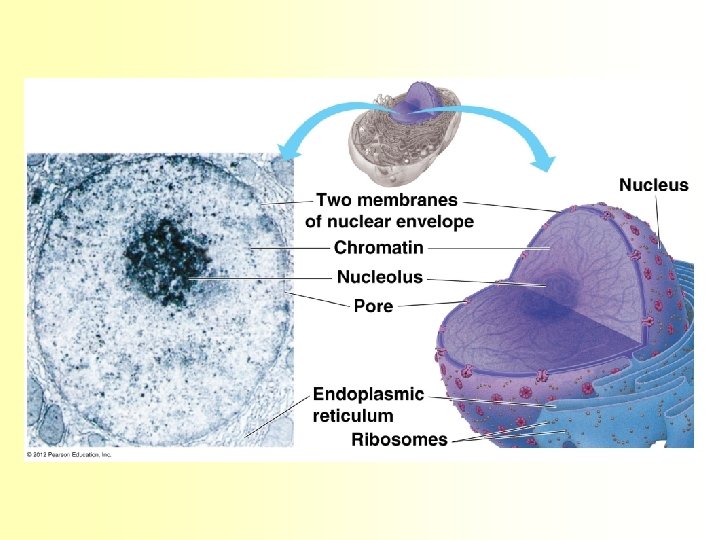
The Nucleus • Contains DNA, controls the cell’s activities, directs protein synthesis • Surrounded by nuclear envelope – a network of protein filaments that maintain structure (nuclear lamina and nuclear matrix) • DNA + proteins = chromatin – chromatin coils up during cell division to become chromosomes • Humans= 46 chromosomes • sex cells= 23 chromosomes
• Nucleolus is located in the nucleus- densely stained fibers and granules adjoining chromatin – ribosomal RNA (r. RNA) is synthesized and assembled with proteins from the cytoplasm to form ribosomal subunits – subunits pass through the nuclear pores to the cytoplasm, where they combine to form ribosomes • The nucleus directs protein synthesis by synthesizing messenger RNA (m. RNA). The m. RNA travels to the cytoplasm through the nuclear pores and combines with ribosomes to translate its genetic message into the primary structure of a specific polypeptide.
Ribosomes - containing r. RNA and protein, are the organelles that carry out protein synthesis – Some free: suspended in the cytosol and synthesize proteins that function within the cytosol – Some bound: attached to the outside of the endoplasmic reticulum or nuclear envelope and synthesize proteins that are either included in membranes or exported from the cell
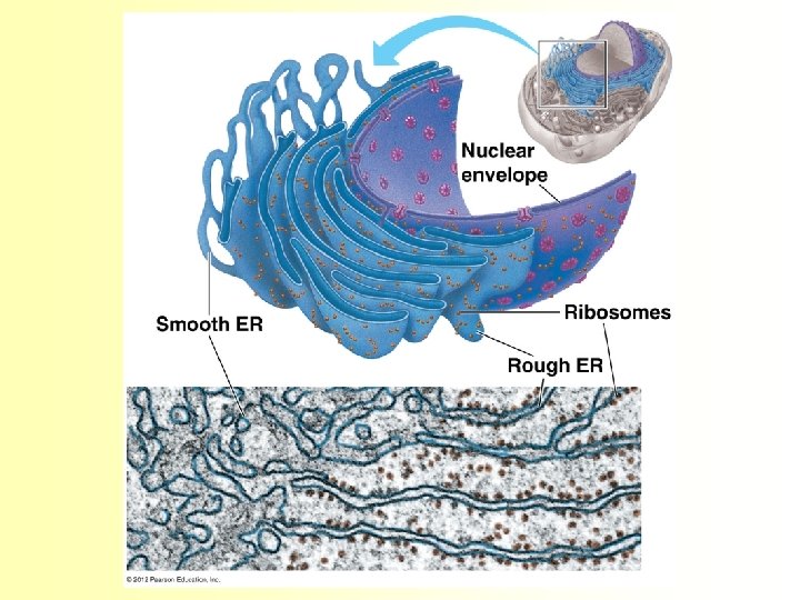
Endoplasmic Reticulum • manufactures membranes and performs many other biosynthetic functions – network of membranous tubules and internal, fluidfilled spaces called cisternae – continuous with the nuclear envelope, and the cisternal space of the ER is continuous with the space between the two membranes of the nuclear envelope
• There are two connected regions of ER that differ in structure and function. • Smooth ER – Lacks ribosomes – Synthesizes lipids, process toxins, stores Ca++ • Rough ER – Has ribosomes imbedded in membrane – Makes more membrane, modifies proteins
Smooth ER rich in enzymes and plays a role in a variety of metabolic processes • Enzymes synthesize lipids, phospholipids, and steroids – In the smooth ER of the liver, enzymes help detoxify poisons and drugs such as alcohol and barbiturates – Metabolizes Carbohydrates • Stores calcium ions • Muscle cells have a specialized smooth ER that pumps calcium ions from the cytosol and stores them in its cisternal space (lumen) Rough ER Millions of ribosomes cover its surface • Protein Generation - production and processing of specific proteins at ribosomal sites that are later exported • Protein Folding -folded into the right three dimensional shapes and carbohydrates may be added. Once the folding aided by chaperonin is complete, they are ready for delivery. • Protein Transport - transport proteins to the sites where they are required • Protein Quality Check - checked for correct ordering and structure and if it doesn't match the exact requirement, it is rejected
The Golgi Apparatus • Flattened sacs (looks like pita) • Sacs not interconnected • Synthesizes lysosomes • Functions: – Serves as a molecular warehouse and finishing factory – Vesicles move from sac to sac as the products get modified
Golgi Functions cis face (“receiving” side of Golgi apparatus) 1 Vesicles move 2 Vesicles coalesce to 6 Vesicles also form new cis Golgi cisternae from ER to Golgi transport certain Cisternae proteins back to ER 3 Cisternal maturation: Golgi cisternae move in a cisto-trans direction 4 Vesicles form and leave Golgi, carrying specific proteins to other locations or to the plasma memtrans face brane for secretion 5 Vesicles transport specific (“shipping” side of proteins backward to newer Golgi apparatus) Golgi cisternae 0. 1 0 µm
Lysosomes • Found in animal cells • Hydrolytic enzyme sac used to digest macromolecules • Aids in the recycling of organic matter • Helps to destroy cells
• Lysosomes carry out intracellular digestion by – Phagocytosis 1 µm Nucleus Lysosome contains active hydrolytic enzymes Food vacuole fuses with lysosome Hydrolytic enzymes digest food particles Digestive enzymes Lysosome Plasma membrane Digestion Food vacuole Figure 6. 14 A (a) Phagocytosis: lysosome digesting food 30
• Lysosomes can play a role in recycling of the cell’s organelles and macromolecules – Autophagy • The degradation of cytoplasmic proteins and organelles by their enclosure in vesicles from the endoplasmic reticulum that fuse with lysosomes. Figure 6. 14 B Lysosome containing two damaged organelles 1µm Mitochondrion fragment Peroxisome fragment Lysosome fuses with vesicle containing damaged organelle Hydrolytic enzymes digest organelle components Lysosome Digestion Vesicle containing damaged mitochondrion 31 (b) Autophagy: lysosome breaking down damaged organelle
• Lysosomes play a critical role in the programmed destruction of cells in multicellular organisms – apoptosis
Abnormal lysosomes can cause fatal diseases • Tay-sachs disease – Lysosomes lack an enzyme needed to break down a lipid abundant in nerve cell membranes – Damages nerve cells (in the brain)
Vacuoles • Membranous sacs • Come in various shapes, sizes, and functions • Function is to aid in intracellular digestion and release cell waste products
• Food vacuoles – Are formed by phagocytosis • Contractile vacuoles – Pump excess water out of protist cells • Central vacuoles – Are found in plant cells – Hold reserves of important organic compounds and water – storage of pigments – allows for a large surface area to volume ratio Central vacuole Cytosol Nucleus Cell wall Chloroplast Tonoplast Central vacuole 5 µm
Energy Conversion • Mitochondria and chloroplasts change energy from one form to another • Mitochondria – Are the sites of cellular respiration • Chloroplasts – Found only in plants, are the sites of photosynthesis
Mitochondria • Carry out cellular respiration in nearly all eukaryotic cells • Converts sugars to ATP (energy currency) • Compartmentalization due to double membrane
Mitochondria • Two membranes – A smooth outer membrane – An inner membrane folded into cristae • cristae have enzymes that aid in ATP production • cristae also increase SA for ATP production
Chloroplasts • Photosynthesis takes place in chloroplasts in algae and higher plants – Conversion of light energy to chemical energy • Internal membranes, 3 compartments
Chloroplasts • Contain chlorophyll – color – light trapping molecules – dominant type: a • Double outer membrane – thylakoids house stacks of grana • produce ATP and NADPH 2 – fuel carbon fixing reactions in Calvin cycle (carbon fixation occurs in stroma converting CO 2 to carbs)
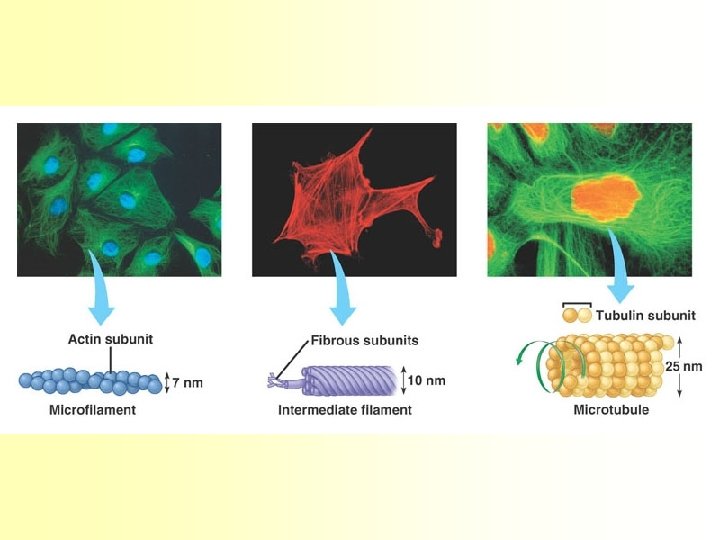
Cytoskeleton • Meshwork of protein fibers • Provides structural support, also allow cellular movement (organelles, etc. ) • Three types: – Microfilaments: actin, solid, twisted double chain, just inside cell membrane – Intermediate filaments: fibrous proteins, ropelike structure, reinforce cell shape, anchor organelles – Microtubules: straight, hollow, tubulins, scaffolding within cells to maintain shape, allow organelle movement
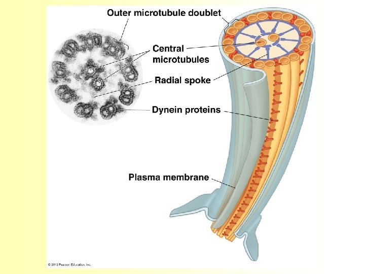
Cilia and Flagella • Cilia: short, numerous, act like oars • Flagella: few, long, act like corkscrew
Plant Cell Surfaces • Extracellular structure of plant cells distinguish them from animal cells – Are made of cellulose fibers embedded in other polysaccharides and protein – May have multiple layers Central vacuole of cell Plasma membrane Secondary cell wall Primary cell wall Central vacuole of cell Middle lamella 1 µm Central vacuole Cytosol Plasma membrane Plant cell walls Figure 6. 28 Plasmodesmata
Plant Cell Surfaces • Cellulose cell walls – Provides skeletal support – 10 - 100 x thicker than plasma membrane – Multilayered, stuck together with glues • Connections between plant cells: – Plasmodesmata: allows materials to pass from one cell to another lamella
Animal Cell Surfaces • Extracellular matrix (ECM) – Support, adhesion, protection – glycoproteins, fibronectin, integrins
Connections between animal cells: Tight junctions Anchoring junctions Gap junctions
Organelle Functional Groups • Genetic Control – Nucleus, ribosomes • Manufacturing, Distribution, and Breakdown – ER, Golgi, Lysosomes, peroxisomes, vacuoles • Energy processing – Chloroplasts, mitochondria • Support, Movement, and Communication – Cytoskeleton, cell walls, extracellular matrix, cell junctions