Chapter 6 7 Lab Cartilage and Bones Axial

Chapter 6 -7 Lab Cartilage and Bones Axial Skeleton

Functions of cartilage • Support soft tissue • keep trachea open • cushions vertebrae • Provide soft, gliding surface at articulations (joints) • Provides model formation of new bone • Comes in 3 types • hyaline cartilage • fibrocartilage • elastic cartilage

Supporting connective tissue • Cartilage and bone are considered connective tissue • protect and support soft body tissues • Cartilage has firm, gel-like extracellular matrix • protein fibers and ground substance • chondrocytes are mature cartilage cells • sit in spaces called lacunae (sing. lacuna) • collagen fibers give strength; stronger than other connective tissue • elastic fibers give resilience • avascular (nutrients absorbed by diffusion)

Hyaline cartilage • most common type; found in nose, trachea, larynx, ribs, at ends of long bones • covers articulating surfaces in joints • weakest type • looks clear and glassy under microscope • forms most of fetal skeleton LM 180 x
 sit in lacunae (sing.")
Hyaline cartilage Table 4. 11 a-1 • Chondrocytes (cartilage cells) sit in lacunae (sing. lacuna) • Dividing chondrocytes often seen sharing lacuna • Extracellular matrix surrounds cells • Collagen fibers are very fine; can’t be seen in microscope Lacuna Chondrocyte Extracellular matrix LM 250 x

Fibrocartilage • numerous, easily visible fibers in irregular bundles • chondrocytes are large • not much ground substance • very durable • acts as shock absorber • found in intervertebral discs, public symphysis, menisci of knee joint LM 80 x Collagen fibers

Table 4. 11 a-2 Fibrocartilage Intervertebral disc • At higher magnification, lacunae with large chondrocytes clearly visible; collagen fibers clear Collagen fibers Lacuna Chondrocyte LM 250 x

Table 4. 11 b-1 Elastic cartilage External ear • contains lots of elastic fibers • very resilient and flexible • surrounded by perichondrium Chondrocytes • found in epiglottis and external ear Elastic fibers LM 200 x

Fig. 6. 1 Cartilage in external ear Epiglottis Larynx Trachea Extracellular matrix Cartilages in nose Lung Respiratory tract cartilages in the lungs, trachea, and larynx Lacuna (with chondrocyte) Articular cartilage of a joint Costal cartilage Cartilage of intervertebral disc LM 180 x (b) Hyaline cartilage Lacunae (with chondrocytes) Pubic symphysis Extracellular matrix Collagen fibers LM 80 x (c) Fibrocartilage Meniscus (padlike cartilage in knee joint) Elastic fibers Lacunae (with chondrocytes) Articular cartilage of a joint (a) Hyaline cartilage Fibrocartilage Elastic cartilage Extracellular matrix LM 200 x (d) Elastic cartilage

Bone • Also a supporting connective tissue • 1/3 organic components (collagen fibers and other proteincarbohydrate molecules) • 2/3 inorganic compounds (mostly calcium phosphate) • very strong, not brittle • most surfaces covered in periosteum (irregular connective tissue)

Components of Bone • Compact bone • appears solid • hard outer shell of bone • Spongy bone • interior of bones • contains latticework of strong, mineralized fibers • provides strength without too much weight
 bone • formed of osteons (AKA Haversian systems) • cylindrical structures •")
Cortical (compact) bone • formed of osteons (AKA Haversian systems) • cylindrical structures • parallel to shaft of long bones • osteons have concentric rings called lamellae • lamellae surround central canal • where blood vessels and nerves travel • osteocytes are bone cells • communicate with each other through canaliculi • branching network throughout compact bone Osteon Lamellae of osteon Central canal Canaliculi Osteocyte in lacuna LM 160 x
 bone LM 160 x")
Cortical (compact) bone LM 160 x
 bone Osteocyte Trabecula (actual bone structure) Bone marrow")
Cancellous (spongy) bone Osteocyte Trabecula (actual bone structure) Bone marrow
 bone • Less dense than compact bone • Bone connective tissue forms")
Spongy (cancellous) bone • Less dense than compact bone • Bone connective tissue forms latticework • Very strong, but lightweight • Located in interior of bones (surrounded by compact bone)

Copyright © Mc. Graw-Hill Education. Permission required for reproduction or display. Fig. 6. 3 Types of bones • Flat bones • have flat, thin surfaces, mostly same thickness across bone • sandwich of spongy bone between compact bone layers • large surfaces for muscle attachment Flat bone (frontal bone)

Copyright © Mc. Graw-Hill Education. Permission required for reproduction or display. Fig. 6. 3 Types of bones • Short bones • nearly equal length and width • sort of similar to cuboidal cells • exterior of compact bone • interior of spongy bone • Include sesamoid bones • tiny, seed-shaped bones along some tendons Short bone (tarsal bone)

Copyright © Mc. Graw-Hill Education. Permission required for reproduction or display. Fig. 6. 3 Types of bones • Irregular bones • irregular shapes • don’t fit in other categories • compact bone with spongy bone inside Irregular bone (vertebra)

Copyright © Mc. Graw-Hill Education. Permission required for reproduction or display. Fig. 6. 3 Types of bones • Longer than wide • cylindrical shaft • increase length (grow) at ends Long bone (femur)
")
Fig. 6. 4 Proximal epiphysis Metaphysis Parts of long bones • Epiphysis Diaphysis (shaft) • knobby region • strengthens bone • provides surface for bone-to-bone articulation • joint surface covered by layer of hyaline cartilage called articular cartilage • provides surface for tendon and ligament attachment Metaphysis Distal epiphysis (a) Anterior view
")
Fig. 6. 4 Proximal epiphysis Metaphysis Parts of long bones • Metaphysis Diaphysis (shaft) • Contains region of growth (in growing bone) called epiphyseal (growth) plate • In adults, growing region epiphyseal plate becomes layer of compact bone called epiphyseal line • Diaphysis • Shaft of bone Metaphysis Distal epiphysis (a) Anterior view
 Epiphyseal line Parts")
Fig. 6. 4 Articular cartilage Spongy bone (contains red bone marrow) Epiphyseal line Parts of long bones Proximal epiphysis Metaphysis Compact bone • Ends of bone contain red bone marrow in spongy bone • Cavity in shaft called medullary cavity Medullary cavity (contains yellow bone marrow in adult) Nutrient artery and vein through nutrient foramen Diaphysis • red marrow in children • yellow marrow in adults Epiphyseal line (b) Sectional view (c) Articular cartilage Metaphysis Distal epiphysis

Fig. 6. 4 Parts of long bones Proximal epiphysis Metaphysis • Internal surfaces of bone (such as medullary cavity) lined with endosteum • • Endosteum incomplete layer of cells contains bone stem cells contains cells for dissolving and rebuilding bone active during bone growth, repair, and remodeling Diaphysis Metaphysis Distal epiphysis

Bone markings: Holes and hole-like things • foramen = hole • fissure = elongate hole or crack

Bone markings: Holes and hole-like things • meatus = tube-like opening • canal = tube-like opening • sinus = cavity or hollow space in bone

Bone markings Flat things and grooves • fossa = depression • often indentation that receives articulating bone • means “ditch” • sulcus = elongated depression or groove • means “trench”

Bone markings Rounded bumps • condyle = knuckle-like rounded bump that fits into a joint • means knuckle • epicondyle = bump near a condyle • for muscle attachment • trochanter = very large bump for muscle attachment • means runner

Bone markings Other bumps • tuberosity = oblong, raised bump • for muscle attachment • tuber means bump or lump • tubercle = small tuberosity • head = distinct endpiece of long bone • neck = narrow portion of longbone usually below head

Bone markings Projections • process = any bone projection or significantly raised area • spine = sharp, pointed process • AKA spinous process • for muscle attachment
 • angle")
Bone markings notch • ramus = branching horn (like a ram’s horn) • angle = inside or outside corner around boundary of bone • border = edge of bone • notch = v-like cut of margin or flat area • body = main part of bone

Bone markings Flat things and grooves • crest = moderately raised ridge • usually site for muscle attachment

Bone markings Flat things and grooves • facet = flat surface that forms a joint with another facet or flat bone • means “little face” facet
 Parietal bone Frontal bone Coronal suture Temporal bone Ethmoid")
Saggital suture (between parietal bones) Parietal bone Frontal bone Coronal suture Temporal bone Ethmoid bone Occipital bone Sphenoid bone Lambdoid Squamous suture

Fig. 7. 10 Coronal suture Glabella Superciliary arch Orbital part Supraorbital margin Zygomatic process Supraorbital foramen (notch) Frontal bone, anterior view

Fig. 7. 11 Sagittal suture Parietal foramen Superior temporal line Coronal suture Lambdoid suture Inferior temporal line Squamous suture Parietal bone, lateral view

Fig. 7. 12 Squamous suture Squamous part External acoustic meatus Zygomatic process Articular tubercle Mastoid process Mandibular fossa Styloid process Right temporal bone, external (lateral) view

Fig. 7. 12 Squamous suture Zygomatic process Internal acoustic meatus Styloid process Mastoid process (b) Right temporal bone, internal (medial) view

Fig. 7. 13 Basilar part Hypoglossal canal Foramen magnum Occipital condyle Condylar canal External occipital crest Inferior nuchal line Superior nuchal line External occipital protuberance (a) Occipital bone, external (inferior) view
")
Fig. 7. 13 Basilar part Jugular notch Hypoglossal canal Foramen magnum Lambdoid suture (b) Occipital bone, internal (superior) view

Bone markings Flat things and grooves • 3 fossae inside inferior cranium • anterior cranial fossa • middle cranial fossa • posterior cranial fossa

Fig. 7. 17 Frontal lobe of cerebrum Temporal lobe of cerebrum Cerebellum Posterior cranial fossa (a) Lateral view Anterior cranial fossa Middle cranial fossa

Fig. 7. 17 Cribriform plate Anterior cranial fossa Middle cranial fossa Carotid canal Lesser wing of sphenoid Sella turcica Foramen ovale Jugular foramen Foramen magnum Posterior cranial fossa (b) Superior view
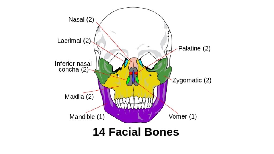

Sphenoid bone Ethmoid bone Inferior nasal concha Vomer Palatine bone Anterior view Lateral view Ethmoid bone Sphenoid bone Inferior nasal concha Vomer Palatine bone Ethmoid bone Vomer Palatine bone

Lesser wing Greater wing Optic canal Foramen rotundum Sella turcica Foramen ovale

Lesser wing Greater wing Superior orbital fissure
 Ethmoid bone, superior")
Cribriform plate Crista galli Ethmoidal sinuses Cribriform plate Orbital plate (a) Ethmoid bone, superior view Superior nasal concha Orbital plate Perpendicular plate (b) Ethmoid bone, anterior view
 Maxillary process Temporal process Right")
Fig. 7. 18 Frontal process Orbital surface (partially obscured) Maxillary process Temporal process Right zygomatic bone, lateral view

Zygomatic arch Zygomatic process of temporal bone Temporal process of zygomatic bone

Ala Vomer, anterior view Vomer, lateral view Posterior Vertical plate Anterior

Right palatine bone, anterior view Right palatine bone, medial view Orbital process Perpendicular plate Horizontal plate

Right maxilla, lateral view Orbital surface Frontal process Infraorbital foramen Anterior nasal spine Zygomatic process Alveolar process

Orbital surface Frontal process Right maxilla, medial view Maxillary sinus Anterior nasal spine Palatine process Alveolar process

Mandible, lateral view mandibular fossa of temporal bone Coronoid process Mandibular foramen Mandibular notch Condylar process Alveolar process Body Mental foramen Angle of mandible

Hyoid bone, anterior view Body
")
Optic canal Superior orbital fissure Inferior orbital fissure (between sphenoid and zygomatic bones)

Temporal bone Sphenoid bone Ethmoid bone Lacrimal bone Nasal bone Perpendicular plate of ethmoid bone Vomer Inferior nasal concha

Parietal bone Coronal suture Frontal bone Squamous suture Lambdoid suture Sutural bone Temporal bone Occipital bone External acoustic meatus Styloid process Zygomatic arch Zygomatic process of temporal bone Temporal process of zygomatic bone Sphenoid bone (greater wing) Nasal bone Lacrimal bone Ethmoid bone Zygomatic bone Maxilla Mandible Mental foramen

Hard palate Maxilla Palatine bone Temporal process of zygomatic bone Zygomatic process of temporal bone Styloid process Mandibular fossa Occipital condyle Vomer Sphenoid bone Foramen ovale Jugular foramen Carotid canal Zygomatic arch Hypoglossal canal Foramen magnum Occipital bone Lambdoid suture External occipital protuberance
- Slides: 59