Chapter 5 Tissues Histology Body tissues are divided
 Body tissues are divided into 4 categories: 1. Connective")
Chapter 5 – Tissues (Histology) Body tissues are divided into 4 categories: 1. Connective 2. Nerve 3. Muscle 4. Epithelial
, defense, and storage (fat) -cells are")
1. Connective Tissue -Functions: Connect, support, transportation (blood), defense, and storage (fat) -cells are surrounded by some type of non-living matrix -classified by their structure and the type of matrix -there are four major classes of connective tissue with subcategories

� I. Fibrous, II. Bone, III. Cartilage, IV. Blood
 a) Loose- the stretchable fascia between organslike an elastic")
I. Fibrous (have extracellular fibers) a) Loose- the stretchable fascia between organslike an elastic glue spread between organs
 Adipose- fat storage under the skin -important for protection and insulation")
b) Adipose- fat storage under the skin -important for protection and insulation
 reticular- web like fibers of the lymphatic (immune) system - helps filter blood")
c) reticular- web like fibers of the lymphatic (immune) system - helps filter blood - contains white blood cells to destroy dangerous invaders
 dense- tendons and ligaments –flexible but strong -tendon- connects muscle to bone -ligament-")
d) dense- tendons and ligaments –flexible but strong -tendon- connects muscle to bone -ligament- connects bone to bone

II. Bone cells are called osteocytes -osteoblast- bone building cell -osteoclast- bone destroying cell � bone � the 2 cells above work together and can reshape bone “unit” of bone is known as the Haversian System (see fig 5 -20) �a � the cells are squeezed between the hard matrix of mineral salts (mostly calcium) bones are called membranous bones (skull, pelvis) � flat

Osteocytes Ha ver sys sian tem Cross-section of long bone

III. Cartilage � chondrocytes are cartilage producing cells � cartilage is avascular (no blood vessels) � there are 3 subcategories of cartilage
 hyaline cartilage- shiny and smooth, at the ends of bones (linings of joints)")
a) hyaline cartilage- shiny and smooth, at the ends of bones (linings of joints) chondrocytes
 Fibrocartilage- strongest and most durable, “shock absorbers” (intervertebral discs, meniscus of the knee")
b) Fibrocartilage- strongest and most durable, “shock absorbers” (intervertebral discs, meniscus of the knee joint)
 Elastic cartilage- most flexible cartilage (ears, nose, larynx)")
c) Elastic cartilage- most flexible cartilage (ears, nose, larynx)

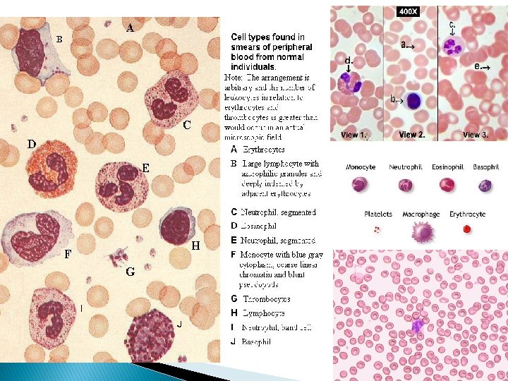
IV. Blood � Considered the bones a connective tissue because it is made in � Hematopoiesis- the process of blood production in the red marrow of the bones � has a liquid matrix called plasma � There are 3 main types of blood cells: 1) Erythrocytes- red blood cells: carry O 2 (some CO 2) 2) Leukocytes- white cells: kill invaders 3) Thrombocytes- responsible for blood clotting by producing platelets

2. Muscle Tissues All are responsible for movement -May be voluntary or involuntary -May be striated or non-striated

ØVoluntary= may be “willed” to move ØInvoluntary= not controlled by thought or will ØStriated= visible “stripes” on tissue ØNon-striated= no visible “stripes

I. Skeletal muscle � Striated and voluntary � Cells are very long and thin with multiple nuclei

II. Smooth Muscle � Non-striated and involuntary � Cells are shorter and each with one nucleus
 � Striated and involuntary � Cells connect at dark “disks”")
III. Cardiac muscle (heart) � Striated and involuntary � Cells connect at dark “disks” and only one nucleus per cell.

3. Nerve Tissue -Rapidly regulate and integrate body activities -2 basic kinds of cells 1. neurons=conducting cells 2. glial cells= support cells
")
Important parts of a neuron: � Soma: main body of cell (nucleus is here) � Dendrites: short thin branches that carry signal towards the soma � Axon: long thin “wire” carrying signal away from the soma � Synapse: where neurons connect to transmit signal

One Very Important Glial Cell: � Schwann Cells- wrap around the axon to insulate the “wiring” to speed nerve signal transmision - Responsible for the fatty white matter of the nervous system

4. Epithelial Tissue -Cells are tightly packed into sheets with no “matrix” between cells -sheets of cells attached to a “basement membrane” -cells are attached to each other by “tight junction” making a tight seal - one side is “exposed” to either internal or external environment -2 main types: membranous glandular

Membranous Epithelium � Classification is based on shapes and layers:
 Cuboidal -(cubic) Columnar -(tall narrow column)")
Description of Cell Shapes: Squamous -(flat and scaly) Cuboidal -(cubic) Columnar -(tall narrow column) Pseudostratified columnar -(odd columns, not all reach the surface)
 Stratified (several layer of same cell type) Transitional")
Description of Layers: Simple (one layer) Stratified (several layer of same cell type) Transitional (many layers transitioning to different cell types)

Simple Squamous - VERY thin. Where quick diffusion is necessary lining of blood vessels, tiny air sacs in lungs, parts of kidney, etc. “flat” Cross-section

Stratified Squamous - areas that receive friction skin, mouth, esophagus Cross-section

Cuboidal - lines many ducts and tubules Cross-section
 Non ciliated - lines")
Columnar epithelium - may be ciliated with tiny “hairs” (cilia) Non ciliated - lines gastrointestinal tract Ciliated - lines respiratory tract

Transitional - bladder – allows for expansion Cross-section

Glandular Epithelium � Synthesizes and secretes a chemical product o. Endocrine gland- secrete directly to bloodstream o. Exocrine gland- secrete into collecting ducts o. Unicellular gland- single cells called goblet cells that produce mucous

Pituitary: Example of an endocrine gland

� Exocrine glands come in a wide variety of shapes:

Exocrine Secretion: � Merocrine- hormones released through cell membrane (sweat & salivary glands, mucous producing goblet cells) � Apocrine- part of cell containing hormones pinches off (mammary gland) � Holocrine- cell bursts to secrete products and must be replaced (sebaceous gland)
- Slides: 38