Chapter 5 The Working Cell Turning on the


 by attaching to the exoskeleton of")


- Slides: 45
Chapter 5 The Working Cell
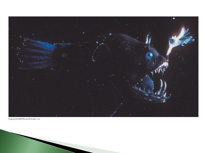
Turning on the Lights to Be Invisible
Energy-Converting Reactions Take place in cell organelles Light producing cells Enzymes embedded in membranes of the organelle Control the reaction
Enzymes: How they Function Membranes: Structure and Function Transport: Through Membranes
ENERGY AND THE CELL Energy is the capacity to perform work 5. 10 All organisms require energy It is the CURRENCY of LIFE
Cellular Metabolism 5 -12 ◦ Sum of endo and exergonic reactions carried out by every working cell in every organism Endergonic reaction◦ Requires net input of energy Exergonic reaction◦ Chemical rxn that release energy
5. 13 ATP shuttles chemical energy and drives cellular work ATP- Adenosine Triphosphate ATP powers nearly all forms of cellular work The energy in an ATP molecule is in the bonds between its phosphate groups ATP is the pocketbook!
Adenosine diphosphate Adenosine Triphosphate Phosphate groups H 2 O P P Adenine P Hydrolysis P Ribose ATP Figure 5. 4 A ADP P + Energy
◦ ATP drives reactions by phosphorylation Transferring a phosphate group to make molecules more reactive ATP Mechanical work Chemical work P Motor protein + Transport work Membrane protein Solute P Reactants P Product Molecule formed Protein moved Figure 5. 4 B ADP + P P Solute transported
◦ Cellular work can be sustained: ◦ “ATM Machine” ◦ ATP is a renewable resource that cells regenerate Figure 5. 4 C Hydrolysis Energy from exergonic reactions Phosphorylation ATP ADP + P Energy for endergonic reactions
HOW ENZYMES FUNCTION 5. 14 Enzymes speed up the cell’s chemical reactions by lowering energy barriers ◦ Energy of Activation- Amount of Energy reactants must absorb before a rxn can begin ◦ Reactants Products ◦ Protiens, DNA, carbohydrates, phospholipids are rich in “potential energy”
A protein catalyst called an enzyme decreases the energy of activation needed to begin a reaction enzyme- a protein molecule that functions as a biological catalyst, increasing the rate of a reaction without itself being changed into a different molecule
Enzyme EA barrier Reactants 1 Figure 5. 5 A Products 2
EA without enzyme EA with enzyme Energy Reactants Net change in energy Products Figure 5. 5 B Progress of the reaction
5. 15 A specific enzyme catalyzes each cellular reaction ◦ Enzymes have unique three-dimensional shapes that determine which chemical reactions occur in a cell ◦ Substrate- specific reactant that an enzyme acts on ◦ Active site- region of enzyme that the substrate fits into ◦ Induced fit- enzyme changes shape slightly to fit the substrate best
◦ The catalytic cycle of an enzyme 1 Enzyme available with empty active site Substrate (sucrose) Active site 2 Glucose Substrate binds to enzyme with induced fit Enzyme (sucrase) Fructose H 2 O 4 Products are released Figure 5. 6 3 Substrate is converted to products
5. 16 Enzyme inhibitors block enzyme action Inhibitors interfere with an enzyme’s activity ◦ A competitive inhibitor Takes the place of a substrate in the active site ◦ A noncompetitive inhibitor Alters an enzyme’s function by changing its shape • Feedback Inhibition • Metabolic rxn is blocked by products
Substrate Active site Enzyme Normal binding of substrate Competitive inhibitor Figure 5. 8 Noncompetitive inhibitor Enzyme inhibition
5. 1 Membranes are a fluid mosaic of phospholipids and proteins Membranes are composed of phospholipids and proteins – Membranes are commonly described as a fluid mosaic – This means that the surface appears mosaic because of the proteins embedded in the phospholipids and fluid because the proteins can drift about in the phospholipids Copyright © 2009 Pearson Education, Inc.
Carbohydrate of glycoprotein Glycolipid Integrin Phospholipid Microfilaments of cytoskeleton fluid mosaic Cholesterol
MEMBRANE STRUCTURE AND FUNCTION ◦ The plasma membrane of the cell is selectively permeable Controlling the flow of substances into or out of the cell
TEM 200, 000 Outside of cell Cytoplasm Figure 5. 10
5. 1 Membrane phospholipids form a bilayer ◦ Phospholipids: ◦ 1 phosphate group and 2 fatty acids Have a hydrophilic head and two hydrophobic tails Are the main structural components of membranes ◦ Phospholipids form a two-layer sheet phospholipid bilayer, with the heads facing outward and the tails facing inward Figure 5. 11 A
Hydrophilic head Phosphate group CH 3 + N CH 2 CH 3 CH 2 O P O O– O CH CH 2 O C CH 2 O O CH 2 CH 2 CH 2 CH 2 CH CH 2 CH 2 Symbol CH CH 2 CH 2 CH 2 CH 3 Hydrophobic tails
Hydrophilic heads Water Hydrophobic tails Hydrophilic heads Figure 5. 11 B Water
Phospholipid bilayer Hydrophobic regions of protein Hydrophilic regions of protein
Membrane Proteins Perform 6 Functions 1. Support (integrins) by attaching to the exoskeleton of a cell 2. Cell-cell recognition (for sorting) 3. Intercellular junctions (where cells connect) 4. Enzymes 5. Signal transduction 6. Transport
5. 2 EVOLUTION CONNECTION: Phospholipids, the key component of biological membranes, naturally assemble into simple membranes – Formation of a membrane that encloses collections of molecules necessary for life was a critical step in evolution – Can be demonstrated in vitro – Allows cells to regulate chemical exchanges with the environment – Basic requirement for life Copyright © 2009 Pearson Education, Inc.
Water-filled bubble made of phospholipids
5. 3 Passive transport is diffusion across a membrane Diffusion- the tendency for particles of any kind to spread out evenly in an available space, moving from where they are more concentrated to regions where they are less concentrated Concentration Gradient: High Concentration Low Concentration Travel down concentration gradient until equilibrium is obtained Multiple substances diffuse independently
Molecules of dye Membrane Equilibrium
◦ In passive transport- substances diffuse through membranes without work by the cell ◦ Ex) O 2 and Co 2 move in and out of our red blood cells in our lung ◦ Small, nonpolar molecules such as O 2 and CO 2 diffuse easily across the phospholipid bilayer ◦ What about large molecules, ions or polar molecules?
5. 6 Facilitated Diffusion Still passive transport- no energy required: 1. Transport protein provides a pore for solute to pass 2. Transport protein binds to solute, changes shape and releases it on the other side Solute examples: Sugars, amino acids, ions and water
Transport proteins may facilitate diffusion across membranes ◦ Many kinds of molecules do not diffuse freely across membranes ◦ Charge, size, polarity ◦ transport proteins Provide passage across membranes through a process called facilitated diffusion Still passive transport- no energy required transport protein provides a pore for solute to pass Figure 5. 15
Solute Molecule Transport Protein
5. 4 Osmosis is the diffusion of water across a membrane ◦ In osmosis Water travels from a solution of lower solute concentration to one of higher solute concentration Water is used to “balance out” different solute concentrations to equilibrium “waters down” the side with “too much” solute
Lower concentration of solute Higher concentration of solute Equal concentration of solute Solute molecule H 2 O Selectively permeable membrane Water molecule Solute molecule with cluster of water molecules Net flow of water
5. 5 Water balance osmoregulation- the control of water balance Isotonic- solution = in solute concentration to the cell Hypotonic - solution with solute concentration lower than the cell Hypertonic- solution with solute concentration greater than the cell Osmosis causes cells to: shrink in hypertonic solutions swell in hypotonic solutions
Isotonic solution H 2 O Hypotonic solution Hypertonic solution H 2 O Animal cell (1) Normal H 2 O (2) Lysed H 2 O (3) Shriveled Plasma membrane H 2 O Plant cell (4) Flaccid (5) Turgid (6) Shriveled (plasmolyzed)
5. 8 Cells expend energy for active transport ◦ Transport proteins can move solutes against a concentration gradient ◦ To the side with the most solute ◦ Through active transport, which requires ATP ◦ Cell work is not ALWAYS about balance ◦ Ex) The cell needs more K+ and less Na+ than its’ external environment (Na+/K+ PUMP) to generate nerve signals
Transport protein ATP Solute 1 Solute binding P ADP 2 Phosphorylation P Protein changes shape Phosphate detaches 3 4 Transport P Protein reversion
5. 9 Exocytosis and endocytosis transport large molecules ◦ To move large molecules or particles through a membrane ◦ Exocytosis ◦ A vesicle may fuse with the membrane and expel its contents ◦ Endocytosis ◦ Membranes may fold inward enclosing material from the outside
Vesicle Protein
Vesicle forming Figure 5. 19 B