Chapter 5 The Structure Function of Macromolecules Slide


 –")


n 3 -7")


 Glycerol Phosphate group-PO 4 Negative charge TAILS (PHOBIC) 2 fatty acids")


 •")


 STABLIZE 3")


, protein synthesis (RNA)")


 ~ double-ring Pyrimidines (Cytosine,")
 Nitrogen base attached to sugar at C-1 Phosphate attached to sugar")
 is the physical carrier of inheritance for 99% of living")
 Deoxyribose sugar Nitrogeneous bases: A, C, G and T DOUBLE HELIX")
 Strands run antiparallel http: //www. biology. arizona. edu/biochemistry/problem_sets/large_molecules/06 t. html")
 Complementary strands H bonds ~ between paired bases van der Waals")

 Ribose sugar Nitrogeneous bases: A, C, G, and U SINGLE STRANDED")

 Ribonucleic acid (RNA) DNA → RNA → protein")
")

- Slides: 79
Chapter 5 The Structure & Function of Macromolecules Slide show modified from Kim Foglia @ http: //www. explorebiology. com
4 MAJOR MACROMOLECULES • Carbohydrates • Lipids • Proteins • Nucleic acids
POLYMERS Large molecule made by linking smaller subunits together – Monomers (small subunits) – Covalent bonds Image by Riedell
SEE AN ANIMATION
SEE AN ANIMATION
CARBOHYDRATES http: //www. graphic-design. com/Type/sugar/index. html http: //www. ifr. ac. uk/SPM/images/Starch%20 products. jpg
MONOSACCHARIDES Simple sugar molecules Composed of C, H, O (CH 2 O)n 3 -7 carbons Name often ends in –ose C 6 H 12 O 6 http: //www. cybercolloids. net/library/sugars/glyceraldehyde. gif http: //www. estrellamountain. edu/faculty/farabee/biobk/Bio. Book. CHEM 2. html http: //217. 60. 75. 10/llt/biokemi/images/galactose. jpg http: //www. estrellamountain. edu/faculty/farabee/biobk/Bio. Book. CHEM 2. html D-glyceraldehyde C 3 H 6 O 3 C 5 H 10 O 5
NUMBERING • Carbons are numbered • Carbon with carbonyl group is #1
Is it D or L ? • For sugars with more than one chiral center, the D or L designation refers to the asymmetric carbon farthest from the aldehyde or keto group. • Most naturally occurring sugars are D isomers. • D & L sugars are mirror images with same name.
Pentoses and hexoses can cyclize in water See animation • Carbons can be numbered • Carbon with carbonyl group is #1
CARBOHYDRATES SUPPLY ENERGY Cells burn glucose and store the energy released as ATP Images from: http: //www. miranda. com/library. en/Images/Pictures/girls-runners. jpg http: //www. estrellamountain. edu/faculty/farabee/biobk/Bio. Book. CHEM 2. html
Disaccharides • Use dehydration synthesis to join TWO sugar molecules • covalent bond between 2 monosaccharides = GLYCOSIDIC linkage EX: Sucrose (table sugar) • most common disaccharide http: //faculty. clintoncc. suny. edu/faculty/michael. gregory/files/Bio%20101%20 Lectures/Biochemistry/bioche 1. gif http: //www. biotech. iastate. edu/lab_protocols/HSSB-TLC_images/sucrose. gif
DISACCHARIDES Glucose + Fructose → Sucrose + H 20 Glucose + Glucose → Maltose + H 20 Glucose + Galactose → Lactose + H 20
http: //www. district 87. org/biology 87/apbio/biochem/Activity 6_notes. pdf POLYSACCHARIDES~ “many sugars” Ex: STARCH • polymer of αlpha glucose • linked by α 1 -4 glycosidic linkages Function: Energy storage in PLANTS Most animals have the enzymes to hydrolyze starch, too http: //www. langara. bc. ca/biology/mario/Assets/Amylopectin. jpg
POLYSACCHARIDES~ “many sugars” TWO KINDS OF STARCH: amylose = unbranched starch amylopectin = branched starch http: //www. langara. bc. ca/biology/mario/Assets/Amylopectin. jpg
POLYSACCHARIDES~ “many sugars” EX: GLYCOGEN alpha 1 -4 glycosidic bonds like starch More branched than amylopectin FUNCTION: Energy storage in ANIMALS Stored in liver and muscle tissue http: //www. abcbodybuilding. com/magazine 04/scientific. htm
POLYSACCHARIDES~ “many sugars” FUNCTION: Structural PLANTS ~ CELLULOSE Major component in cell walls Most abundant organic compound on Earth beta (ß) 1 -4 glycosidic linkages
• Enzymes that digest starch by hydrolyzing alpha linkages can’t hydrolyze beta linkages in cellulose • Cellulose in human food passes through the digestive tract as insoluble fiber • Some microbes use enzymes to digest cellulose • Many herbivores, from cows to termites, have symbiotic relationships with these microbes
POLYSACCHARIDES FUNCTION: Structural EX: CHITIN Structural polysaccharide made from ß glucose with a NITROGEN containing group attached
Major component of: Exoskeletons in Arthropods Cell walls in Fungi Dissolvable surgical thread
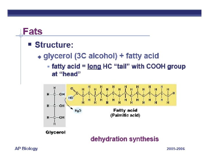
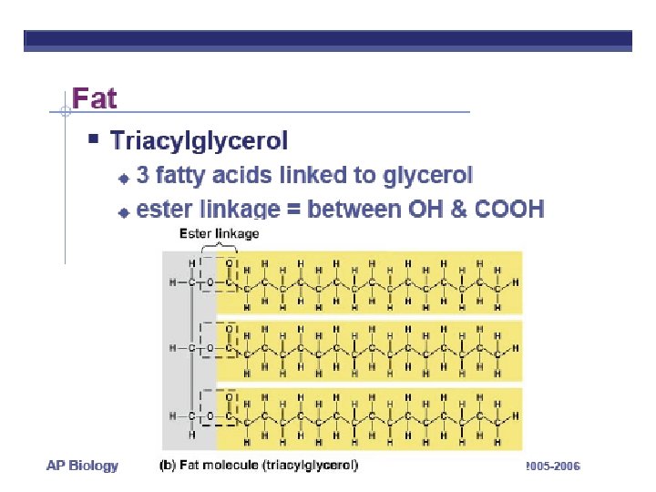
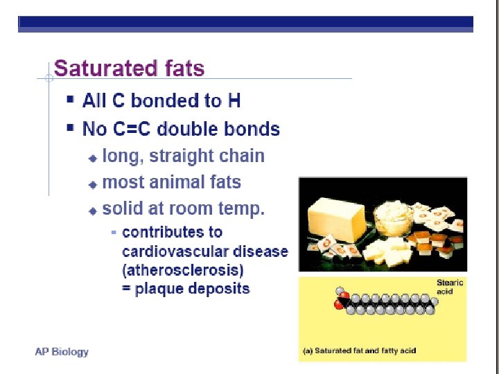
FATTY ACIDS USED CAN : • • Be same or different in one molecule Vary in length Vary in number/location of double bonds Saturated (single bonds) vs. unsaturated fats (double bonds) Kink in chain wherever a cis double bond occurs
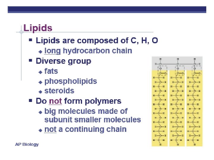
FATS LONG HC chain • NON-POLAR • HYDROPHOBIC FUNCTION: • Energy storage very rich 2 X energy in carbos • Cushions organs • Insulates body Think whale blubber!
Lipids, II
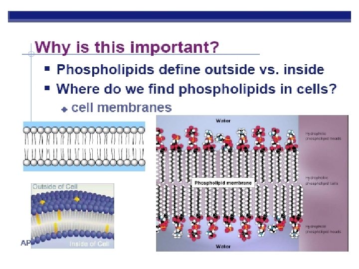
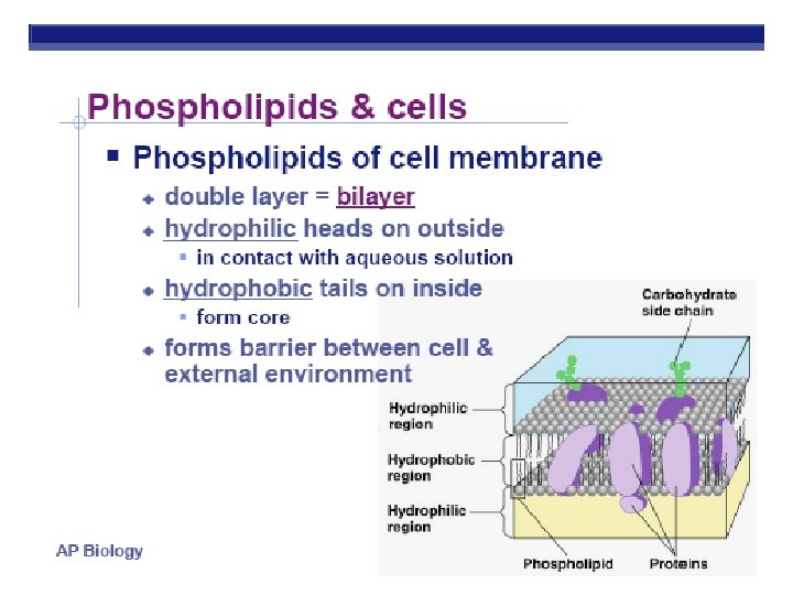
Phospholipids HEAD (PHILIC) Glycerol Phosphate group-PO 4 Negative charge TAILS (PHOBIC) 2 fatty acids instead of 3
SEE A MOVIE !
PROTEINS http: //images. foodnetwork. com/webfood/images/gethealthy/nutritionalallstars/Lean. Proteins_header. jpg
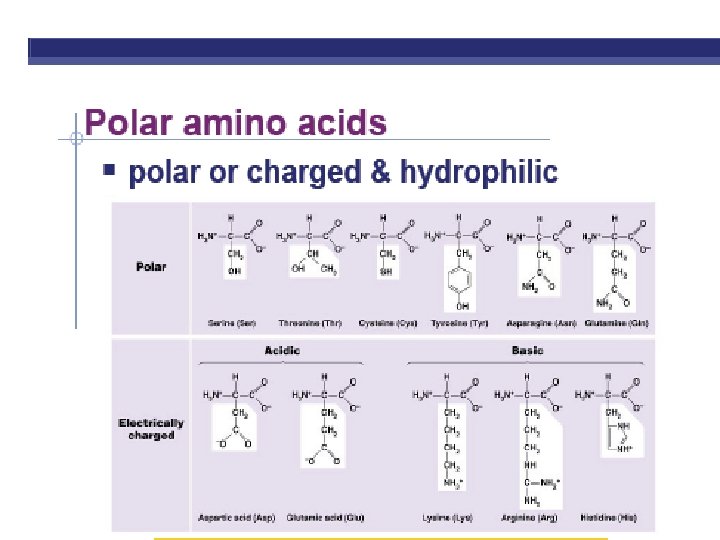
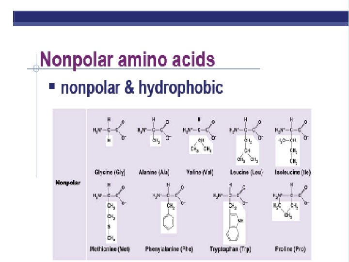
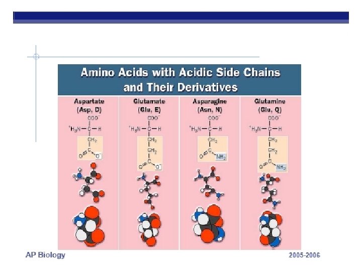
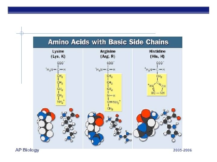
PROTEINS ARE MADE FROM AMINO ACID SUBUITS • Structure – Central carbon – Amino group – Carboxyl group – R group (side chain) • Variable group • Confers unique chemical properties • polar (hydrophilic), nonpolar (hydrophobic), acid or base • Join via DEHYDRATION SYNTHESIS reactions
R GROUPS Each kind of amino acid has a different R group 20 different amino acids are used by cells to make proteins (There a few other aa’s, but rare)
See an animation
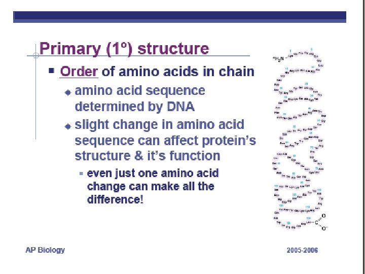
POLYPEPTIDES • POLYMERS OF AMINO ACIDS ARE CALLED POLYPEPTIDES • DNA determines the amino acid sequence http: //www. emc. maricopa. edu/faculty/farabee/BIOBK/Bio. Book. CHEM 2. html http: //www. cherishedtimedesigns. com/images/Bali. Charm. Bracelet. Graduation 500. jpg
A functional PROTEIN is not just the polypeptide chain. A PROTEIN consists of one or more polypeptide chains twisted, folded, and coiled into a unique molecular shape What determines the shape? Image from: http: //www. tvdsb. on. ca/saunders/courses/online/SBI 3 C/Cells/Protein-Structure 03. jpg SEE AN ANIMATION
PROTEIN STRUCTURE & FUNCTION Function depends on structure • 4 levels of organization • result in 3 -D structure
Primary Structure Amino acid substitution: in hemoglobin code sickle-cell anemia A T
Secondary Structure folding along short sections • Due to: R group interactions (phobic/philic) • Alpha Helix: coiling; • ß Pleated Sheet: parallel; • Hydrogen bonds between adjacent amino acids hold shape
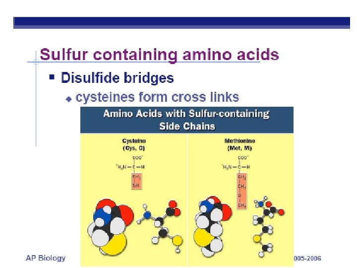
Tertiary Structure interactions between side chains Conformation: irregular contortions from R group bonding √ hydrophobic √ disulfide bridges √ hydrogen bonds √ ionic bonds
Functional Groups • Sulfhydral Group • Called: thiols • http: //www. mun. ca/biology/scarr/Disulfide_bridge. htm
DISULFIDE BRIDGES BETWEEN nearby CYSTEINE amino acids (Notice name change when bonded) STABLIZE 3 -D SHAPE http: //sandwalk. blogspot. com/2007/02/disulfide-bridges-stabilize-folded. html
Quaternary Structure • Conformation: 2 or more polypeptide chains aggregated into one macromolecule √ collagen (connective tissue) √ hemoglobin
See an animation
WHAT DO PROTEINS DO? * See page 78 in Campbell for other examples
ENZYMES http: //www. biologie. uni-hamburg. de/b-online/library/cat-removed/enzyme_. gif Enzymes are protein catalysts that accelerate chemical reactions in living things SEE ANIMATION of AMYLASE Enzymes reduce activation energy required for reaction Enzymes are specific and fit substrate like a lock and key. LEARN MORE Enzymes are not changed by reaction and are reusable. http: //www. grand-illusions. com/images/articles/toyshop/trick_lock/mainimage. jpg
PROTEIN CONFORMATION ALSO DEPENDS ON PHYSICAL ENVIRONMENT LEARN MORE • p. H • Salt concentration • Temperature See a movie Choose narrated http: //www. nealbrownstudio. com/adm/photo/163_nb_fried_egg. jpg http: //www. desktopfotos. de/Downloads/melt_cd. jpg
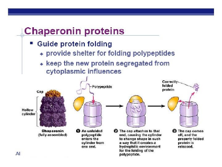
Proteins that have denatured are biologically inactive Once conditions change, protein may need help returning to its functional shape. Facilitation of folding
NUCLEIC ACIDS
Nucleic Acids The main functions of nucleotides are: information storage (DNA), protein synthesis (RNA) energy transfers (ATP and NAD).
Nucleic Acids Nucleic acids are polymers composed of units known as nucleotides. The main functions of nucleotides are: information storage (DNA), protein synthesis (RNA) energy transfers (ATP and NAD).
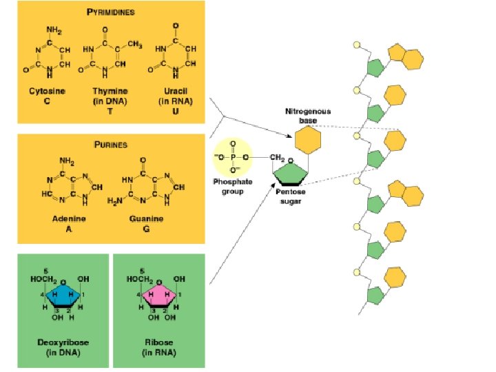
Nucleic Acids Nucleic acids are polymers composed of units known as nucleotides. Nucleotides consist of a pentose (5 C) sugar, a nitrogenous base, and a phosphate. Sugar and phosphate alone = nucleoside
Nucleic Acids The sugars are either: OR deoxyribose
Nucleic Acids Nitrogeneous bases can be: Purines (Adenine and Guanine) ~ double-ring Pyrimidines (Cytosine, Thymine and Uracil) ~ single-ring
Deoxyribonucleic acid (DNA) Nitrogen base attached to sugar at C-1 Phosphate attached to sugar at C-5 Phosphate attached to next nucleoside at C-1 by phosphodiester linkage Each strand has a 3’ and 5’ end http: //staff. um. edu. mt/acus 1/3 Molgen. htm
DNA Deoxyribonucleic acid (DNA) is the physical carrier of inheritance for 99% of living organisms. Image from: http: //sbchem. sunysb. edu/msl/dna. gif
Deoxyribonucleic acid (DNA) Deoxyribose sugar Nitrogeneous bases: A, C, G and T DOUBLE HELIX sugar & phosphates make up sides of ladder nitrogen bases form steps
Deoxyribonucleic acid (DNA) Strands run antiparallel http: //www. biology. arizona. edu/biochemistry/problem_sets/large_molecules/06 t. html
Deoxyribonucleic acid (DNA) Complementary strands H bonds ~ between paired bases van der Waals ~ between stacked bases http: //staff. um. edu. mt/acus 1/3 Molgen. htm
Nucleic Acids • Inheritance based on DNA replication • Double helix (Watson & Crick - 1953) • Based on Rosalind Franklin’s Xray crystallograpy
Ribonucleic acid (RNA) Ribose sugar Nitrogeneous bases: A, C, G, and U SINGLE STRANDED http: //www. biology. arizona. edu/biochemistry/problem_sets/large_molecules/06 t. html
RNA functions in protein synthesis. There are three types of RNA: Messenger RNA (m. RNA) ~ blueprint for construction of a protein. Ribosomal RNA (r. RNA) ~ construction site where the protein is made. Transfer RNA (t. RNA) ~ truck delivering the proper amino acid to the site at the right time.
Deoxyribonucleic acid (DNA) Ribonucleic acid (RNA) DNA → RNA → protein
NUCLEOTIDES can transfer and store energy Adenosine triphosphate (ATP)
NUCLEOTIDES can transfer and store energy NAD+ NADP+ FAD Coenzyme A Energy and electron carriers used in photosynthesis and respiration More on this next unit!