Chapter 5 Membrane Structure and Function Use 5

Chapter 5 Membrane Structure and Function
 EQ: How do different")
Use 5 left pages (221, 223, 225, 227, and 229) EQ: How do different molecules pass through the plasma membrane of a cell?

What You Must Know: Why membranes are selectively permeable. The role of phospholipids, proteins, and carbohydrates in membranes. How water will move if a cell is placed in an isotonic, hypertonic, or hypotonic solution and be able to predict the effect of different environments on the organism. How electrochemical gradients and proton gradients are formed and function in cells.

Cell Membrane A. Plasma membrane is selectively permeable Allows some substances to cross more easily than others B. Fluid Mosaic Model Fluid: membrane held together by weak interactions Mosaic: phospholipids, proteins, carbs
 Davson/Danielli – Sandwich model phospholipid bilayer between 2 protein layers")
Early membrane model (1935) Davson/Danielli – Sandwich model phospholipid bilayer between 2 protein layers Problems: varying chemical composition of membrane, hydrophobic protein parts

The freeze-fracture method: revealed the structure of membrane’s interior

Fluid Mosaic Model

This is on a left page - don’t draw

Phospholipids Bilayer Amphipathic = hydrophilic head, hydrophobic tail Hydrophobic barrier: keeps hydrophilic molecules out
 Cholesterol resists changes")
Membrane fluidity Low temps: phospholipids w/unsaturated tails (kinks prevent close packing) Cholesterol resists changes by: limit fluidity at high temps hinder close packing at low temps Adaptations: bacteria in hot springs (unusual lipids); winter wheat ( unsaturated phospholipids)

Membrane Proteins Integral Proteins Peripheral Proteins Embedded in membrane Determined by freeze fracture Transmembrane with hydrophilic heads/tails and hydrophobic middles Extracellular or cytoplasmic sides of membrane NOT embedded Held in place by the cytoskeleton or ECM Provides stronger framework

Integral & Peripheral proteins

Transmembrane protein structure Hydrophobic interior Hydrophilic ends
")
Some functions of membrane proteins (YOU NEED THESE!)

Carbohydrates Function: cell-cell recognition; developing organisms Glycolipids, glycoproteins Eg. blood transfusions are type-specific

Synthesis and sidedness of membranes
 cross easily (hydrocarbons, hydrophobic molecules, CO 2,")
Selective Permeability Small molecules (polar or nonpolar) cross easily (hydrocarbons, hydrophobic molecules, CO 2, O 2) Hydrophobic core prevents passage of ions, large polar molecules
 needed! Diffusion down concentration gradient (high low concentration) Eg.")
Passive Transport NO ENERGY (ATP) needed! Diffusion down concentration gradient (high low concentration) Eg. hydrocarbons, CO 2, H 2 O

Diffusion

Osmosis: diffusion of H 2 O

External environments can be hypotonic, isotonic or hypertonic to internal environments of cell
")
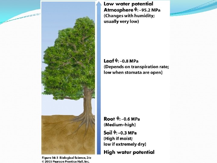
Understanding Water Potential (YOU MUST KNOW THIS CONCEPT AND BE ABLE TO CALCULATE IT)
: H 2 O moves from high ψ low ψ")
Water Potential Water potential (ψ): H 2 O moves from high ψ low ψ potential Water potential equation: ψ = ψS + ψP Water potential (ψ) = free energy of water Solute potential (ψS) = solute concentration (osmotic potential) Pressure potential (ψP) = physical pressure on solution; turgor pressure (plants) Pure water: ψP = 0 MPa Plant cells: ψP = 1 MPa
 ψS = -i. CRT • • i = ionization constant")
Calculating Solute Potential (ψS) ψS = -i. CRT • • i = ionization constant (# particles made in water) C = molar concentration R = pressure constant (0. 0831 liter bars/mole-K) T = temperature in K (273 + 0 C) The addition of solute to water lowers the solute potential (more negative) and therefore decreases the water potential.

Where will WATER move? From an area of: higher ψ lower ψ (more negative ψ) low solute concentration high solute concentration high pressure low pressure

1. 2. 3. 4. Which chamber has a lower water potential? Which chamber has a lower solute potential? In which direction will osmosis occur? If one chamber has a Ψ of -2000 k. Pa, and the other -1000 k. Pa, which is the chamber that has the higher Ψ?

Sample Problem 1. Calculate the solute potential of a 0. 1 M Na. Cl solution at 25°C. 2. If the concentration of Na. Cl inside the plant cell is 0. 15 M, which way will the water diffuse if the cell is placed in the 0. 1 M Na. Cl solution?
 help hydrophilic substances cross § Two")
Facilitated Diffusion Transport proteins (channel or carrier proteins) help hydrophilic substances cross § Two ways: § Provide hydrophilic channel § Loosely bind/carry molecule across § Eg. ions, polar molecules (H 2 O, glucose)

Aquaporin: channel protein that allows passage of H 2 O
")
Glucose Transport Protein (carrier protein)
 Proteins transport substances against concentration gradient (low high conc.")
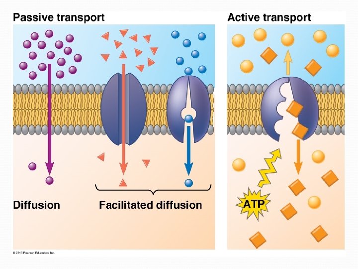
Active Transport Requires ENERGY (ATP) Proteins transport substances against concentration gradient (low high conc. ) Eg. Na+/K+ pump, proton pump

Electrogenic Pumps: generate voltage across membrane Na+/K+ Pump Na+ out, K+ into cell Nerve transmission Proton Pump Push protons (H+) across membrane Eg. mitochondria (ATP production)

Cotransport: membrane protein enables “downhill” diffusion of one solute to drive “uphill” transport of other Eg. sucrose-H+ cotransporter (sugar-loading in plants)

Passive vs. Active Transport Little or no Energy High low concentrations DOWN the concentration gradient eg. diffusion, osmosis, facilitated diffusion (w/transport protein) Requires Energy (ATP) Low high concentrations AGAINST the concentration gradient eg. pumps, exo/endocytosis

Osmoregulation Control solute & water balance Contractile vacuole: “bilge pump” forces out fresh water as it enters by osmosis Eg. paramecium caudatum – freshwater protist

Bulk Transport of proteins, polysaccharides, large molecules Endocytosis: take in macromolecules, form new vesicles Exocytosis: vesicles fuse with cell membrane, expel contents

Types of Endocytosis Phagocytosis: “cellular eating” - solids Pinocytosis: “cellular drinking” - fluids Receptor-Mediated Endocytosis: Ligands bind to specific receptors on cell surface
- Slides: 39