Chapter 5 Cartilage and bone One Cartilage Structure

Chapter 5 Cartilage and bone

One. Cartilage

Structure: Cells: chondrocytes Extensive extracellular matrix Functions ◘ Bear mechanical stresses without permanent disortion. ◘ Support soft tissues (nose, ear, trachea). ◘ Shock-absorbing and sliding areas for joints, facilitate bone movements. ◘ Essential for the development and growth of long bones.

Cartilage composition Cells Chondrocytes: located within matrix; lacunae Chondroblasts: located at periphery; secrete l extracellular matrix l Fibers Collagen and elastic fibers l Ground substance Rich in glycosaminoglycans and proteoglycans

More Features of Cartilage l l l Avascular; Nutrients diffusion from perichondrium or synovial fluid; No innervation; No lymphatic vessels; Chondrocytes have low metabolic activity

Classification: Hyaline cartilage: most common, much type II collagen Elastic cartilage: elastic fibers, type II collagen Fibrocatilage: dense network of type I collagen; high stress and weight bearing

Hyaline Cartilage l l Blue-white in color In embryo it serves as skeleton until replacement by bone, acts as template Epiphyseal plate in long bone growth Joint surfaces, nose, larynx, trachea, bronchi, ends of ribs adjacent to sternum

· Structure: Matrix Chondrocytes Perichondrium
 Hyaline cartilage matrix is produced by chondrocytes and contains three major")
1. Matrix (1) Hyaline cartilage matrix is produced by chondrocytes and contains three major classes of molecules: ①Collagen molecules ②Proteoglycans ③Nocollagenous proteins fibronectin, chondronectin
 Hyaline cartilage matrix is highly hydrated to permit diffusion of small metabolites and")
(2) Hyaline cartilage matrix is highly hydrated to permit diffusion of small metabolites and resilience (3) Grounds substance components of hyaline cartilage matrix are not distributed uniformly Capsusle/territorial matrix (TM) Interterritorial matrix (IM)

2. Perichondrium is a layer of dense irregular connective tissue. It is essential for the growth and maintenance of cartilage. Outer cellular layer Inner fibrous layer: give rise to new cartilage cells l Found around all HC except joint HC l Rich in collagen type I fibers and contains numerous chondroblasts.
 In hyaline cartilage, chondrocytes are distributed either singly or in cluster")
2. Chondrocyte (1) In hyaline cartilage, chondrocytes are distributed either singly or in cluster
 chondrocytes are specialized cells that produce and maintain the extracellular matrix")
(2) chondrocytes are specialized cells that produce and maintain the extracellular matrix

Histogenesis and Growth of Hyaline cartilage · Histogenesis Cartilage derives from mesenchyme. (图basice 7 -6)

· Growth Interstitial growth: ‘growth from within’ mitotic division of existing chondrocytes and production of matrix Appositional growth: ‘growth from the surface’ differentiation of new chondrocytes from perichondrial cells and production of matrix at surface

Elastic Cartilage • Auricle of ear, external auditory canal, eustachian tube, epiglottis, cuneiform cartilage of larynx • Similar to HC, many elastic fibers • Perichondrium

Elastic cartilage

Fibrocartilage • Found in Intervetebral disks, tendon and ligament attachment to bone, symphysis pubis • Combination of HC and dense regular connective tissue • Chondrocytes often in rows or groups • Matrix acidophilic due to high collagen • No perichondrium

Fibrocartilage

summary • Cartilage is a semi-rigid form of connective tissue • 3 types • Growth of cartilage occurs by interstitial growth from within and appositional growth at the periphery

Two. Bone

Overview of bone · Bone is a specialized form of connective tissue. Cells Extracellular matrix: mineralization · Functions of bone: Support: Provides attachment for tendons of skeletal muscles Protection: Protects internal organs Storage: calcium , phosphate

1. Matrix · Organic matter type I Collagen: 90% Ground substance: 10% · Inorganic matter The inorganic matter is calcium phosphate in the form of hydroxyapatite crystle [Ca 10(PO 4)6(OH)2]

2. Cells osteoprogenitor, osteoblast, osteocyte and osteoclast.

A. osteoprogenitor cell -- Resting cell that can transform into osteoblast. -- Found on the external and internal surfaces of bones. -- Only bone cells that undergoes cell division, daughter cells becomes osteoblasts

B. osteoblast -- secretes organic compounds for bone matrix. -- located at the surfaces of bone.

C. osteocyte -- mature cell and is enclosed by bone matrix -- osteocytes are found in matrix. Lacuna Canaliculi

D. osteoclast -- responsible for bone resorption. -- rest directly on bone where resorption is taking place.

3. Structure of long bones epiphysis diaphysis epiphysis

Compact bone --has no cavities. --forms the outside of the bone. Spongy bone --has numerous interconnecting cavities. --forms the interior of the bone.

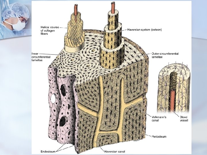
Compact Bone n Functional Unit is the Osteon or Haversian System—concentric rings of lamellae around a Central Canal, with connecting Peripheral Canals

Histogenesis l Intramembranous ossification: flat bone l Endochondral ossification: short and long bone

Intramembranous Ossification

Endochondral Ossification

Endochondral Ossification

Epiphyseal cartilage

Summary · Matrix: organic matter, inorganic matter and a little water · Cells: osteoprogenitor cell, osteoblast, osteocyte and osteoclast · Structure of long bones periosteum and endosteum, epiphysis, diaphysis ossification

Questions: 1. What’s the function of cartilage? 2. Review the structure of bone. 3. Describe the two processes of ossification.
- Slides: 39